

The DOI matches its Crossref record; 8 of the 8 references it cites were found in a registry or an index.
Peripheral Treatment with Enoxaparin, a Low Molecular Weight Heparin, Reduces Plaques and β-Amyloid Accumulation in a Mouse Model of Alzheimer's Disease
Abstract
We investigated the effect of long-term, peripheral treatment with enoxaparin, a low molecular weight heparin, in transgenic mice overexpressing human amyloid precursor protein 751 . Enoxaparin (6 IU per mouse intraperitoneally, three times a week for 6 months) significantly lowered the number and the area occupied by cortical β-amyloid deposits and the total β-amyloid (1-40) cortical concentration. Immunocytochemical analysis of glial fibrillary acid protein-positive cells showed that enoxaparin markedly reduced the number of activated astrocytes surrounding β-amyloid deposits. In vitro , the drug dose-dependently attenuated the toxic effect of β-amyloid on neuronal cells. Enoxaparin dose-dependently reduced the ability of β-amyloid to activate complement and contact systems, two powerful effectors of inflammatory response in AD brain. By reducing the β-amyloid load and cytotoxicity and proinflammatory activity, enoxaparin offers promise as a tool for slowing the progression of Alzheimer's disease.
Overview
The claims and statistics of this paper haven’t been checked yet.
Run the check — free, no account needed. It reads the open-access text and checks the sources it cites.
Integrity signals
Registry
Cites
Statistics
Figures
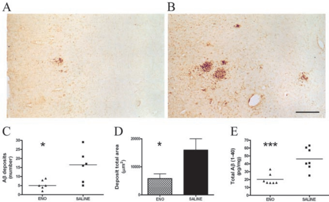
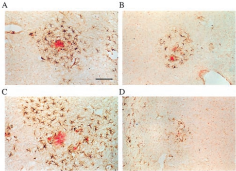
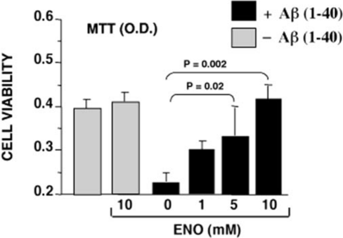
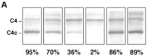
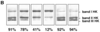
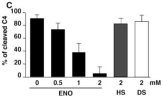
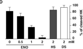
Verification
Claims the paper makes
The claims and statistics of this paper haven’t been checked yet.
Run the check — free, no account needed. It reads the open-access text and checks the sources it cites.
Claims other papers make about this one
Each is checked against this paper’s own text: does it say what the citing paper says it does?
The cited source says what the claim attributes to it.
In the citing paper
“Bergamaschini et al. demonstrated that the effect of long-term, peripheral treatment with a low-molecular-weight heparin significantly decreased Aβ concentration and deposition in the brain of APP transgenic mice (Bergamaschini et al., 2004).”
From Heparan Sulphate Proteoglycan and the Low-Density Lipoprotein Receptor-Related Protein 1 Constitute Major Pathways for Neuronal Amyloid-β Uptake (2011) · cites this paper as Bergamaschini et al., 2004
Self-check
Percentages, sums and reported differences re-computed from the paper’s own numbers.
The self-check needs the paper’s full text.
Citations
Cites
Immunization with amyloid-β attenuates Alzheimer-disease-like pathology in the PDAPP mouse
Schenk, D., Barbour, R., Dunn, W., Gordon, G., Grajeda, H., Guido, T., Hu, K., Huang, J., Johnson-Wood, K., Khan, K., et al. (1999) · Nature · doi:10.1038/22124 · Cited by 3,160
Bard, F., Cannon, C., Barbour, R., Burke, R.-L., Games, D., Grajeda, H., Guido, T., Hu, K., Huang, J., Johnson-Wood, K., et al. (2000) · Nature Medicine · doi:10.1038/78682 · Cited by 2,012
Two amyloid precursor protein transgenic mouse models with Alzheimer disease-like pathology
Sturchler-Pierrat, C., Abramowski, D., Duke, M., Wiederhold, K.-H., Mistl, C., Rothacher, S., Ledermann, B., Bürki, K., Frey, P., Paganetti, P. A., et al. (1997) · Proceedings of the National Academy of Sciences · doi:10.1073/pnas.94.24.13287 · Cited by 1,507
DeMattos, R. B., Bales, K. R., Cummins, D. J., Dodart, J.-C., Paul, S. M., & Holtzman, D. M. (2001) · Proceedings of the National Academy of Sciences · doi:10.1073/pnas.151261398 · Cited by 1,298
Immunization reverses memory deficits without reducing brain Aβ burden in Alzheimer's disease model
Dodart, J.-C., Bales, K. R., Gannon, K. S., Greene, S. J., DeMattos, R. B., Mathis, C., DeLong, C. A., Wu, S., Wu, X., Holtzman, D. M., et al. (2002) · Nature Neuroscience · doi:10.1038/nn842 · Cited by 858
Neuron loss in APP transgenic mice
Calhoun, M. E., Wiederhold, K.-H., Abramowski, D., Phinney, A. L., Probst, A., Sturchler-Pierrat, C., Staufenbiel, M., Sommer, B., & Jucker, M. (1998) · Nature · doi:10.1038/27351 · Cited by 502
Mackic, J. B., Stins, M., McComb, J. G., Calero, M., Ghiso, J., Kim, K. S., Yan, S. D., Stern, D., Schmidt, A. M., Frangione, B., et al. (1998) · Journal of Clinical Investigation · doi:10.1172/jci2029 · Cited by 242
Martel, C. L., Mackic, J. B., Matsubara, E., Governale, S., Miguel, C., Miao, W., McComb, J. G., Frangione, B., Ghiso, J., & Zlokovic, B. V. (1997) · Journal of Neurochemistry · doi:10.1046/j.1471-4159.1997.69051995.x · Cited by 149
Research works printed but not linked: 45
- 2002, J Neurosci, 22, 7873, 10.1523/JNEUROSCI.22-18-07873.2002 · DOI · Paper page
- 10.1073/pnas.2332818100 · DOI
- 10.1021/bi025817e · DOI
- 10.1016/S0047-6374(01)00311-6 · DOI
- 2001, Neurobiol Aging, 22, 63, 10.1016/S0197-4580(00)00174-3 · DOI
- 10.1016/S0197-4580(02)00003-9 · DOI · Paper page
- 1986, Blood, 68, 455, 10.1182/blood.V68.2.455.455 · DOI
- 2000, Ann NY Acad Sci, 908, 260, 10.1111/j.1749-6632.2000.tb06653.x · DOI
- 10.1002/jnr.10286 · DOI
- 10.1046/j.1460-9568.2000.00140.x · DOI
- 10.1002/ana.10253 · DOI
- 10.1016/S0197-4580(01)00255-X · DOI
- 10.4049/jimmunol.170.11.5764 · DOI
- 10.1074/jbc.273.45.29719 · DOI
- 10.1159/000022146 · DOI
- 10.1016/S0197-4580(02)00098-2 · DOI
- 1997, Ann NY Acad Sci, 826, 117, 10.1111/j.1749-6632.1997.tb48465.x · DOI
- 10.1038/nm0295-143 · DOI
- 10.1056/NEJMoa025313 · DOI
- 1994, NeuroReport, 5, 1389
- 10.1046/j.1471-4159.1998.70020736.x · DOI
- 2000, J Neurosci, 20, 5709, 10.1523/JNEUROSCI.20-15-05709.2000 · DOI
- 10.1016/S0049-3848(02)00050-6 · DOI
- 2003, J Neurosci, 23, 29, 10.1523/JNEUROSCI.23-01-00029.2003 · DOI
- 10.1016/S0197-4580(01)00289-5 · DOI
- 10.1016/S1471-4914(02)02422-X · DOI
- 10.1074/jbc.M203193200 · DOI
- 10.1016/S0165-5728(00)00291-5 · DOI
- 10.1016/S0304-3940(00)00764-3 · DOI
- 10.1016/S0002-9440(10)61715-4 · DOI
- 2000, J Neuropathol Exp Neurol, 59, 29, 10.1093/jnen/59.1.29 · DOI
- 10.1016/S0197-4580(98)00049-9 · DOI
- 1999, Adv Pharmacol, 46, 151, 10.1016/S1054-3589(08)60471-8 · DOI
- 10.1111/j.1749-6632.2003.tb03138.x · DOI
- 10.1016/S1474-4422(03)00484-8 · DOI
- 2003, Acta Neuropathol (Berl), 105, 135, 10.1007/s00401-002-0624-7 · DOI · Paper page
- 10.1038/nm0197-77 · DOI
- 10.1016/S0014-2999(02)01759-4 · DOI
- 10.1074/jbc.272.50.31617 · DOI
- 1997, Am J Pathol, 150, 1531
- 10.1016/S0197-4580(97)00042-0 · DOI
- 10.1073/pnas.162350199 · DOI · Paper page
- 10.1016/0006-8993(94)90484-7 · DOI
- 2001, Mol Med, 7, 517, 10.1007/BF03401857 · DOI
- 10.1006/bbrc.1993.2582 · DOI · Paper page
Cited by
Sorted by citations
Blennow, K., de Leon, M. J., & Zetterberg, H. (2006) · The Lancet · doi:10.1016/s0140-6736(06)69113-7 · Cited by 3,134
The role of the anaphylatoxins in health and disease
Klos, A., Tenner, A. J., Johswich, K.-O., Ager, R. R., Reis, E. S., & Köhl, J. (2009) · Molecular Immunology · doi:10.1016/j.molimm.2009.04.027 · Cited by 730
ApoE and Aβ in Alzheimer’s Disease: Accidental Encounters or Partners?
Kanekiyo, T., Xu, H., & Bu, G. (2014) · Neuron · doi:10.1016/j.neuron.2014.01.045 · Cited by 608
Fibrinogen in neurological diseases: mechanisms, imaging and therapeutics
Petersen, M. A., Ryu, J. K., & Akassoglou, K. (2018) · Nature reviews. Neuroscience · doi:10.1038/nrn.2018.13 · Cited by 527
Natural products as a source of Alzheimer's drug leads
Williams, P., Sorribas, A., & Howes, M.-J. R. (2011) · Nat. Prod. Rep. · doi:10.1039/c0np00027b · Cited by 404
Metadata
- Authors
- Bergamaschini, L., Rossi, E., Storini, C., Pizzimenti, S., Distaso, M., Perego, C., De Luigi, A., Vergani, C., & Grazia De Simoni, M.
- Venue
- The Journal of Neuroscience
- Publisher
- Society for Neuroscience
- Published
- 2004-04-28
- Volume
- 24
- Issue
- 17
- Pages
- 4181-4186
- ISSN
- 0270-6474
- 1529-2401
- Cited by
- 146
- References
- 8
- Language
- en
About these checks
Registry, retraction, Cites and statistics results are computed, not judged, with one exception: a printed reference that no index lists is read once by a model before it counts as a citation error. Claims are extracted by a model and kept only when their quote appears verbatim in the text; each cited source is then checked, with a model’s help, for whether it says what the claim relies on. Those verdicts can be wrong — read the claim’s quote and the cited source before relying on one.
Statistics and arithmetic are recomputed, not judged.
Confidence bands are shown only for fields calibrated on our annotated test set. None is calibrated yet, so no band is shown.
What counts as a claim ›How we measure confidence ›How we check this ›
Are you an author, or do you see a problem with this page? Request a review
