

The DOI matches its Crossref record; 91 of the 91 references it cites were found in a registry or an index.
Differentiation and Characterization of Excitatory and Inhibitory Synapses by Cryo-electron Tomography and Correlative Microscopy
Open accessAbstract
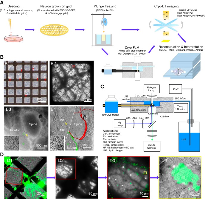
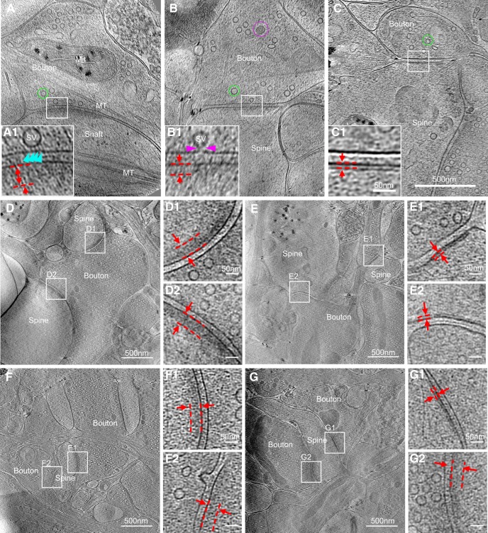
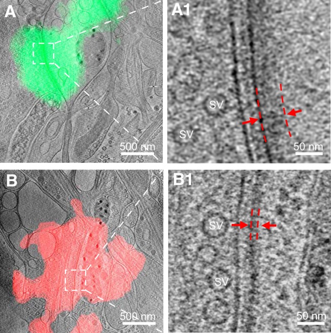
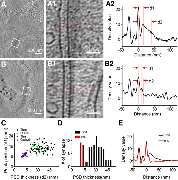
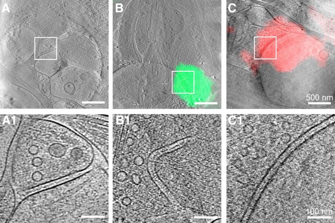
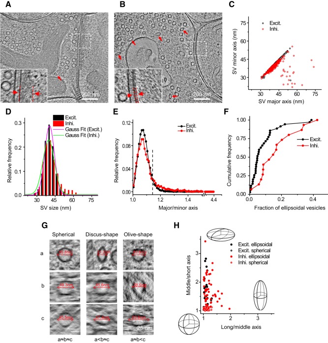
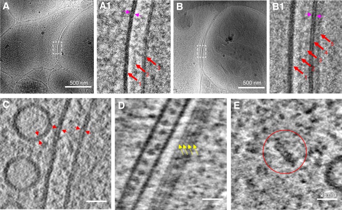
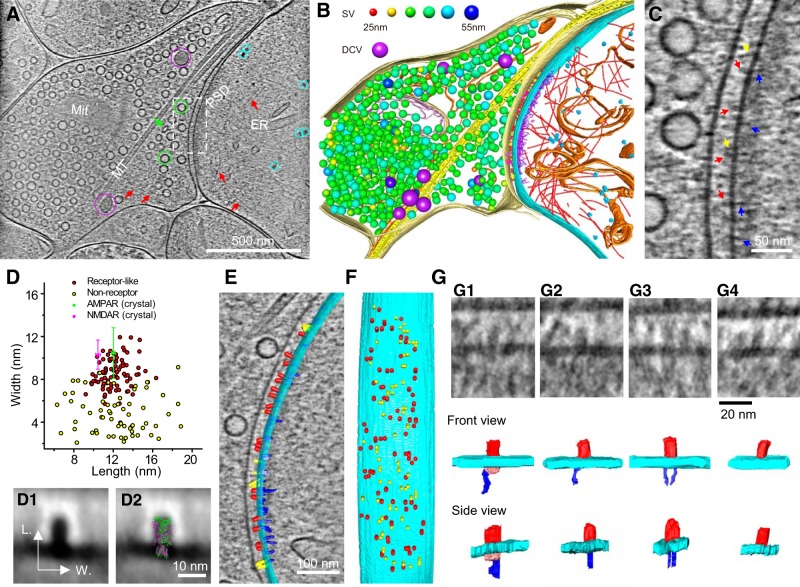
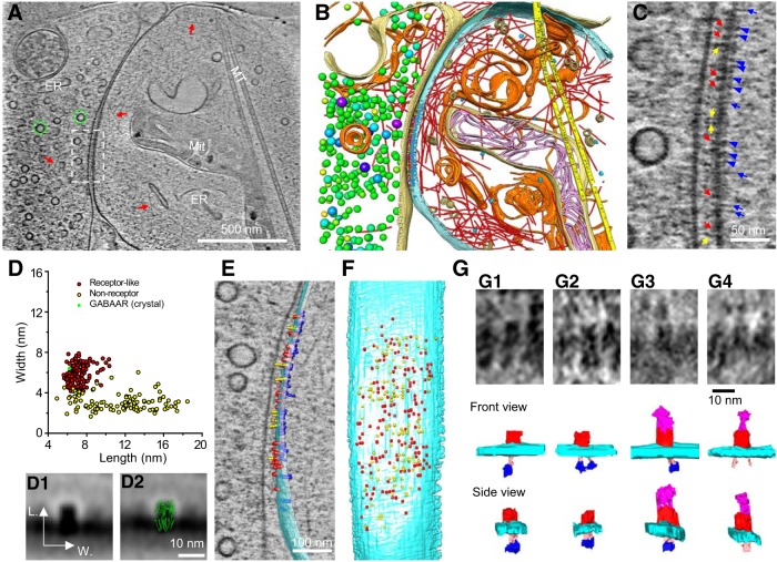
As key functional units in neural circuits, different types of neuronal synapses play distinct roles in brain information processing, learning, and memory. Synaptic abnormalities are believed to underlie various neurological and psychiatric disorders. Here, by combining cryo-electron tomography and cryo-correlative light and electron microscopy, we distinguished intact excitatory and inhibitory synapses of cultured hippocampal neurons, and visualized the in situ 3D organization of synaptic organelles and macromolecules in their native state. Quantitative analyses of >100 synaptic tomograms reveal that excitatory synapses contain a mesh-like postsynaptic density (PSD) with thickness ranging from 20 to 50 nm. In contrast, the PSD in inhibitory synapses assumes a thin sheet-like structure ∼12 nm from the postsynaptic membrane. On the presynaptic side, spherical synaptic vesicles (SVs) of 25–60 nm diameter and discus-shaped ellipsoidal SVs of various sizes coexist in both synaptic types, with more ellipsoidal ones in inhibitory synapses. High-resolution tomograms obtained using a Volta phase plate and electron filtering and counting reveal glutamate receptor-like and GABA A receptor-like structures that interact with putative scaffolding and adhesion molecules, reflecting details of receptor anchoring and PSD organization. These results provide an updated view of the ultrastructure of excitatory and inhibitory synapses, and demonstrate the potential of our approach to gain insight into the organizational principles of cellular architecture underlying distinct synaptic functions. SIGNIFICANCE STATEMENT To understand functional properties of neuronal synapses, it is desirable to analyze their structure at molecular resolution. We have developed an integrative approach combining cryo-electron tomography and correlative fluorescence microscopy to visualize 3D ultrastructural features of intact excitatory and inhibitory synapses in their native state. Our approach shows that inhibitory synapses contain uniform thin sheet-like postsynaptic densities (PSDs), while excitatory synapses contain previously known mesh-like PSDs. We discovered “discus-shaped” ellipsoidal synaptic vesicles, and their distributions along with regular spherical vesicles in synaptic types are characterized. High-resolution tomograms further allowed identification of putative neurotransmitter receptors and their heterogeneous interaction with synaptic scaffolding proteins. The specificity and resolution of our approach enables precise in situ analysis of ultrastructural organization underlying distinct synaptic functions.
Overview
The claims and statistics of this paper haven’t been checked yet.
Run the check — free, no account needed. It reads the open-access text and checks the sources it cites.
Integrity signals
Registry
Cites
Statistics
Figures
Verification
Claims the paper makes
The claims and statistics of this paper haven’t been checked yet.
Run the check — free, no account needed. It reads the open-access text and checks the sources it cites.
Claims other papers make about this one
Each is checked against this paper’s own text: does it say what the citing paper says it does?
The cited source says what the claim attributes to it.
In the citing paper
“In vertebrate EM datasets, human annotators can see differences in the vesicles for excitatory and inhibitory synapses.16,35,36,37,109,110”
From Neurotransmitter classification from electron microscopy images at synaptic sites in Drosophila melanogaster (2024) · cites this paper as [36]
Self-check
Percentages, sums and reported differences re-computed from the paper’s own numbers.
The self-check needs the paper’s full text.
Citations
Cites
UCSF Chimera—A visualization system for exploratory research and analysis
Pettersen, E. F., Goddard, T. D., Huang, C. C., Couch, G. S., Greenblatt, D. M., Meng, E. C., & Ferrin, T. E. (2004) · Journal of Computational Chemistry · doi:10.1002/jcc.20084 · Cited by 46,047
Shrestha, S., & Offer, S. M. (2016) · Medical Epigenetics · doi:10.1159/000444713 · Cited by 8,951
Computer Visualization of Three-Dimensional Image Data Using IMOD
Kremer, J. R., Mastronarde, D. N., & McIntosh, J. R. (1996) · Journal of Structural Biology · doi:10.1006/jsbi.1996.0013 · Cited by 5,530
Automated electron microscope tomography using robust prediction of specimen movements
Mastronarde, D. N. (2005) · Journal of Structural Biology · doi:10.1016/j.jsb.2005.07.007 · Cited by 5,136
Bi, G.-Q., & Poo, M.-M. (1998) · The Journal of Neuroscience · doi:10.1523/jneurosci.18-24-10464.1998 · Cited by 4,744
Self-citationDirect Least Square Fitting of Ellipses
Fitzgibbon, A., Pilu, M., & Fisher, R. B. (1999) · IEEE Transactions on Pattern Analysis and Machine Intelligence · doi:10.1109/34.765658 · Cited by 2,797
Ginsborg, B. L. (1964) · Quarterly Journal of Experimental Physiology and Cognate Medical Sciences · doi:10.1113/expphysiol.1964.sp001756 · Cited by 1,917
Li, X.-M., Mooney, P., Zheng, S., Booth, C., Braunfeld, M. B., Gubbens, S., Agard, D., & Cheng, Y.-F. (2013) · Nature Methods · doi:10.1038/nmeth.2472 · Cited by 1,866
Axo-somatic and axo-dendritic synapses of the cerebral cortex: an electron microscope study.
Eg, G. (1959) · PubMed · Cited by 1,768
Automated molecular microscopy: The new Leginon system
Suloway, C. J. M., Pulokas, J., Fellmann, D., Cheng, A., Guerra, F., Quispe, J. D., Stagg, S. M., Potter, C. S., & Carragher, B. O. (2005) · Journal of Structural Biology · doi:10.1016/j.jsb.2005.03.010 · Cited by 1,695
Cited by
Sorted by citations
Items 101–200 of 207
Inhibitory postsynaptic density from the lens of phase separation
Bai, G., & Zhang, M. (2022) · Oxford Open Neuroscience · doi:10.1093/oons/kvac003 · Cited by 8
On the cause of sleep: Protein fragments, the concept of sentinels, and links to epilepsy
Varshavsky, A. J. (2019) · Proceedings of the National Academy of Sciences · doi:10.1073/pnas.1904709116 · Cited by 8
Molecular composition of developing glutamatergic synapses
Sell, G. L., Barrow, S. L., & McAllister, A. K. (2020) · Elsevier eBooks · doi:10.1016/b978-0-12-823672-7.00001-6 · Cited by 8
Integrated Light and Electron Microscopy
Koning, R. I., Raja, A. S., Lane, R. I., Koster, A. J., & Hoogenboom, J. P. (2019) · Wiley eBooks · doi:10.1002/9781119086420.ch7 · Cited by 8
Structural properties and peptide ligand binding of the capsid homology domains of human ArcPreprint
Hallin, E. I., Bramham, C. R., & Kursula, P. (2020) · bioRxiv (Cold Spring Harbor Laboratory) · doi:10.1101/2020.12.17.423319 · Cited by 8
Cho, Y., Kim, S., Cho, H. J., Ham, H., Lee, K. E., Kang, H. W., Jeong, Y. W., Ahn, J., Choi, N., Kang, J. H., et al. (2024) · Small Structures · doi:10.1002/sstr.202400314 · Cited by 8
Cryo-electron tomographic investigation of native hippocampal glutamatergic synapsesPreprint
Matsui, A., Spangler, C. J., Elferich, J., Shiozaki, M., Jean, N., Zhao, X., Qin, M., Zhong, H., Yu, Z., & Gouaux, E. (2024) · eLife · doi:10.7554/elife.98458.3 · Cited by 40
Automatic classification and neurotransmitter prediction of synapses in electron microscopy
Zhang, A., Shailja, S., Borba, C., Miao, Y., Goebel, M., Ruschel, R., Ryan, K., Smith, W., & Manjunath, B. S. (2022) · Biological Imaging · doi:10.1017/s2633903x2200006x · Cited by 7
Fast, synchronous neurotransmitter release: Past, present and future
Gundersen, C. B. (2019) · Neuroscience · doi:10.1016/j.neuroscience.2019.04.030 · Cited by 7
Recent technical advances in cellular cryo-electron tomography
Zheng, T., & Cai, S. (2024) · The International Journal of Biochemistry & Cell Biology · doi:10.1016/j.biocel.2024.106648 · Cited by 6
Phase separation in the multi-compartment organization of synapses
Zhu, S., Shen, Z., Wu, X., & Zhang, M. (2025) · Current Opinion in Neurobiology · doi:10.1016/j.conb.2025.102975 · Cited by 7
Dynamic nanoscale architecture of synaptic vesicle fusion in mouse hippocampal neurons
Kroll, J., Kravčenko, U., Sadeghi, M., Diebolder, C. A., Ivanov, L., Lubas, M., Sprink, T., Schacherl, M., Kudryashev, M., & Rosenmund, C. (2025) · Nature Communications · doi:10.1038/s41467-025-67291-6 · Cited by 6
Human mood disorder risk gene Synaptotagmin-14 contributes to mania-like behaviors in mice
Zhang, Y., Zhang, C., Yuan, J., Jiang, H., Sun, P., Hui, L., Xu, L., Yu, L., Guo, Z., Wang, L., et al. (2025) · Molecular Psychiatry · doi:10.1038/s41380-025-02933-1 · Cited by 6
Yan, X., Zhao, F., Zhang, S., Lei, F., Wang, W., & Zheng, Y. (2019) · Environmental Toxicology and Pharmacology · doi:10.1016/j.etap.2019.03.002 · Cited by 5
A mammalian system for high-resolution imaging of intact cells by cryo-electron tomography
Li, X., Park, D., Chang, Y., Radhakrishnan, A., Wu, H., Wang, P., & Liu, J. (2020) · Progress in Biophysics and Molecular Biology · doi:10.1016/j.pbiomolbio.2020.09.005 · Cited by 5
Cryo-Electron Tomography and Automatic Segmentation of Cultured Hippocampal Neurons
Hylton, R. K., Seader, V. H., & Swulius, M. T. (2020) · Methods in molecular biology · doi:10.1007/978-1-0716-0966-8_2 · Cited by 5
Mesophasic Assembly of Inhibitory Postsynaptic Density
Bai, G., & Zhang, M. (2020) · Neuroscience Bulletin · doi:10.1007/s12264-020-00623-2 · Cited by 5
Digitalizing neuronal synapses with cryo-electron tomography and correlative microscopy
Liu, Y.-T., & Tao, C.-L. (2022) · Current Opinion in Neurobiology · doi:10.1016/j.conb.2022.102595 · Cited by 7
Self-citationZhao, W., & Jensen, G. J. (2022) · bioRxiv (Cold Spring Harbor Laboratory) · doi:10.1101/2022.02.17.480955 · Cited by 5
Li, Z., Zhao, P., Fu, J., Qiao, Y., Hou, W., Liu, C., & Min, W. (2025) · Food Bioscience · doi:10.1016/j.fbio.2025.107214 · Cited by 4
Assessing the Potential of NGF-Differentiated PC12 Cells as a Model for Synaptic Transmission
Ott, G., Leuenberger, J., Ntamati, N. R., Ivanov, A., Nevian, T., Rostami, I., & Zuber, B. (2025) · Molecular Neurobiology · doi:10.1007/s12035-025-05562-5 · Cited by 5
A New In Situ Neuronal Model for Cryo-ET
Kim, J., Yang, S. Z., Wildonger, J., & Wright, E. (2020) · Microscopy and Microanalysis · doi:10.1017/s1431927620013513 · Cited by 4
Subcellular Structures in Native Hippocampal Synapses Revealed by Cryo-electron Tomography
Tian, C.-L., Qi, L., Lu, Z.-H., Liu, S., Gu, M., Huang, W.-L., Yan, Y.-T., Liu, Y., Wu, J., Wang, P., et al. (2026) · Neuroscience Bulletin · doi:10.1007/s12264-025-01569-z · Cited by 4
Self-citationKumar, I., Paudyal, A., Kádková, A., Stewart, M. E., Sørensen, J. B., & Radecke, J. (2023) · International Journal of Molecular Sciences · doi:10.3390/ijms242015191 · Cited by 3
Neuronal synaptic architecture revealed by cryo-correlative light and electron microscopy
Wang, P., Tian, B., Xu, X., Luan, H., Zhang, Y., Sun, W., Hu, L., Li, Y., Yao, Y., Li, W., et al. (2024) · Biophysics Reports · doi:10.52601/bpr.2024.240035 · Cited by 3
Benson, D. L. (2019) · Journal of Neuroscience · doi:10.1523/jneurosci.0743-19.2019 · Cited by 3
Al-Yaari, M., Onogi, C., Yamada, R., Adachi, R., Kondo, D., & Kuba, H. (2021) · Journal of Neuroscience · doi:10.1523/jneurosci.0884-21.2021 · Cited by 3
Gogoi, P., Shiozaki, M., & Gouaux, E. (2022) · PLoS ONE · doi:10.1371/journal.pone.0271799 · Cited by 2
Brain organoids for addressing COVID-19 challenge
Yu, J. S., Wang, K., & Zheng, D. (2022) · Frontiers in Neuroscience · doi:10.3389/fnins.2022.1055601 · Cited by 3
Maiellano, G., Scandella, L., & Francolini, M. (2023) · Frontiers in Cellular Neuroscience · doi:10.3389/fncel.2023.1153593 · Cited by 3
The supramolecular landscape of growing human axonsPreprint
Hoffmann, P. C., Giandomenico, S. L., Ganeva, I., Wozny, M. R., Sutcliffe, M. A., Kukulski, W., & Lancaster, M. A. (2020) · bioRxiv (Cold Spring Harbor Laboratory) · doi:10.1101/2020.07.23.216622 · Cited by 3
The molecular infrastructure of glutamatergic synapses in the mammalian forebrainPreprint
Peukes, J., Lovatt, C., Leistner, C., Boulanger, J., Morado, D. R., Fuller, M. J., Kukulski, W., Zhu, F., Komiyama, N. H., Briggs, J. A. G., et al. (2024) · eLife · doi:10.7554/elife.100335 · Cited by 5
The molecular infrastructure of glutamatergic synapses in the mammalian forebrain
Peukes, J., Lovatt, C., Leistner, C., Boulanger, J., Morado, D. R., Fuller, M. J., Kukulski, W., Zhu, F., Komiyama, N. H., Briggs, J. A. G., et al. (2025) · eLife · doi:10.7554/elife.100335.3 · Cited by 2
Calcium dynamics in small spaces: Lessons learned from modeling in dendritic spines
McCabe, K. J., Mesa, M. H., & Rangamani, P. (2025) · Biophysical Journal · doi:10.1016/j.bpj.2025.09.038 · Cited by 2
Jung, M., Ko, G., Lim, D., Kim, S., Kim, S., Kim, Y., Choi, M., & Roh, S. (2025) · Nature Communications · doi:10.1038/s41467-025-64431-w · Cited by 4
Memory engram synapse 3D macromolecular architecture visualized by cryoCLEM-guided cryoET
Lovatt, C., O’Sullivan, T., Luis, C. O. S., Ryan, T. J., & Frank, R. A. W. (2025) · Structure · doi:10.1016/j.str.2025.10.014 · Cited by 3
Martínez-Sánchez, A., & Lučić, V. (2012) · Focus on structural biology · doi:10.1007/978-3-031-51171-4_11 · Cited by 2
Targeting TRP channels: The emerging role of cryogenic electron microscopy in drug discovery
Lopez, K. E., & Horn, W. D. V. (2024) · Elsevier eBooks · doi:10.1016/b978-0-443-18653-0.00010-1 · Cited by 2
Wei, Z., Zhang, S., Bai, K., Liu, Y., Luan, Y., Hu, Z., Li, Y., Qu, Z., Hu, X., Ding, X., et al. (2025) · Development · doi:10.1242/dev.204532 · Cited by 2
Self-citationVolumetric Analysis of the Blood–Brain Barrier After Ischemic Stroke by Electron Tomography in Mice
Kotchetkov, P., & Lacoste, B. (2025) · Microcirculation · doi:10.1111/micc.70025 · Cited by 2
Tran, H. T., Lucas, M. S., Ishikawa, T., Shahmoradian, S. H., & Padeste, C. (2021) · Frontiers in Neuroscience · doi:10.3389/fnins.2021.726763 · Cited by 2
A new method for isolation and purification of fusion-competent inhibitory synaptic vesicles
Gopal, N., Leitz, J., Wang, C., Esquivies, L. R., Pfuetzner, R. A., & Brünger, A. T. (2024) · Current Research in Physiology · doi:10.1016/j.crphys.2024.100121 · Cited by 2
Inhibitory synaptic vesicles have unique dynamics and exocytosis propertiesPreprint
Park, C., Chen, X., Tian, C.-L., Park, G. N., Chenouard, N., Lee, H., Yeo, X. Y., Jung, S., Bi, G., Tsien, R. W., et al. (2020) · bioRxiv (Cold Spring Harbor Laboratory) · doi:10.1101/2020.09.21.289314 · Cited by 2
Self-citationLycas, M. D., Morado, D. R., Gether, U., Briggs, J. A. G., & Erlendsson, S. (2024) · bioRxiv (Cold Spring Harbor Laboratory) · doi:10.1101/2024.04.15.589543 · Cited by 2
Cryo-electron tomographic investigation of native hippocampal glutamatergic synapsesPreprint
Matsui, A., Spangler, C. J., Elferich, J., Shiozaki, M., Jean, N., Zhao, X., Qin, M., Zhong, H., Yu, Z., & Gouaux, E. (2024) · eLife · doi:10.7554/elife.98458.1 · Cited by 2
The molecular infrastructure of glutamatergic synapses in the mammalian forebrainPreprint
Peukes, J., Lovatt, C., Leistner, C., Boulanger, J., Morado, D. R., Fuller, M. J., Kukulski, W., Zhu, F., Komiyama, N. H., Briggs, J. A. G., et al. (2024) · eLife · doi:10.7554/elife.100335.1 · Cited by 2
Chen, X., Pan, D., Liu, J., & Yang, Y. (2024) · eLife · doi:10.7554/elife.102792 · Cited by 2
Orozco-Borunda, D. H., Papantoniou, C., Brose, N., & Lučić, V. (2024) · bioRxiv (Cold Spring Harbor Laboratory) · doi:10.1101/2024.12.18.629213 · Cited by 2
CoVET: Cryo-Electron Tomography guided by optical electrophysiologyPreprint
Jung, M., Ko, G., Lim, D., Kim, S., Kim, S., Kim, Y., Choi, M., & Roh, S. (2025) · bioRxiv (Cold Spring Harbor Laboratory) · doi:10.1101/2025.05.14.653951 · Cited by 2
Visualizing Postsynaptic Density in Excitatory Synapses with Electron Tomography
Sun, R., & Zhou, Q. (2026) · Advances in neurobiology · doi:10.1007/978-3-032-12594-1_10 · Cited by 2
Self-citationA correlative workflow for synaptic imaging by cryo-electron tomography
Do, T. T., Siegert, A., Domart, F., Hahn, F., Zeising, C., Muth, S., Pape, C., Kusch, K., Dresbach, T., Rizzoli, S. O., et al. (2026) · Structure · doi:10.1016/j.str.2026.02.017 · Cited by 2
Liu, X., Wang, Y., Yang, P., Yang, C., Chen, S. F., Wen, C., Wong, L. E., & Chang, S. (2026) · Nature Communications · doi:10.1038/s41467-026-72234-w · Cited by 3
Hastings, M. H., Gilbert, J. P., Huo, Y., & Man, H. (2023) · Springer eBooks · doi:10.1007/978-3-031-20792-1_5 · Cited by 1
Nartsissov, Y. R., & Zagubnaya, O. A. (2023) · AIP conference proceedings · doi:10.1063/5.0162989 · Cited by 1
Epon Post Embedding Correlative Light and Electron Microscopy
Wang, S., Xiong, H., Chang, Q., Zhuang, X., Wu, Y., Wang, X., Wu, C., & Fu, Z. (2024) · Journal of Visualized Experiments · doi:10.3791/66141 · Cited by 1
Cryo-ET Workflow for Understanding Ion Channels Localization on the Nodes of Ranvier
Wang, J. (2020) · Journal of Biosciences and Medicines · doi:10.4236/jbm.2020.82005 · Cited by 1
Digging Deeper: Advancements in Visualization of Inhibitory Synapses in Neurodegenerative Disorders
Radulović, S., Sunkara, S., Maurer, C., & Leitinger, G. (2021) · International Journal of Molecular Sciences · doi:10.3390/ijms222212470 · Cited by 1
Synaptosomal Preparation and Its Application in Preclinical Studies
Feligioni, M., Buccarello, L., Hassanzadeh, K., & Corbo, M. (2022) · Neuromethods · doi:10.1007/978-1-0716-2569-9_2 · Cited by 1
Brown, N. K., Roche, J. K., Farmer, C. B., & Roberts, R. C. (2023) · Journal of Neural Transmission · doi:10.1007/s00702-023-02593-x · Cited by 1
Kawano, H., Kakazu, Y., Iwabuchi, S., & Harata, N. C. (2020) · bioRxiv (Cold Spring Harbor Laboratory) · doi:10.1101/2020.06.30.180810 · Cited by 1
Automatic Detection and Neurotransmitter Prediction of Synapses in Electron MicroscopyPreprint
Zhang, A., Shailja, S., Borba, C., Miao, Y., Goebel, M. G., Ruschel, R., Ryan, K., Smith, W. C., & Manjunath, B. S. (2021) · bioRxiv (Cold Spring Harbor Laboratory) · doi:10.1101/2021.11.02.467022 · Cited by 1
Editorial: Quantifying and controlling the nano-architecture of neuronal synapses
Chen, X., Kuner, T., & Blanpied, T. A. (2022) · Frontiers in Synaptic Neuroscience · doi:10.3389/fnsyn.2022.1024073 · Cited by 1
Sohn, J. (2024) · eLife · doi:10.7554/elife.98458.1.sa2 · Cited by 1
Cryo-electron tomographic investigation of native hippocampal glutamatergic synapsesPreprint
Matsui, A., Spangler, C. J., Elferich, J., Shiozaki, M., Jean, N., Zhao, X., Qin, M., Zhong, H., Yu, Z., & Gouaux, E. (2024) · eLife · doi:10.7554/elife.98458.2 · Cited by 2
The molecular infrastructure of glutamatergic synapses in the mammalian forebrainPreprint
Peukes, J., Lovatt, C., Leistner, C., Boulanger, J., Morado, D. R., Fuller, M. J., Kukulski, W., Zhu, F., Komiyama, N. H., Briggs, J. A. G., et al. (2025) · eLife · doi:10.7554/elife.100335.2 · Cited by 1
Zhou, J. (2018) · Electronics Letters · doi:10.1049/el.2018.5136
Castejón, O. J. (2019) · American Journal of Biomedical Science & Research · doi:10.34297/ajbsr.2019.01.000514
Viard, J., Loe-Mie, Y., Daudin, R., Khelfaoui, M., Plançon, C., Boland, A., Tejedor, F. J., Huganir, R. L., Kim, E., Kinoshita, M., et al. (2019) · bioRxiv (Cold Spring Harbor Laboratory) · doi:10.1101/2019.12.11.872606
Yu, Y., Yadav-Samudrala, B. J., Meeker, R. B., Fitting, S., & Kourkoutis, L. F. (2020) · Microscopy and Microanalysis · doi:10.1017/s143192762002259x
Frontiers of electron microscopy for biomedical research
Zhao, J., Zhang, X., Hua, H. A. N., Tao, C.-L., Bi, G., & Liang, W. (2020) · Scientia Sinica Vitae · doi:10.1360/ssv-2019-0094
Self-citationUsing Maskless Photopatterning for Cryo-ET of Primary Drosophila Melanogaster Neurons
Kim, J. Y., Sibert, B. S., Yang, J., Yang, S. Z., Mitchell, J., Wildonger, J., & Wright, E. (2021) · Microscopy and Microanalysis · doi:10.1017/s1431927621009855
Liang, J. (2022) · Utrecht University Repository (Utrecht University) · doi:10.33540/1112
A role for synapsin 2a oligomerization in maintaining a reserve pool of synaptic vesicles
Zhang, M. (2022) · DR-NTU (Nanyang Technological University) · doi:10.32657/10356/159292
Scheefhals, N. (2022) · Utrecht University Repository (Utrecht University) · doi:10.33540/1224
Revealing ultrastructural changes that determine the development of a CNS synapse
Hintze, A. (2023) · eDiss (Georg-August-Universität Göttingen) · doi:10.53846/goediss-9828
Morphological Comparison of Primary Neurons Cryo-Preserved Under Varied Conditions
Kim, J. Y., Yang, J., Mitchell, J. W., English, L. A., Wildonger, J., Dent, E. W., & Wright, E. (2023) · Microscopy and Microanalysis · doi:10.1093/micmic/ozad067.477
Correlative Cryo-FIB and Cryo-ET of Dendritic Spines and Synaptic Connections
Anderson, E. D., Cronkite, C. A., Tolias, K. F., & Ludtke, S. J. (2023) · Microscopy and Microanalysis · doi:10.1093/micmic/ozad067.559
Matsui, A., Spangler, C. J., Elferich, J., et al. (2024) · eLife · doi:10.7554/elife.98458.1.sa0
Matsui, A., Spangler, C. J., Elferich, J., et al. (2024) · eLife · doi:10.7554/elife.98458.1.sa1
Jin, M., Yasunaga, T., Cui, X., Miyazawa, A., Yanagisawa, H., Mayanagi, K., Fukunaga, Y., Nishino, Y., & Kamiya, R. (2024) · SSRN Electronic Journal · doi:10.2139/ssrn.4901799
Ultrastructure of immature synaptic inputs in the lateral superior olive of rodent brainstemPreprint
Ma, S., Watanabe, S., & Gillespie, D. C. (2024) · bioRxiv (Cold Spring Harbor Laboratory) · doi:10.1101/2024.08.09.606139
A Technique to Perform Structure–Function Analysis of Synapses
Wichmann, C., & Michanski, S. (2024) · Neuromethods · doi:10.1007/978-1-0716-4019-7_5
Chen, X., Pan, D., Liu, J., & Yang, Y. (2025) · eLife · doi:10.7554/elife.102792.4
Ultrastructural analysis of the brain endothelium by electron tomographyPreprint
Kotchetkov, P., & Lacoste, B. (2024) · bioRxiv (Cold Spring Harbor Laboratory) · doi:10.1101/2024.09.09.612128
Chen, X., Pan, D., Liu, J., & Yang, Y. (2024) · eLife · doi:10.7554/elife.102792.1
A correlative workflow streamlines synaptic imaging by cryo-electron tomographyPreprint
Do, T. T., Siegert, A., Domart, F., Hahn, F., Zeising, C., Muth, S., Pape, C., Kusch, K., Dresbach, T., Rizzoli, S. O., et al. (2025) · bioRxiv (Cold Spring Harbor Laboratory) · doi:10.1101/2025.03.01.640558
Chen, X., Pan, D., Liu, J., & Yang, Y. (2025) · eLife · doi:10.7554/elife.102792.2
Chen, X., Pan, D., Liu, J., & Yang, Y. (2025) · eLife · doi:10.7554/elife.102792.3
Camporesi, E., Lashley, T., Gobom, J., Lantero‐Rodriguez, J., Hansson, O., Zetterberg, H., Blennow, K., & Becker, B. (2021) · Figshare · doi:10.6084/m9.figshare.13684508
Camporesi, E., Lashley, T., Gobom, J., Lantero‐Rodriguez, J., Hansson, O., Zetterberg, H., Blennow, K., & Becker, B. (2021) · Figshare · doi:10.6084/m9.figshare.13684505
Synaptic Cell Adhesion: A Structural Perspective
Bandekar, S. J., Kordon, S. P., & Araç, D. (2026) · Advances in neurobiology · doi:10.1007/978-3-032-12594-1_6 · Cited by 1
In-tissue structural biology of the brain: A focus on Alzheimer’s disease pathology
Jenkins, J., Gilbert, M. A. G., Fatima, N., O’Sullivan, T. J., Thompson, W. G., & Frank, R. A. W. (2026) · Methods in enzymology on CD-ROM/Methods in enzymology · doi:10.1016/bs.mie.2026.02.001
Ultrastructural and molecular analysis of cortico-striatal dopamine hub synapses
Lapios, P. (2024) · theses.fr (ABES) · doi:10.70675/527646a6zd9b5z4935z80ebz0950bba9d08e
ER-associated control of axonal vesicle trafficking during neuronal developmentPreprint
Subra, M., Meehl, J. B., Abrisch, R. G., & Voeltz, G. K. (2026) · bioRxiv (Cold Spring Harbor Laboratory) · doi:10.64898/2026.04.23.720437
Lycas, M. D., Morado, D. R., Gether, U., Briggs, J. A. G., & Erlendsson, S. (2026) · The Journal of Cell Biology · doi:10.1083/jcb.202503059 · Cited by 3
Atomic insights into the physiological and functional diversity of NMDA receptors
(134879), K. X., Wu, S., & Zhu, S. (2026) · Trends in Neurosciences · doi:10.1016/j.tins.2026.05.004 · Cited by 1
Lv, Z.-Y., Lu, Z.-H., Liu, S., Zhou, P., Lau, P.-M., Bi, G., & Tao, C.-L. (2026) · Neuroscience Bulletin · doi:10.1007/s12264-026-01646-x
Self-citation
Metadata
- Authors
- Tao, C.-L., Liu, Y.-T., Sun, R., Zhang, B., Qi, L., Shivakoti, S., Tian, C.-L., Zhang, P., Lau, P.-M., Zhou, Z. H., & Bi, G.-Q.
- Affiliations
- Chinese Academy of Sciences (CN)
- Centre for Human Genetics (GB)
- California NanoSystems Institute (US)
- University of Science and Technology of China (CN)
- Corresponding author
- Zhou, Z. Hong
- Bi, Guo-Qiang
- ORCID
- Venue
- The Journal of Neuroscience
- Publisher
- Society for Neuroscience
- Published
- 2018-01-08
- Volume
- 38
- Issue
- 6
- Pages
- 1493-1510
- ISSN
- 0270-6474 (ISSN-L)
- 1529-2401
- PMC
- PMC5815350
- OpenAlex
- W2783481291
- Cited by
- 207
- References
- 91
- Citations by year
- 2026: 18 · 2025: 24 · 2024: 30 · 2023: 14 · 2022: 27 · 2021: 40 · 2020: 30 · 2019: 17 · 2018: 5
- FWCI
- 10.48
- Type
- journal article
- Language
- en
- Open access
- hybrid
- doi.org
- Locations
- Journal of Neuroscience (publishedVersion, cc-by)
- PubMed Central (publishedVersion, cc-by)
- PubMed (publishedVersion)
- eScholarship (California Digital Library) (submittedVersion)
- eScholarship (California Digital Library) (submittedVersion, cc-by)
- Funders
- National Science Foundation
- Wellcome Trust (206422/Z/17/Z)
- National Natural Science Foundation of China (2009CB941300, 31070935, PSD-95)
- National Institutes of Health (1s10od018111-01)
- Biotechnology and Biological Sciences Research Council (BB/S003339/1)
- National Institute of General Medical Sciences (R01GM071940)
- National Center for Research Resources (S10RR023057)
- Topics
- Life Sciences › Neuroscience › Cellular and Molecular Neuroscience › Neuroscience and Neuropharmacology Research
- Life Sciences › Biochemistry, Genetics and Molecular Biology › Structural Biology › Advanced Electron Microscopy Techniques and Applications
- Life Sciences › Biochemistry, Genetics and Molecular Biology › Cell Biology › Cellular transport and secretion
- Keywords
- excitatory synapses, inhibitory synapses, synaptic ultrastructure, postsynaptic density, synaptic vesicles, glutamate receptors, GABAA receptor, synaptic scaffolding proteins, adhesion molecules, hippocampal neurons, cryo-electron tomography, correlative light and electron microscopy
- MeSH terms
- Animals, Female, Image Processing, Computer-Assisted, Inhibition, Psychological, Neurons, Pregnancy, Receptors, GABA-A, Synapses, Synaptic Vesicles, Tomography, Cell Adhesion Molecules, Receptors, Glutamate, Excitatory Postsynaptic Potentials, Cryoelectron Microscopy, Rats, Post-Synaptic Density
About these checks
Registry, retraction, Cites and statistics results are computed, not judged, with one exception: a printed reference that no index lists is read once by a model before it counts as a citation error. Claims are extracted by a model and kept only when their quote appears verbatim in the text; each cited source is then checked, with a model’s help, for whether it says what the claim relies on. Those verdicts can be wrong — read the claim’s quote and the cited source before relying on one.
Statistics and arithmetic are recomputed, not judged.
Confidence bands are shown only for fields calibrated on our annotated test set. None is calibrated yet, so no band is shown.
What counts as a claim ›How we measure confidence ›How we check this ›
Are you an author, or do you see a problem with this page? Request a review
