

The DOI matches its Crossref record; 91 of the 91 references it cites were found in a registry or an index.
Differentiation and Characterization of Excitatory and Inhibitory Synapses by Cryo-electron Tomography and Correlative Microscopy
Open accessAbstract
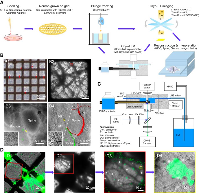
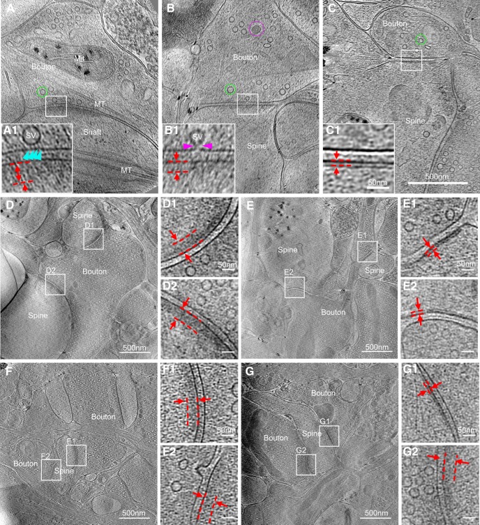
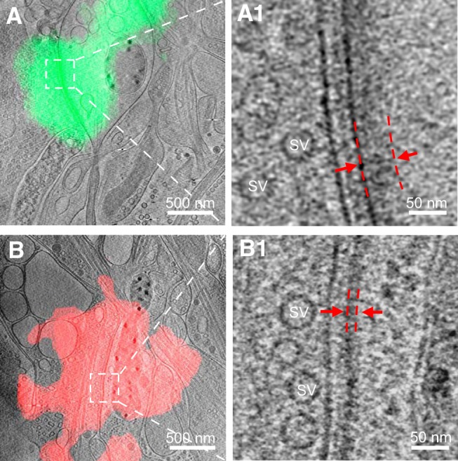
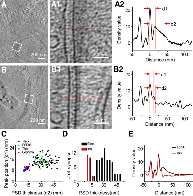
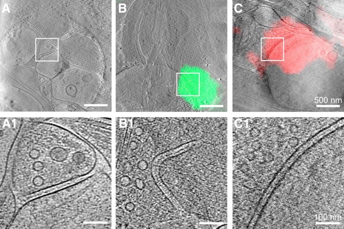
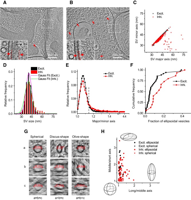
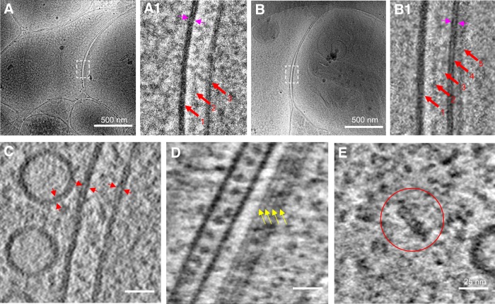
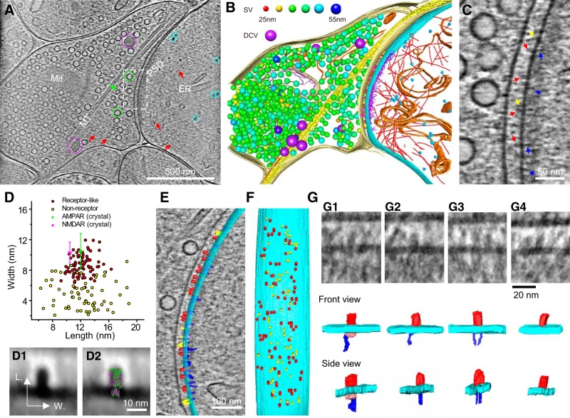
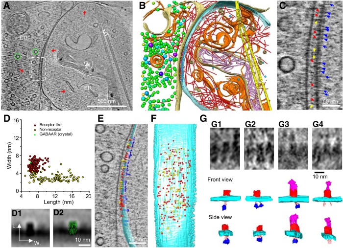
As key functional units in neural circuits, different types of neuronal synapses play distinct roles in brain information processing, learning, and memory. Synaptic abnormalities are believed to underlie various neurological and psychiatric disorders. Here, by combining cryo-electron tomography and cryo-correlative light and electron microscopy, we distinguished intact excitatory and inhibitory synapses of cultured hippocampal neurons, and visualized the in situ 3D organization of synaptic organelles and macromolecules in their native state. Quantitative analyses of >100 synaptic tomograms reveal that excitatory synapses contain a mesh-like postsynaptic density (PSD) with thickness ranging from 20 to 50 nm. In contrast, the PSD in inhibitory synapses assumes a thin sheet-like structure ∼12 nm from the postsynaptic membrane. On the presynaptic side, spherical synaptic vesicles (SVs) of 25–60 nm diameter and discus-shaped ellipsoidal SVs of various sizes coexist in both synaptic types, with more ellipsoidal ones in inhibitory synapses. High-resolution tomograms obtained using a Volta phase plate and electron filtering and counting reveal glutamate receptor-like and GABA A receptor-like structures that interact with putative scaffolding and adhesion molecules, reflecting details of receptor anchoring and PSD organization. These results provide an updated view of the ultrastructure of excitatory and inhibitory synapses, and demonstrate the potential of our approach to gain insight into the organizational principles of cellular architecture underlying distinct synaptic functions. SIGNIFICANCE STATEMENT To understand functional properties of neuronal synapses, it is desirable to analyze their structure at molecular resolution. We have developed an integrative approach combining cryo-electron tomography and correlative fluorescence microscopy to visualize 3D ultrastructural features of intact excitatory and inhibitory synapses in their native state. Our approach shows that inhibitory synapses contain uniform thin sheet-like postsynaptic densities (PSDs), while excitatory synapses contain previously known mesh-like PSDs. We discovered “discus-shaped” ellipsoidal synaptic vesicles, and their distributions along with regular spherical vesicles in synaptic types are characterized. High-resolution tomograms further allowed identification of putative neurotransmitter receptors and their heterogeneous interaction with synaptic scaffolding proteins. The specificity and resolution of our approach enables precise in situ analysis of ultrastructural organization underlying distinct synaptic functions.
Overview
The claims and statistics of this paper haven’t been checked yet.
Run the check — free, no account needed. It reads the open-access text and checks the sources it cites.
Integrity signals
Registry
Cites
Statistics
Figures
Verification
Claims the paper makes
The claims and statistics of this paper haven’t been checked yet.
Run the check — free, no account needed. It reads the open-access text and checks the sources it cites.
Claims other papers make about this one
Each is checked against this paper’s own text: does it say what the citing paper says it does?
The cited source says what the claim attributes to it.
In the citing paper
“In vertebrate EM datasets, human annotators can see differences in the vesicles for excitatory and inhibitory synapses.16,35,36,37,109,110”
From Neurotransmitter classification from electron microscopy images at synaptic sites in Drosophila melanogaster (2024) · cites this paper as [36]
Self-check
Percentages, sums and reported differences re-computed from the paper’s own numbers.
The self-check needs the paper’s full text.
Citations
Cites
UCSF Chimera—A visualization system for exploratory research and analysis
Pettersen, E. F., Goddard, T. D., Huang, C. C., Couch, G. S., Greenblatt, D. M., Meng, E. C., & Ferrin, T. E. (2004) · Journal of Computational Chemistry · doi:10.1002/jcc.20084 · Cited by 46,061
Shrestha, S., & Offer, S. M. (2016) · Medical Epigenetics · doi:10.1159/000444713 · Cited by 8,951
Computer Visualization of Three-Dimensional Image Data Using IMOD
Kremer, J. R., Mastronarde, D. N., & McIntosh, J. R. (1996) · Journal of Structural Biology · doi:10.1006/jsbi.1996.0013 · Cited by 5,530
Automated electron microscope tomography using robust prediction of specimen movements
Mastronarde, D. N. (2005) · Journal of Structural Biology · doi:10.1016/j.jsb.2005.07.007 · Cited by 5,136
Bi, G.-Q., & Poo, M.-M. (1998) · The Journal of Neuroscience · doi:10.1523/jneurosci.18-24-10464.1998 · Cited by 4,744
Self-citationDirect Least Square Fitting of Ellipses
Fitzgibbon, A., Pilu, M., & Fisher, R. B. (1999) · IEEE Transactions on Pattern Analysis and Machine Intelligence · doi:10.1109/34.765658 · Cited by 2,797
Ginsborg, B. L. (1964) · Quarterly Journal of Experimental Physiology and Cognate Medical Sciences · doi:10.1113/expphysiol.1964.sp001756 · Cited by 1,917
Li, X.-M., Mooney, P., Zheng, S., Booth, C., Braunfeld, M. B., Gubbens, S., Agard, D., & Cheng, Y.-F. (2013) · Nature Methods · doi:10.1038/nmeth.2472 · Cited by 1,866
Axo-somatic and axo-dendritic synapses of the cerebral cortex: an electron microscope study.
Eg, G. (1959) · PubMed · Cited by 1,768
Automated molecular microscopy: The new Leginon system
Suloway, C. J. M., Pulokas, J., Fellmann, D., Cheng, A., Guerra, F., Quispe, J. D., Stagg, S. M., Potter, C. S., & Carragher, B. O. (2005) · Journal of Structural Biology · doi:10.1016/j.jsb.2005.03.010 · Cited by 1,695
Cited by
Sorted by citations
Tang, J., Yuan, F., Shen, X., Wang, Z., Rao, M., He, Y., Sun, Y., Li, X., Zhang, W., Li, Y., et al. (2019) · Advanced Materials · doi:10.1002/adma.201902761 · Cited by 850
Self-citationIsotropic reconstruction for electron tomography with deep learning
Liu, Y.-T., Zhang, H., Wang, H., Tao, C.-L., Bi, G., & Zhou, Z. H. (2022) · Nature Communications · doi:10.1038/s41467-022-33957-8 · Cited by 397
Self-citationEckstein, N., Bates, A. S., Champion, A., Du, M., Yin, Y., Schlegel, P., Lu, A. K.-Y., Rymer, T., Finley-May, S., Paterson, T., et al. (2024) · Cell · doi:10.1016/j.cell.2024.03.016 · Cited by 277
“Among clear core vesicle, human experts can distinguish excitatory and inhibitory transmitters by how elliptical their synaptic vesicles are (Tao et al., 2018; Atwood et al., 1972; Uchizono, 1965), though they have not yet to our knowledge been automatically predicted at scale in these species.”
Fluid Biomarkers for Synaptic Dysfunction and Loss
Camporesi, E., Nilsson, J., Brinkmalm, A., Becker, B., Ashton, N. J., Blennow, K., & Zetterberg, H. (2020) · Biomarker Insights · doi:10.1177/1177271920950319 · Cited by 165
MemBrain v2: an end-to-end tool for the analysis of membranes in cryo-electron tomography
Lamm, L., Zufferey, S., Zhang, H., Righetto, R. D., Waltz, F., Wietrzyñski, W., Yamauchi, K. A., Burt, A., Liu, Y., Martínez-Sánchez, A., et al. (2026) · Nature Methods · doi:10.1038/s41592-026-03178-8 · Cited by 179
Self-citationEckstein, N., Bates, A. S., Champion, A., Du, M., Yin, Y., Schlegel, P., Lu, A. K.-Y., Rymer, T., Finley-May, S., Paterson, T., et al. (2020) · doi:10.1101/2020.06.12.148775 · Cited by 115
Gastric Parietal Cell Physiology and Helicobacter pylori–Induced Disease
Yao, X., & Smolka, A. J. (2019) · Gastroenterology · doi:10.1053/j.gastro.2019.02.036 · Cited by 120
Dynamin is primed at endocytic sites for ultrafast endocytosis
Imoto, Y., Raychaudhuri, S., Ma, Y., Fenske, P., Sandoval, E., Itoh, K., Blumrich, E., Matsubayashi, H. T., Mamer, L., Zarebidaki, F., et al. (2022) · Neuron · doi:10.1016/j.neuron.2022.06.010 · Cited by 103
NG2 glia-derived GABA release tunes inhibitory synapses and contributes to stress-induced anxiety
Zhang, X., Liu, Y., Hong, X., Li, X., Meshul, C. K., Moore, C., Yang, Y., Han, Y., Li, W., Qi, X., et al. (2021) · Nature Communications · doi:10.1038/s41467-021-25956-y · Cited by 115
Self-citationMesophasic organization of GABAA receptors in hippocampal inhibitory synapses
Liu, Y.-T., Tao, C.-L., Zhang, X., Xia, W., Shi, D., Qi, L., Xu, C., Sun, R., Li, X., Lau, P.-M., et al. (2020) · Nature Neuroscience · doi:10.1038/s41593-020-00729-w · Cited by 103
Self-citationPhase separation drives decision making in cell division
Liu, X., Liu, X., Wang, H., Dou, Z., Ruan, K., Hill, D. L., Li, L., Shi, Y., & Yao, X. (2020) · Journal of Biological Chemistry · doi:10.1074/jbc.rev120.011746 · Cited by 88
A cross-species proteomic map reveals neoteny of human synapse development
Wang, L., Pang, K., Zhou, L., Cebrián-Silla, A., González-Granero, S., Wang, S., Bi, Q., White, M. L., Ho, B., Li, J., et al. (2023) · Nature · doi:10.1038/s41586-023-06542-2 · Cited by 82
A cross-species proteomic map reveals neoteny of human synapse development
Wang, L., Pang, K., Zhou, L., Cebrián-Silla, A., González-Granero, S., Wang, S., Bi, Q., White, M. L., Ho, B., Li, J., et al. (2022) · doi:10.1101/2022.10.24.513541 · Cited by 82
Gephyrin-mediated formation of inhibitory postsynaptic density sheet via phase separation
Bai, G., Wang, Y., & Zhang, M. (2020) · Cell Research · doi:10.1038/s41422-020-00433-1 · Cited by 80
Towards Visual Proteomics at High Resolution
Bäuerlein, F. J. B., & Baumeister, W. P. (2021) · Journal of Molecular Biology · doi:10.1016/j.jmb.2021.167187 · Cited by 79
Phase separation as a mechanism for assembling dynamic postsynaptic density signalling complexes
Feng, Z., Chen, X., Zeng, M., & Zhang, M. (2018) · Current Opinion in Neurobiology · doi:10.1016/j.conb.2018.12.001 · Cited by 69
A cryo-ET survey of microtubules and intracellular compartments in mammalian axons
Foster, H. E., Ventura Santos, C., & Carter, A. P. (2022) · Journal of Cell Biology · doi:10.1083/jcb.202103154 · Cited by 69
Demixing is a default process for biological condensates formed via phase separation
Zhu, S., Shen, Z., Wu, X., Han, W., Jia, B., Lü, W., & Zhang, M. (2024) · Science · doi:10.1126/science.adj7066 · Cited by 72
Liquid-Liquid Phase Separation in Physiology and Pathophysiology of the Nervous System
Hayashi, Y., Ford, L. K., Fioriti, L., McGurk, L., & Zhang, M. (2021) · Journal of Neuroscience · doi:10.1523/jneurosci.1656-20.2020 · Cited by 73
Imig, C., López-Murcia, F. J., Maus, L., García-Plaza, I. H., Mortensen, L. S., Schwark, M., Schwarze, V., Angibaud, J., Nägerl, U. V., Taschenberger, H., et al. (2020) · Neuron · doi:10.1016/j.neuron.2020.09.004 · Cited by 66
Synapsins and the Synaptic Vesicle Reserve Pool: Floats or Anchors?
Zhang, M., & Augustine, G. J. (2021) · Cells · doi:10.3390/cells10030658 · Cited by 66
Symmetrical arrangement of proteins under release-ready vesicles in presynaptic terminals
Radhakrishnan, A., Li, X., Grushin, K. S., Krishnakumar, S. S., Liu, J., & Rothman, J. E. (2021) · Proceedings of the National Academy of Sciences · doi:10.1073/pnas.2024029118 · Cited by 64
Trans-synaptic assemblies link synaptic vesicles and neuroreceptors
Martínez-Sánchez, A., Laugks, U., Kochovski, Z., Papantoniou, C., Zinzula, L., Baumeister, W. P., & Lučić, V. (2021) · Science Advances · doi:10.1126/sciadv.abe6204 · Cited by 64
Synaptic Vesicle Recycling and the Endolysosomal System: A Reappraisal of Form and Function
Ivanova, D., & Cousin, M. A. (2022) · Frontiers in Synaptic Neuroscience · doi:10.3389/fnsyn.2022.826098 · Cited by 55
Munc13- and SNAP25-dependent molecular bridges play a key role in synaptic vesicle priming
Papantoniou, C., Laugks, U., Betzin, J., Capitanio, C., Ferrero, J. J., Sánchez‐Prieto, J., Schoch, S., Brose, N., Baumeister, W. P., Cooper, B. H., et al. (2023) · Science Advances · doi:10.1126/sciadv.adf6222 · Cited by 55
Understanding the molecular diversity of synapses
van Oostrum, M., & Schuman, E. M. (2025) · Nature Reviews Neuroscience · doi:10.1038/s41583-024-00888-w · Cited by 49
Brockmann, M. M., Zarebidaki, F., Camacho, M., Grauel, M. K., Trimbuch, T., Südhof, T. C., & Rosenmund, C. (2020) · Cell Reports · doi:10.1016/j.celrep.2020.107960 · Cited by 53
Structural basis for cholinergic regulation of neural circuits in the mouse olfactory bulb
Hamamoto, M., Kiyokage, E., Sohn, J., Hioki, H., Harada, T., & Toida, K. (2016) · The Journal of Comparative Neurology · doi:10.1002/cne.24088 · Cited by 52
Held, R. G., Liang, J., & Brünger, A. T. (2024) · Proceedings of the National Academy of Sciences · doi:10.1073/pnas.2403136121 · Cited by 56
Hoffmann, P. C., Giandomenico, S. L., Ganeva, I., Wozny, M. R., Sutcliffe, M., Lancaster, M. A., & Kukulski, W. (2021) · eLife · doi:10.7554/elife.70269 · Cited by 53
Heterogeneity of glutamatergic synapses: cellular mechanisms and network consequences
Wichmann, C., & Kuner, T. (2021) · Physiological Reviews · doi:10.1152/physrev.00039.2020 · Cited by 55
Isotropic Reconstruction of Electron Tomograms with Deep LearningPreprint
Liu, Y.-T., Zhang, H., Wang, H., Tao, C.-L., Bi, G., & Zhou, Z. H. (2021) · bioRxiv (Cold Spring Harbor Laboratory) · doi:10.1101/2021.07.17.452128 · Cited by 47
Self-citationTransient docking of synaptic vesicles: Implications and mechanisms
Kusick, G. F., Ogunmowo, T. H., & Watanabe, S. (2022) · Current Opinion in Neurobiology · doi:10.1016/j.conb.2022.102535 · Cited by 40
Yang, J. E., Larson, M., Sibert, B. S., Shrum, S., & Wright, E. (2021) · Journal of Structural Biology · doi:10.1016/j.jsb.2021.107709 · Cited by 45
Monitoring protein communities and their responses to therapeutics
Budayeva, H. G., & Kirkpatrick, D. S. (2020) · Nature Reviews Drug Discovery · doi:10.1038/s41573-020-0063-y · Cited by 43
Neuroligin-1 in brain and CSF of neurodegenerative disorders: investigation for synaptic biomarkers
Camporesi, E., Lashley, T., Gobom, J., Lantero‐Rodriguez, J., Hansson, O., Zetterberg, H., Blennow, K., & Becker, B. (2021) · Acta Neuropathologica Communications · doi:10.1186/s40478-021-01119-4 · Cited by 43
Rapid and Gentle Immunopurification of Brain Synaptic Vesicles
Bradberry, M. M., Mishra, S., Zhang, Z., Wu, L., McKetney, J. M., Vestling, M. M., Coon, J. J., & Chapman, E. R. (2022) · Journal of Neuroscience · doi:10.1523/jneurosci.2521-21.2022 · Cited by 39
Considerations of Antibody Geometric Constraints on NK Cell Antibody Dependent Cellular Cytotoxicity
Murin, C. D. (2020) · Frontiers in Immunology · doi:10.3389/fimmu.2020.01635 · Cited by 45
Postsynaptic protein organization revealed by electron microscopy
Liu, Y.-T., Tao, C.-L., Lau, P.-M., Zhou, Z., & Bi, G. (2019) · Current Opinion in Structural Biology · doi:10.1016/j.sbi.2019.02.012 · Cited by 38
Self-citationVisualizing insulin vesicle neighborhoods in β cells by cryo–electron tomography
Zhang, X., Carter, S. D., Singla, J., White, K. L., Butler, P. C., Stevens, R. C., & Jensen, G. J. (2020) · Science Advances · doi:10.1126/sciadv.abc8258 · Cited by 37
Molecular architecture of the presynaptic terminal
Zuber, B., & Lučić, V. (2019) · Current Opinion in Structural Biology · doi:10.1016/j.sbi.2019.01.008 · Cited by 33
Structural and functional imaging of brains
Liu, Z., Zhu, Y. J., Zhang, L., Jiang, W., Liu, Y., Tang, Q., Cai, X., Li, J., Wang, L., Tao, C.-L., et al. (2022) · Science China Chemistry · doi:10.1007/s11426-022-1408-5 · Cited by 33
Self-citationFu, X., Ning, J., Zhong, Z., Ambrose, Z., Watkins, S. C., & Zhang, P. (2019) · Scientific Reports · doi:10.1038/s41598-019-55766-8 · Cited by 32
Self-citationAccumulation of Dense Core Vesicles in Hippocampal Synapses Following Chronic Inactivity
Tao, C.-L., Liu, Y.-T., Zhou, Z., Lau, P.-M., & Bi, G. (2018) · Frontiers in Neuroanatomy · doi:10.3389/fnana.2018.00048 · Cited by 31
Self-citation“Kiss-shrink-run” unifies mechanisms for synaptic vesicle exocytosis and hyperfast recycling
Tao, C.-L., Tian, C.-L., Liu, Y.-T., Lu, Z.-H., Qi, L., Li, X., Li, C., Shen, X., Gu, M., Huang, W.-L., et al. (2025) · Science · doi:10.1126/science.ads7954 · Cited by 31
Self-citationXu, C., Liu, H.-J., Qi, L., Tao, C.-L., Wang, Y., Shen, Z., Tian, C.-L., Lau, P.-M., & Bi, G. (2020) · Cell Discovery · doi:10.1038/s41421-019-0139-1 · Cited by 30
Self-citationStructural Analysis of Retrovirus Assembly and Maturation
Krebs, A.-S., Mendonça, L., & Zhang, P. (2021) · Viruses · doi:10.3390/v14010054 · Cited by 29
Self-citationQuantitative Cryo-Electron Tomography
Navarro, P. P. (2022) · Frontiers in Molecular Biosciences · doi:10.3389/fmolb.2022.934465 · Cited by 28
Glynn, C. A., Smith, J. L. R., Case, M. J., Csöndör, R., Katsini, A., Sanita, M. E., Glen, T. S., Pennington, A., & Grange, M. (2025) · Cell Reports Methods · doi:10.1016/j.crmeth.2025.101080 · Cited by 27
The Decade of Super-Resolution Microscopy of the Presynapse
Nosov, G., Kahms, M., & Klingauf, J. (2020) · Frontiers in Synaptic Neuroscience · doi:10.3389/fnsyn.2020.00032 · Cited by 27
Guiding the Patterned Growth of Neuronal Axons and Dendrites Using Anisotropic Micropillar Scaffolds
Fan, S., Qi, L., Li, J., Pan, D., Zhang, Y., Li, R., Zhang, C., Wu, D. Q., Lau, P.-M., Hu, Y., et al. (2021) · Advanced Healthcare Materials · doi:10.1002/adhm.202100094 · Cited by 27
Self-citationCisternas, P., Taylor, X., Perkins, A., Maldonado, O., Allman, E., Cordova, R. A., Marambio, Y. G., Muñoz, B., Pennington, T., Xiang, S., et al. (2020) · Aging Cell · doi:10.1111/acel.13233 · Cited by 26
Mitochondria-ER Tethering in Neurodegenerative Diseases
Raeisossadati, R., & Ferrari, M. (2020) · Cellular and Molecular Neurobiology · doi:10.1007/s10571-020-01008-9 · Cited by 26
Fractional occupancy of synaptic binding sites and the molecular plasticity of inhibitory synapses
Specht, C. G. (2019) · Neuropharmacology · doi:10.1016/j.neuropharm.2019.01.008 · Cited by 25
Structural insights into the contactin 1 – neurofascin 155 adhesion complex
Chataigner, L., Gogou, C., den Boer, M. A., Frias, C. P., Thies-Weesie, D., Granneman, J., Heck, A., Meijer, D., & Janssen, B. (2022) · Nature Communications · doi:10.1038/s41467-022-34302-9 · Cited by 21
Shanks — multidomain molecular scaffolds of the postsynaptic density
Kursula, P. (2019) · Current Opinion in Structural Biology · doi:10.1016/j.sbi.2019.01.007 · Cited by 22
Analytical Techniques: Shedding Light upon Nanometer-Sized Secretory Vesicles
Ranjbari, E., Majdi, S., & Ewing, A. G. (2019) · Trends in Chemistry · doi:10.1016/j.trechm.2019.02.014 · Cited by 23
Park, C., Chen, X., Tian, C.-L., Park, G. N., Chenouard, N., Lee, H., Yeo, X. Y., Jung, S., Tsien, R. W., Bi, G., et al. (2021) · Proceedings of the National Academy of Sciences · doi:10.1073/pnas.2022133118 · Cited by 23
Self-citationThe role of molecular diffusion within dendritic spines in synaptic function
Obashi, K., Taraska, J. W., & Okabe, S. (2021) · The Journal of General Physiology · doi:10.1085/jgp.202012814 · Cited by 24
Auxiliary subunits keep AMPA receptors compact during activation and desensitization
Baranovic, J., & Plested, A. J. R. (2018) · eLife · doi:10.7554/elife.40548 · Cited by 23
Seo, Y., Bang, S., Son, J., Kim, D., Jeong, Y., Kim, P., Yang, J.-H., Eom, J.-H., Choi, N., & Kim, H. N. (2021) · Bioactive Materials · doi:10.1016/j.bioactmat.2021.11.009 · Cited by 22
Nanomachinery Organizing Release at Neuronal and Ribbon Synapses
Chakrabarti, R., & Wichmann, C. (2019) · International Journal of Molecular Sciences · doi:10.3390/ijms20092147 · Cited by 22
Arrangement of Excitatory Synaptic Inputs on Dendrites of the Medial Superior Olive
Callan, A. R., Heß, M., Felmy, F., & Leibold, C. (2020) · Journal of Neuroscience · doi:10.1523/jneurosci.1055-20.2020 · Cited by 21
Postsynaptic Proteins at Excitatory Synapses in the Brain—Relationship with Depressive Disorders
Samojedny, S., Czechowska, E., Pańczyszyn-Trzewik, P., & Sowa-Kućma, M. (2022) · International Journal of Molecular Sciences · doi:10.3390/ijms231911423 · Cited by 22
Neonatal exposures to sevoflurane in rhesus monkeys alter synaptic ultrastructure in later life
Fehr, T., Janssen, W. G. M., Park, J., & Baxter, M. G. (2022) · iScience · doi:10.1016/j.isci.2022.105685 · Cited by 21
The Nanoscopic Organization of Synapse Structures: A Common Basis for Cell Communication
Yang, X., & Annaert, W. G. (2021) · Membranes · doi:10.3390/membranes11040248 · Cited by 22
The postsynaptic density in excitatory synapses is composed of clustered, heterogeneous nanoblocks
Sun, R., Allen, J. P., Mao, Z., Wilson, L., Haider, M., Alten, B., Zhou, Z., Wang, X., & Zhou, Q. (2025) · Journal of Cell Biology · doi:10.1083/jcb.202406133 · Cited by 20
Self-citationGasdermin D in peripheral nerves: the pyroptotic microenvironment inhibits nerve regeneration
Tao, Y., Wang, F., Xu, Z., Lu, X., Yang, Y., Wu, J., Yao, C., Yi, F., Li, J., Huang, Z., et al. (2021) · Cell Death Discovery · doi:10.1038/s41420-021-00529-6 · Cited by 19
Self-citationSun, R., Liu, Y.-T., Tao, C.-L., Qi, L., Lau, P.-M., Zhou, Z. H., & Bi, G. (2019) · Biophysics Reports · doi:10.1007/s41048-019-0092-4 · Cited by 19
Self-citationMeijer, D. H., Frias, C. P., Beugelink, J. W., Deurloo, Y. N., & Janssen, B. J. C. (2022) · The EMBO Journal · doi:10.15252/embj.2020107505 · Cited by 18
Stephens, A. D., Fernández‐Villegas, A., Chung, C. W., Vanderpoorten, O., Pinotsi, D., Mela, I., Ward, E., McCoy, T. M., Cubitt, R., Routh, A. F., et al. (2023) · Communications Biology · doi:10.1038/s42003-023-04884-1 · Cited by 20
Complement drives circuit modulation in the adult brain
Parker, S. E., Bellingham, M. C., & Woodruff, T. M. (2022) · Progress in Neurobiology · doi:10.1016/j.pneurobio.2022.102282 · Cited by 18
Chen, S., Yoo, H., Li, C. H., Park, C., Park, G., Tan, L. Y., Jung, S., & Park, H. (2021) · iScience · doi:10.1016/j.isci.2021.103181 · Cited by 18
SynapseNet: Deep learning for automatic synapse reconstruction
Muth, S., Moschref, F., Freckmann, L., Mutschall, S., García-Plaza, I. H., Bahr, J. N., Petrović, A., Do, T. T., Schwarze, V., Archit, A., et al. (2025) · Molecular Biology of the Cell · doi:10.1091/mbc.e24-11-0519 · Cited by 20
Gephyrin: a scaffold that builds a phase at the inhibitory postsynapses
Hoffmann, C., & Milovanović, D. (2020) · Cell Research · doi:10.1038/s41422-020-00440-2 · Cited by 16
Structural properties and peptide ligand binding of the capsid homology domains of human Arc
Hallin, E. I., Bramham, C. R., & Kursula, P. (2021) · Biochemistry and Biophysics Reports · doi:10.1016/j.bbrep.2021.100975 · Cited by 15
Feng, Z., Wu, X., & Zhang, M. (2021) · Neuropharmacology · doi:10.1016/j.neuropharm.2021.108622 · Cited by 16
Tian, B., Xu, X., Xue, Y., Ji, W., & Xu, T. (2021) · Biophysical Reviews · doi:10.1007/s12551-021-00851-4 · Cited by 16
End-to-end robust joint unsupervised image alignment and clustering
Zeng, X., Howe, G., & Xu, M. (2021) · IEEE/CVF International Conference on Computer Vision (ICCV) · doi:10.1109/iccv48922.2021.00383 · Cited by 15
AMPA receptors in the synapse: Very little space and even less time
Baranovic, J. (2021) · Neuropharmacology · doi:10.1016/j.neuropharm.2021.108711 · Cited by 14
A Survey on Spiking Neural Networks
Han, C. S., & Lee, K. M. (2021) · International Journal of Fuzzy Logic and Intelligent Systems · doi:10.5391/ijfis.2021.21.4.317 · Cited by 13
Liu, Y.-T., Shivakoti, S., Jia, F., Tao, C.-L., Zhang, B., Xu, F., Lau, P.-M., Bi, G., & Zhou, Z. H. (2020) · Cell Discovery · doi:10.1038/s41421-019-0134-6 · Cited by 14
Self-citationNeurons as a model system for cryo-electron tomography
Zuber, B., & Lučić, V. (2022) · Journal of Structural Biology X · doi:10.1016/j.yjsbx.2022.100067 · Cited by 13
Zhou, B., Li, Q., Su, M., Liao, P. S., Luo, Y., Luo, R., Yu, Y.-Q., Luo, M., Fan, L., Li, X., et al. (2025) · Molecular Psychiatry · doi:10.1038/s41380-025-02978-2 · Cited by 14
Genetic Probe for Visualizing Glutamatergic Synapses and Vesicles by 3D Electron Microscopy
Steinkellner, T., Madany, M., Haberl, M. G., Zell, V., Li, C., Hu, J., Mackey, M. R., Ramachandra, R., Adams, S., Ellisman, M. H., et al. (2021) · ACS Chemical Neuroscience · doi:10.1021/acschemneuro.0c00643 · Cited by 12
Studying membrane modulation mechanisms by electron cryo-tomography
Zabeo, D., & Davies, K. M. (2022) · Current Opinion in Structural Biology · doi:10.1016/j.sbi.2022.102464 · Cited by 12
Structural basis for noradrenergic regulation of neural circuits in the mouse olfactory bulb
Horie, S., Kiyokage, E., Hayashi, S., Inoue, K., Sohn, J., Hioki, H., Furuta, T., & Toida, K. (2020) · The Journal of Comparative Neurology · doi:10.1002/cne.25085 · Cited by 10
Viard, J., Loe-Mie, Y., Daudin, R., Khelfaoui, M., Plançon, C., Boland, A., Tejedor, F. J., Huganir, R. L., Kim, E., Kinoshita, M., et al. (2022) · Life Science Alliance · doi:10.26508/lsa.202101205 · Cited by 12
Current molecular approaches to investigate pre‐synaptic dysfunction
Harper, C. B., & Smillie, K. J. (2021) · Journal of Neurochemistry · doi:10.1111/jnc.15316 · Cited by 11
CryoVesNet: A dedicated framework for synaptic vesicle segmentation in cryo-electron tomograms
Khosrozadeh, A., Seeger, R., Witz, G., Radecke, J., Sørensen, J. B., & Zuber, B. (2024) · The Journal of Cell Biology · doi:10.1083/jcb.202402169 · Cited by 10
Handling Difficult Cryo-ET Samples: A Study with Primary Neurons from Drosophila melanogaster
Kim, J. Y., Yang, J. E., Mitchell, J. W., English, L. A., Yang, S. Z., Tenpas, T., Dent, E. W., Wildonger, J., & Wright, E. (2023) · Microscopy and Microanalysis · doi:10.1093/micmic/ozad125 · Cited by 10
Colombo, M. N., Maiellano, G., Putignano, S., Scandella, L., & Francolini, M. (2021) · International Journal of Molecular Sciences · doi:10.3390/ijms22031188 · Cited by 9
Hintze, A., Gültaş, M., Semmelhack, E. A., & Wichmann, C. (2021) · iScience · doi:10.1016/j.isci.2021.102282 · Cited by 11
The use of soft X-ray tomography to explore mitochondrial structure and function
Loconte, V., & White, K. L. (2021) · Molecular Metabolism · doi:10.1016/j.molmet.2021.101421 · Cited by 11
Gephyrin filaments represent the molecular basis of inhibitory postsynaptic densities
Macha, A., Liebsch, F., Bruckisch, E. H. W., Burdina, N., von Stuelpnagel, I., Benting, K., Gunkel, M., Behrmann, E., & Schwarz, G. (2025) · Nature Communications · doi:10.1038/s41467-025-63748-w · Cited by 10
Jung, J. H., Chen, X., & Reese, T. S. (2023) · Frontiers in Synaptic Neuroscience · doi:10.3389/fnsyn.2023.1123564 · Cited by 9
Sun, R., Chen, X., Yin, C.-Y., Qi, L., Lau, P.-M., Han, H., & Bi, G. (2018) · Lab on a Chip · doi:10.1039/c8lc00703a · Cited by 9
Self-citationSu, F., Wei, M., Sun, M., Jiang, L., Dong, Z., Wang, J., & Zhang, C. (2022) · Journal of Neuroscience Methods · doi:10.1016/j.jneumeth.2022.109750 · Cited by 10
The molecular infrastructure of glutamatergic synapses in the mammalian forebrainPreprint
Peukes, J., Lovatt, C., Leistner, C., Boulanger, J., Morado, D. R., Fuller, M. J., Kukulski, W., Zhu, F., Komiyama, N. H., Briggs, J. A. G., et al. (2021) · bioRxiv (Cold Spring Harbor Laboratory) · doi:10.1101/2021.02.19.432002 · Cited by 8
Lapios, P., Anger, R., Paget-Blanc, V., Marza, E., Lučić, V., Fronzes, R., Herzog, É., & Perrais, D. (2025) · Nature Communications · doi:10.1038/s41467-025-66355-x · Cited by 8
Metadata
- Authors
- Tao, C.-L., Liu, Y.-T., Sun, R., Zhang, B., Qi, L., Shivakoti, S., Tian, C.-L., Zhang, P., Lau, P.-M., Zhou, Z. H., & Bi, G.-Q.
- Affiliations
- Chinese Academy of Sciences (CN)
- Centre for Human Genetics (GB)
- California NanoSystems Institute (US)
- University of Science and Technology of China (CN)
- Corresponding author
- Zhou, Z. Hong
- Bi, Guo-Qiang
- ORCID
- Venue
- The Journal of Neuroscience
- Publisher
- Society for Neuroscience
- Published
- 2018-01-08
- Volume
- 38
- Issue
- 6
- Pages
- 1493-1510
- ISSN
- 0270-6474 (ISSN-L)
- 1529-2401
- PMC
- PMC5815350
- OpenAlex
- W2783481291
- Cited by
- 207
- References
- 91
- Citations by year
- 2026: 18 · 2025: 24 · 2024: 30 · 2023: 14 · 2022: 27 · 2021: 40 · 2020: 30 · 2019: 17 · 2018: 5
- FWCI
- 10.48
- Type
- journal article
- Language
- en
- Open access
- hybrid
- doi.org
- Locations
- Journal of Neuroscience (publishedVersion, cc-by)
- PubMed Central (publishedVersion, cc-by)
- PubMed (publishedVersion)
- eScholarship (California Digital Library) (submittedVersion)
- eScholarship (California Digital Library) (submittedVersion, cc-by)
- Funders
- National Science Foundation
- Wellcome Trust (206422/Z/17/Z)
- National Natural Science Foundation of China (2009CB941300, 31070935, PSD-95)
- National Institutes of Health (1s10od018111-01)
- Biotechnology and Biological Sciences Research Council (BB/S003339/1)
- National Institute of General Medical Sciences (R01GM071940)
- National Center for Research Resources (S10RR023057)
- Topics
- Life Sciences › Neuroscience › Cellular and Molecular Neuroscience › Neuroscience and Neuropharmacology Research
- Life Sciences › Biochemistry, Genetics and Molecular Biology › Structural Biology › Advanced Electron Microscopy Techniques and Applications
- Life Sciences › Biochemistry, Genetics and Molecular Biology › Cell Biology › Cellular transport and secretion
- Keywords
- excitatory synapses, inhibitory synapses, synaptic ultrastructure, postsynaptic density, synaptic vesicles, glutamate receptors, GABAA receptor, synaptic scaffolding proteins, adhesion molecules, hippocampal neurons, cryo-electron tomography, correlative light and electron microscopy
- MeSH terms
- Animals, Female, Image Processing, Computer-Assisted, Inhibition, Psychological, Neurons, Pregnancy, Receptors, GABA-A, Synapses, Synaptic Vesicles, Tomography, Cell Adhesion Molecules, Receptors, Glutamate, Excitatory Postsynaptic Potentials, Cryoelectron Microscopy, Rats, Post-Synaptic Density
About these checks
Registry, retraction, Cites and statistics results are computed, not judged, with one exception: a printed reference that no index lists is read once by a model before it counts as a citation error. Claims are extracted by a model and kept only when their quote appears verbatim in the text; each cited source is then checked, with a model’s help, for whether it says what the claim relies on. Those verdicts can be wrong — read the claim’s quote and the cited source before relying on one.
Statistics and arithmetic are recomputed, not judged.
Confidence bands are shown only for fields calibrated on our annotated test set. None is calibrated yet, so no band is shown.
What counts as a claim ›How we measure confidence ›How we check this ›
Are you an author, or do you see a problem with this page? Request a review
