

The DOI matches its Crossref record; 91 of the 91 references it cites were found in a registry or an index.
Differentiation and Characterization of Excitatory and Inhibitory Synapses by Cryo-electron Tomography and Correlative Microscopy
Open accessAbstract
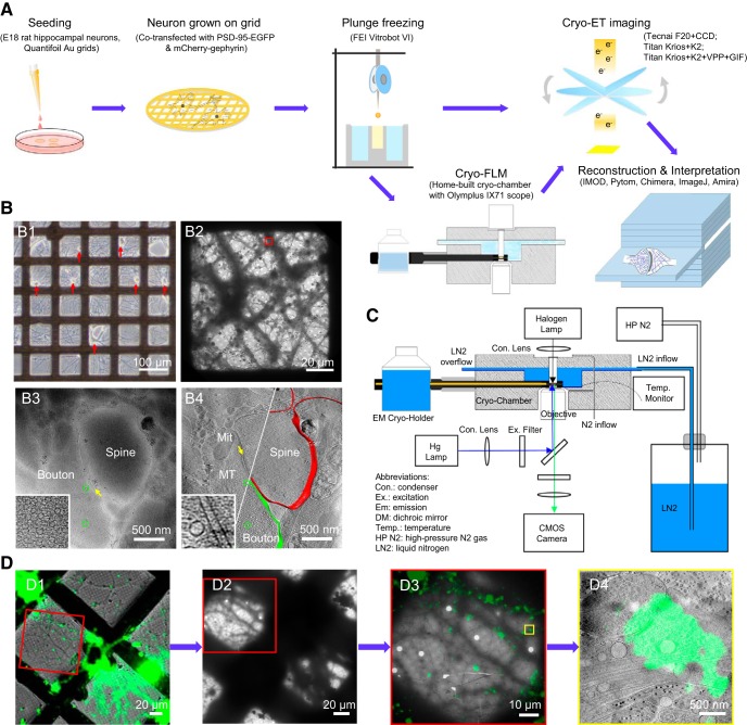
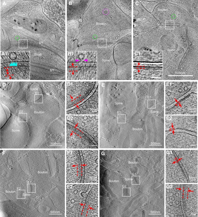
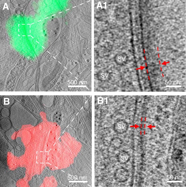
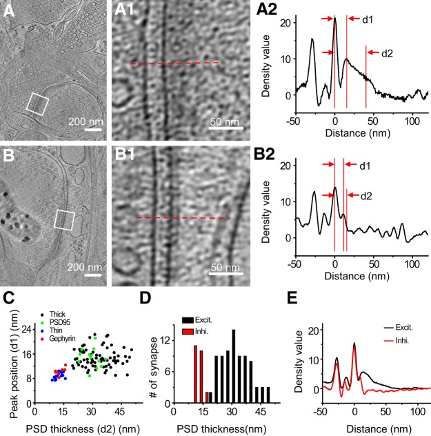
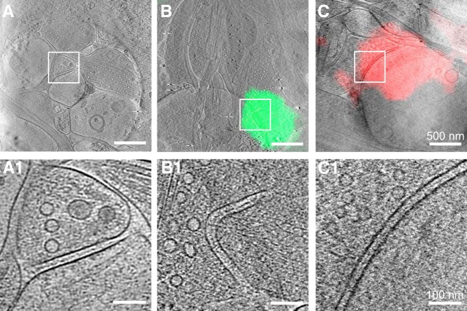
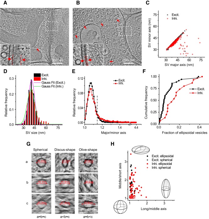
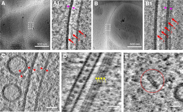
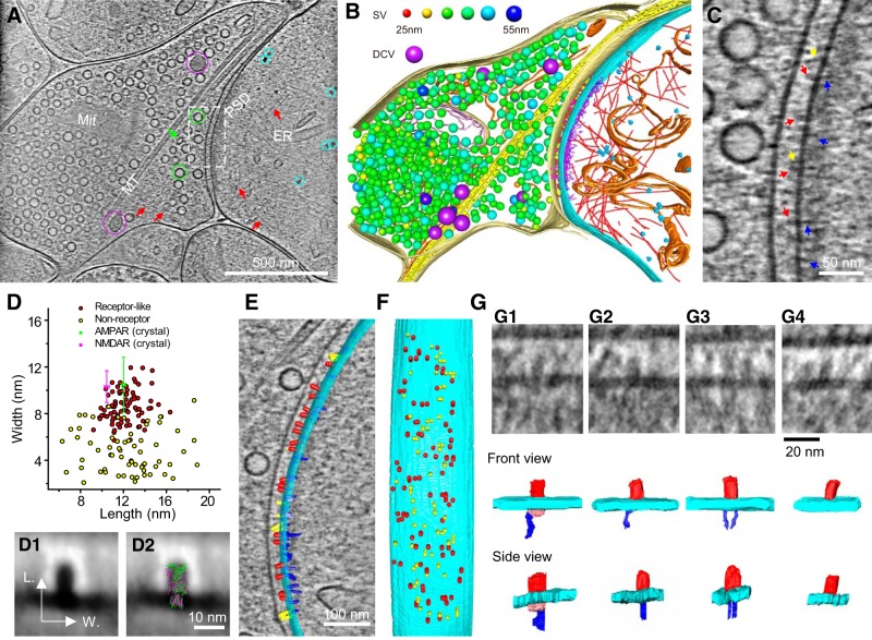
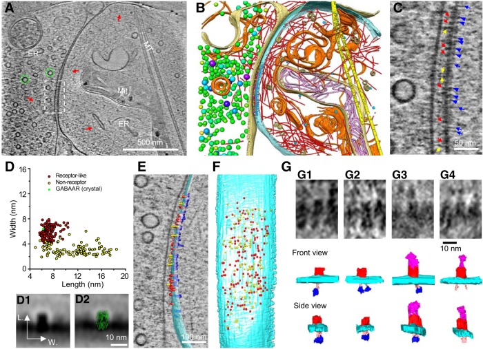
As key functional units in neural circuits, different types of neuronal synapses play distinct roles in brain information processing, learning, and memory. Synaptic abnormalities are believed to underlie various neurological and psychiatric disorders. Here, by combining cryo-electron tomography and cryo-correlative light and electron microscopy, we distinguished intact excitatory and inhibitory synapses of cultured hippocampal neurons, and visualized the in situ 3D organization of synaptic organelles and macromolecules in their native state. Quantitative analyses of >100 synaptic tomograms reveal that excitatory synapses contain a mesh-like postsynaptic density (PSD) with thickness ranging from 20 to 50 nm. In contrast, the PSD in inhibitory synapses assumes a thin sheet-like structure ∼12 nm from the postsynaptic membrane. On the presynaptic side, spherical synaptic vesicles (SVs) of 25–60 nm diameter and discus-shaped ellipsoidal SVs of various sizes coexist in both synaptic types, with more ellipsoidal ones in inhibitory synapses. High-resolution tomograms obtained using a Volta phase plate and electron filtering and counting reveal glutamate receptor-like and GABA A receptor-like structures that interact with putative scaffolding and adhesion molecules, reflecting details of receptor anchoring and PSD organization. These results provide an updated view of the ultrastructure of excitatory and inhibitory synapses, and demonstrate the potential of our approach to gain insight into the organizational principles of cellular architecture underlying distinct synaptic functions. SIGNIFICANCE STATEMENT To understand functional properties of neuronal synapses, it is desirable to analyze their structure at molecular resolution. We have developed an integrative approach combining cryo-electron tomography and correlative fluorescence microscopy to visualize 3D ultrastructural features of intact excitatory and inhibitory synapses in their native state. Our approach shows that inhibitory synapses contain uniform thin sheet-like postsynaptic densities (PSDs), while excitatory synapses contain previously known mesh-like PSDs. We discovered “discus-shaped” ellipsoidal synaptic vesicles, and their distributions along with regular spherical vesicles in synaptic types are characterized. High-resolution tomograms further allowed identification of putative neurotransmitter receptors and their heterogeneous interaction with synaptic scaffolding proteins. The specificity and resolution of our approach enables precise in situ analysis of ultrastructural organization underlying distinct synaptic functions.
Overview
The claims and statistics of this paper haven’t been checked yet.
Run the check — free, no account needed. It reads the open-access text and checks the sources it cites.
Integrity signals
Registry
Cites
Statistics
Figures
Verification
Claims the paper makes
The claims and statistics of this paper haven’t been checked yet.
Run the check — free, no account needed. It reads the open-access text and checks the sources it cites.
Claims other papers make about this one
Each is checked against this paper’s own text: does it say what the citing paper says it does?
The cited source says what the claim attributes to it.
In the citing paper
“In vertebrate EM datasets, human annotators can see differences in the vesicles for excitatory and inhibitory synapses.16,35,36,37,109,110”
From Neurotransmitter classification from electron microscopy images at synaptic sites in Drosophila melanogaster (2024) · cites this paper as [36]
Self-check
Percentages, sums and reported differences re-computed from the paper’s own numbers.
The self-check needs the paper’s full text.
Citations
Cites
UCSF Chimera—A visualization system for exploratory research and analysis
Pettersen, E. F., Goddard, T. D., Huang, C. C., Couch, G. S., Greenblatt, D. M., Meng, E. C., & Ferrin, T. E. (2004) · Journal of Computational Chemistry · doi:10.1002/jcc.20084 · Cited by 46,047
Shrestha, S., & Offer, S. M. (2016) · Medical Epigenetics · doi:10.1159/000444713 · Cited by 8,951
Computer Visualization of Three-Dimensional Image Data Using IMOD
Kremer, J. R., Mastronarde, D. N., & McIntosh, J. R. (1996) · Journal of Structural Biology · doi:10.1006/jsbi.1996.0013 · Cited by 5,530
Automated electron microscope tomography using robust prediction of specimen movements
Mastronarde, D. N. (2005) · Journal of Structural Biology · doi:10.1016/j.jsb.2005.07.007 · Cited by 5,136
Bi, G.-Q., & Poo, M.-M. (1998) · The Journal of Neuroscience · doi:10.1523/jneurosci.18-24-10464.1998 · Cited by 4,744
Self-citationDirect Least Square Fitting of Ellipses
Fitzgibbon, A., Pilu, M., & Fisher, R. B. (1999) · IEEE Transactions on Pattern Analysis and Machine Intelligence · doi:10.1109/34.765658 · Cited by 2,797
Ginsborg, B. L. (1964) · Quarterly Journal of Experimental Physiology and Cognate Medical Sciences · doi:10.1113/expphysiol.1964.sp001756 · Cited by 1,917
Li, X.-M., Mooney, P., Zheng, S., Booth, C., Braunfeld, M. B., Gubbens, S., Agard, D., & Cheng, Y.-F. (2013) · Nature Methods · doi:10.1038/nmeth.2472 · Cited by 1,866
Axo-somatic and axo-dendritic synapses of the cerebral cortex: an electron microscope study.
Eg, G. (1959) · PubMed · Cited by 1,768
Automated molecular microscopy: The new Leginon system
Suloway, C. J. M., Pulokas, J., Fellmann, D., Cheng, A., Guerra, F., Quispe, J. D., Stagg, S. M., Potter, C. S., & Carragher, B. O. (2005) · Journal of Structural Biology · doi:10.1016/j.jsb.2005.03.010 · Cited by 1,695
Cited by
Sorted by citations
Tang, J., Yuan, F., Shen, X., Wang, Z., Rao, M., He, Y., Sun, Y., Li, X., Zhang, W., Li, Y., et al. (2019) · Advanced Materials · doi:10.1002/adma.201902761 · Cited by 850
Self-citationIsotropic reconstruction for electron tomography with deep learning
Liu, Y.-T., Zhang, H., Wang, H., Tao, C.-L., Bi, G., & Zhou, Z. H. (2022) · Nature Communications · doi:10.1038/s41467-022-33957-8 · Cited by 397
Self-citationEckstein, N., Bates, A. S., Champion, A., Du, M., Yin, Y., Schlegel, P., Lu, A. K.-Y., Rymer, T., Finley-May, S., Paterson, T., et al. (2024) · Cell · doi:10.1016/j.cell.2024.03.016 · Cited by 277
“Among clear core vesicle, human experts can distinguish excitatory and inhibitory transmitters by how elliptical their synaptic vesicles are (Tao et al., 2018; Atwood et al., 1972; Uchizono, 1965), though they have not yet to our knowledge been automatically predicted at scale in these species.”
Fluid Biomarkers for Synaptic Dysfunction and Loss
Camporesi, E., Nilsson, J., Brinkmalm, A., Becker, B., Ashton, N. J., Blennow, K., & Zetterberg, H. (2020) · Biomarker Insights · doi:10.1177/1177271920950319 · Cited by 165
MemBrain v2: an end-to-end tool for the analysis of membranes in cryo-electron tomography
Lamm, L., Zufferey, S., Zhang, H., Righetto, R. D., Waltz, F., Wietrzyñski, W., Yamauchi, K. A., Burt, A., Liu, Y., Martínez-Sánchez, A., et al. (2026) · Nature Methods · doi:10.1038/s41592-026-03178-8 · Cited by 179
Self-citation
Metadata
- Authors
- Tao, C.-L., Liu, Y.-T., Sun, R., Zhang, B., Qi, L., Shivakoti, S., Tian, C.-L., Zhang, P., Lau, P.-M., Zhou, Z. H., & Bi, G.-Q.
- Affiliations
- Chinese Academy of Sciences (CN)
- Centre for Human Genetics (GB)
- California NanoSystems Institute (US)
- University of Science and Technology of China (CN)
- Corresponding author
- Zhou, Z. Hong
- Bi, Guo-Qiang
- ORCID
- Venue
- The Journal of Neuroscience
- Publisher
- Society for Neuroscience
- Published
- 2018-01-08
- Volume
- 38
- Issue
- 6
- Pages
- 1493-1510
- ISSN
- 0270-6474 (ISSN-L)
- 1529-2401
- PMC
- PMC5815350
- OpenAlex
- W2783481291
- Cited by
- 207
- References
- 91
- Citations by year
- 2026: 18 · 2025: 24 · 2024: 30 · 2023: 14 · 2022: 27 · 2021: 40 · 2020: 30 · 2019: 17 · 2018: 5
- FWCI
- 10.48
- Type
- journal article
- Language
- en
- Open access
- hybrid
- doi.org
- Locations
- Journal of Neuroscience (publishedVersion, cc-by)
- PubMed Central (publishedVersion, cc-by)
- PubMed (publishedVersion)
- eScholarship (California Digital Library) (submittedVersion)
- eScholarship (California Digital Library) (submittedVersion, cc-by)
- Funders
- National Science Foundation
- Wellcome Trust (206422/Z/17/Z)
- National Natural Science Foundation of China (2009CB941300, 31070935, PSD-95)
- National Institutes of Health (1s10od018111-01)
- Biotechnology and Biological Sciences Research Council (BB/S003339/1)
- National Institute of General Medical Sciences (R01GM071940)
- National Center for Research Resources (S10RR023057)
- Topics
- Life Sciences › Neuroscience › Cellular and Molecular Neuroscience › Neuroscience and Neuropharmacology Research
- Life Sciences › Biochemistry, Genetics and Molecular Biology › Structural Biology › Advanced Electron Microscopy Techniques and Applications
- Life Sciences › Biochemistry, Genetics and Molecular Biology › Cell Biology › Cellular transport and secretion
- Keywords
- excitatory synapses, inhibitory synapses, synaptic ultrastructure, postsynaptic density, synaptic vesicles, glutamate receptors, GABAA receptor, synaptic scaffolding proteins, adhesion molecules, hippocampal neurons, cryo-electron tomography, correlative light and electron microscopy
- MeSH terms
- Animals, Female, Image Processing, Computer-Assisted, Inhibition, Psychological, Neurons, Pregnancy, Receptors, GABA-A, Synapses, Synaptic Vesicles, Tomography, Cell Adhesion Molecules, Receptors, Glutamate, Excitatory Postsynaptic Potentials, Cryoelectron Microscopy, Rats, Post-Synaptic Density
About these checks
Registry, retraction, Cites and statistics results are computed, not judged, with one exception: a printed reference that no index lists is read once by a model before it counts as a citation error. Claims are extracted by a model and kept only when their quote appears verbatim in the text; each cited source is then checked, with a model’s help, for whether it says what the claim relies on. Those verdicts can be wrong — read the claim’s quote and the cited source before relying on one.
Statistics and arithmetic are recomputed, not judged.
Confidence bands are shown only for fields calibrated on our annotated test set. None is calibrated yet, so no band is shown.
What counts as a claim ›How we measure confidence ›How we check this ›
Are you an author, or do you see a problem with this page? Request a review
