

The DOI matches its Crossref record; 65 of the 65 references it cites were found in a registry or an index.
Selective Activation of Microglia Facilitates Synaptic Strength
Open accessAbstract
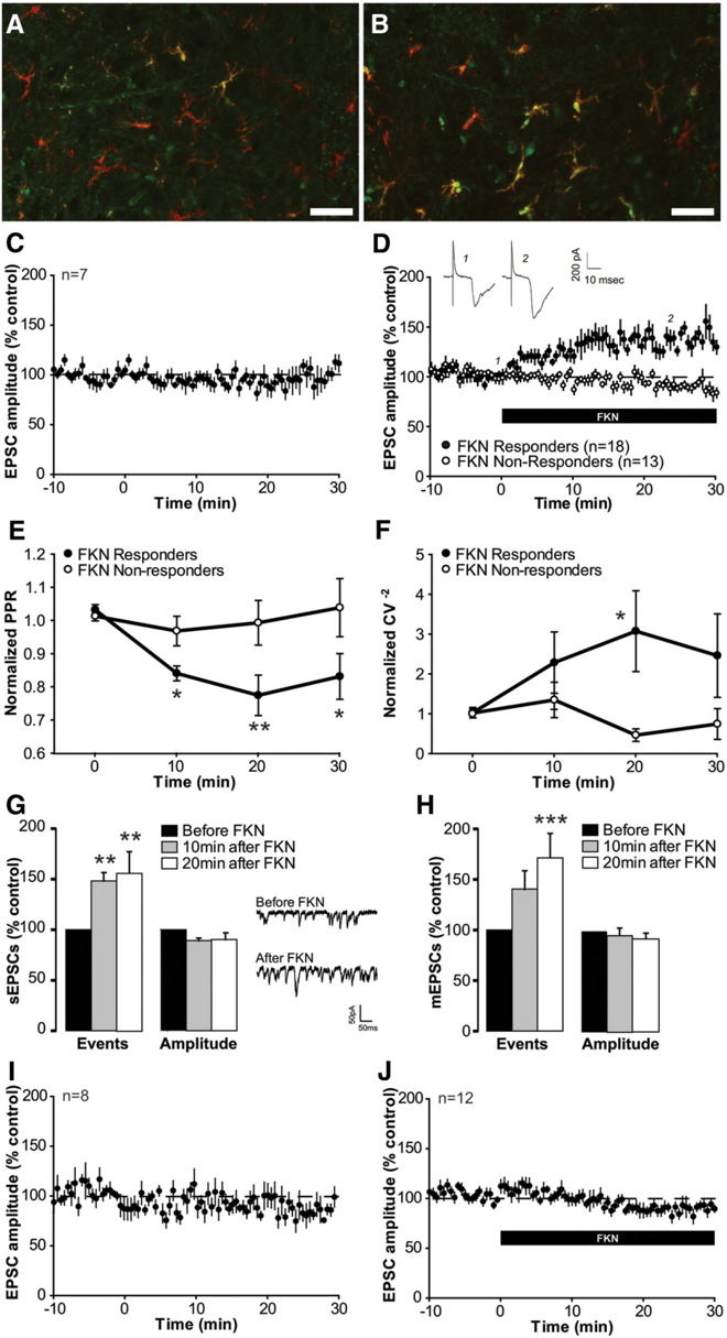
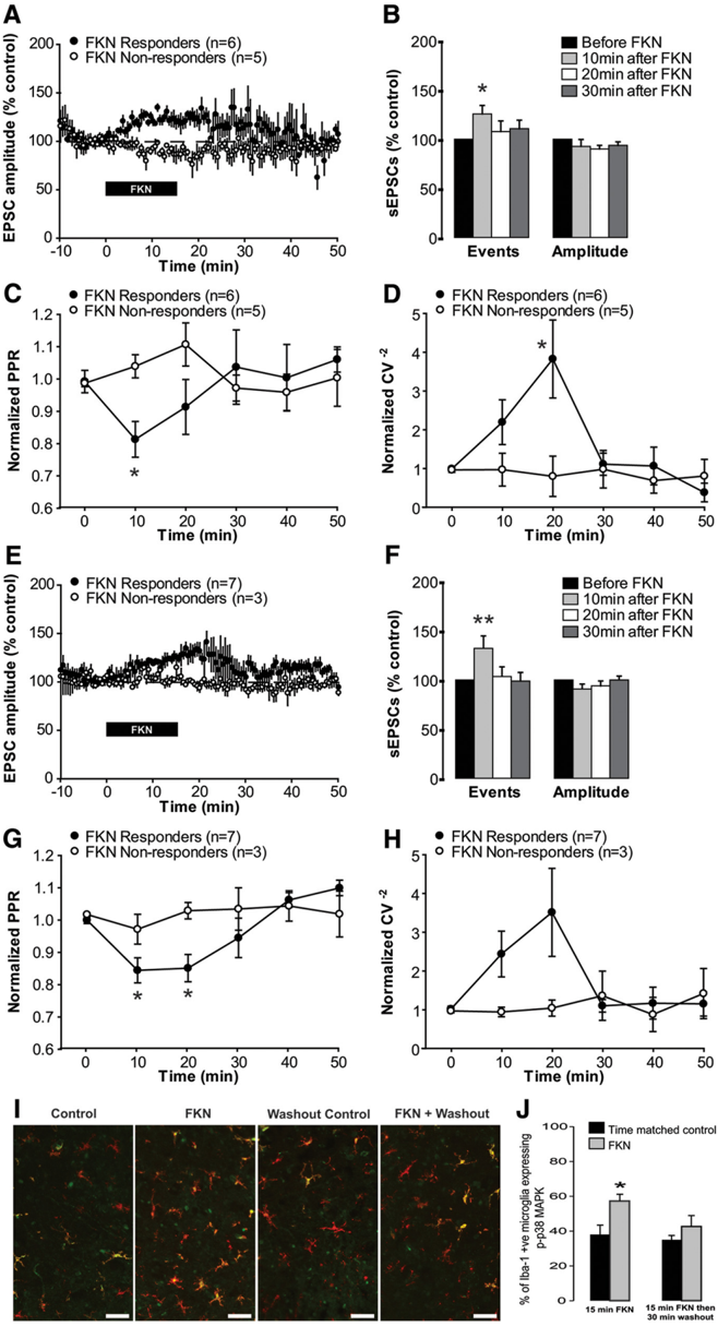
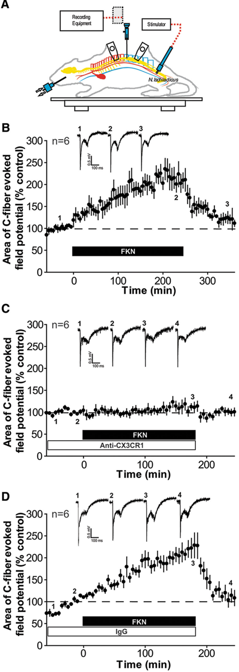
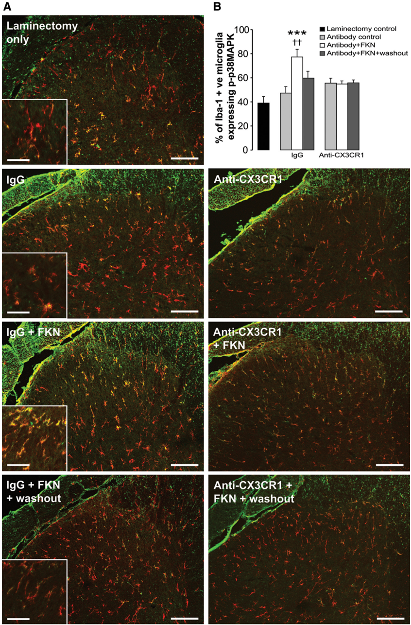
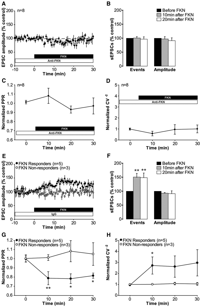
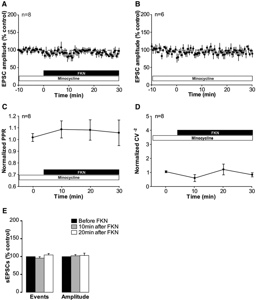
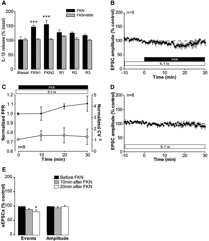
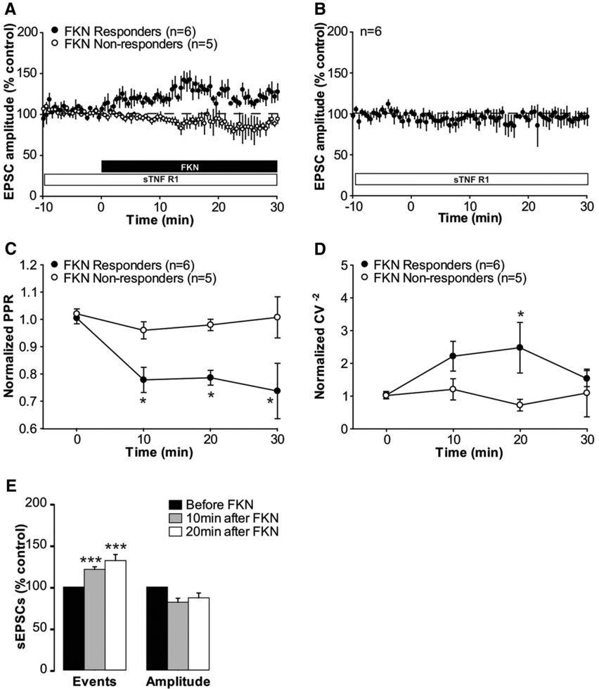
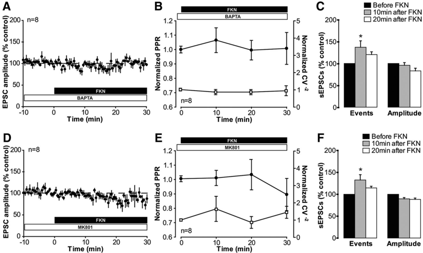
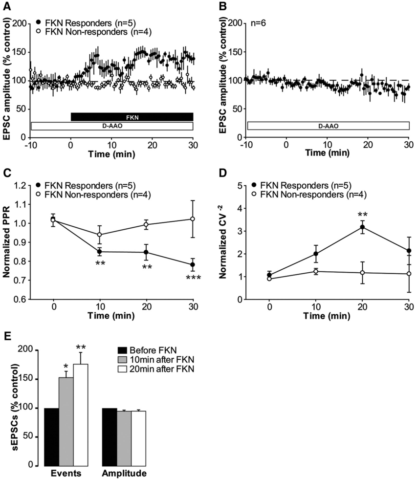
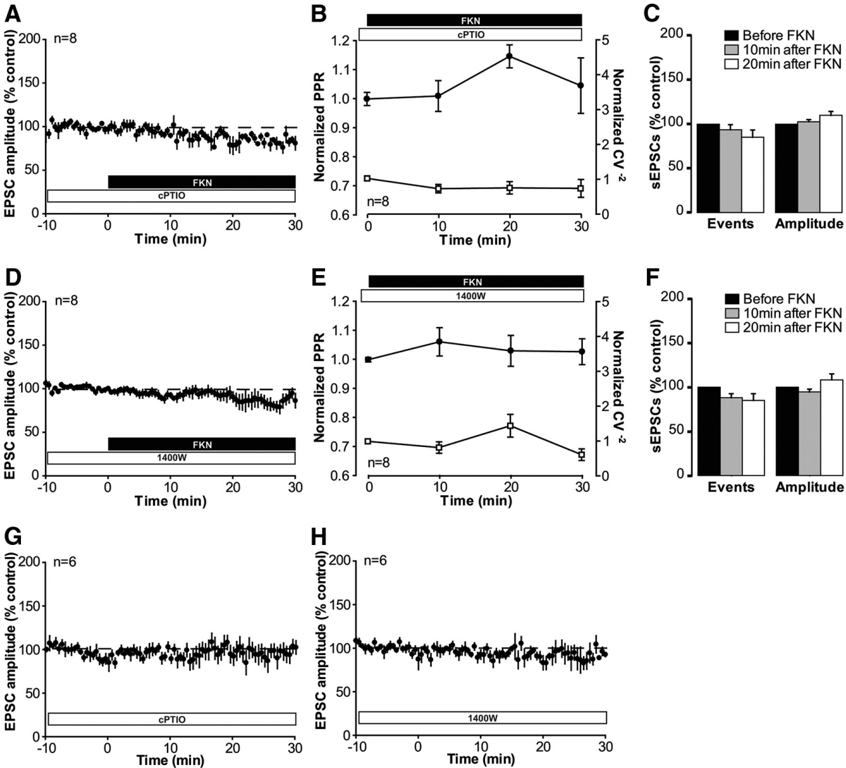
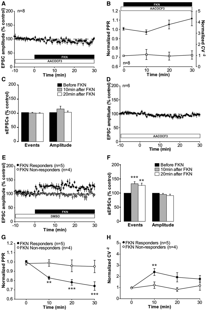
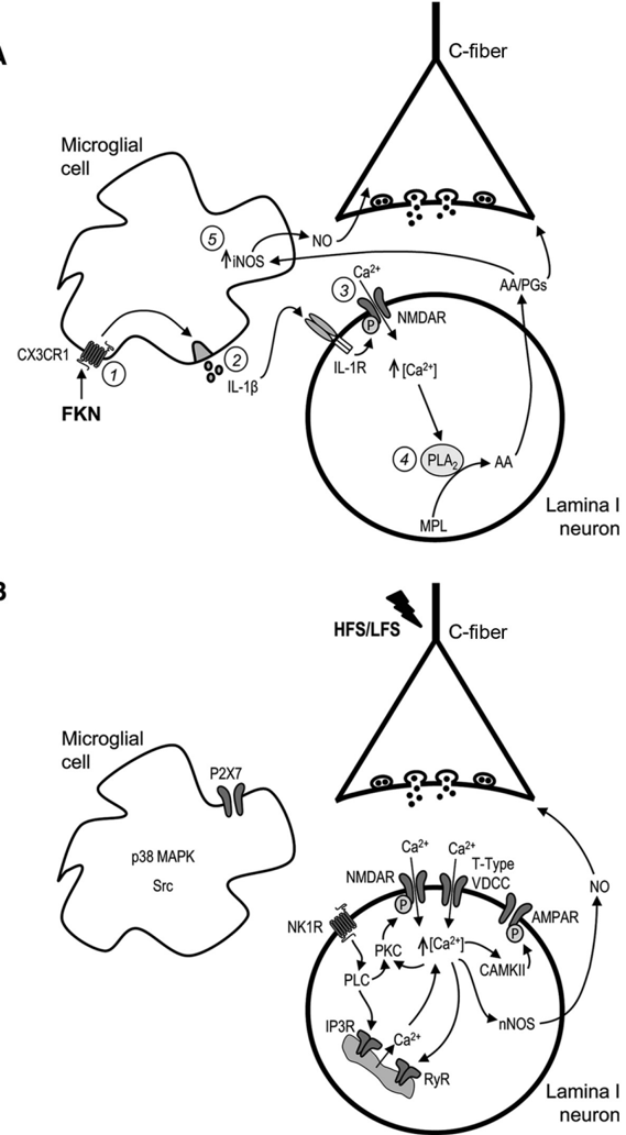
Synaptic plasticity is thought to be initiated by neurons only, with the prevailing view assigning glial cells mere specify supportive functions for synaptic transmission and plasticity. We now demonstrate that glial cells can control synaptic strength independent of neuronal activity. Here we show that selective activation of microglia in the rat is sufficient to rapidly facilitate synaptic strength between primary afferent C-fibers and lamina I neurons, the first synaptic relay in the nociceptive pathway. Specifically, the activation of the CX3CR1 receptor by fractalkine induces the release of interleukin-1β from microglia, which modulates NMDA signaling in postsynaptic neurons, leading to the release of an eicosanoid messenger, which ultimately enhances presynaptic neurotransmitter release. In contrast to the conventional view, this form of plasticity does not require enhanced neuronal activity to trigger the events leading to synaptic facilitation. Augmentation of synaptic strength in nociceptive pathways represents a cellular model of pain amplification. The present data thus suggest that, under chronic pain states, CX3CR1-mediated activation of microglia drives the facilitation of excitatory synaptic transmission in the dorsal horn, which contributes to pain hypersensitivity in chronic pain states.
Overview
The claims and statistics of this paper haven’t been checked yet.
Run the check — free, no account needed. It reads the open-access text and checks the sources it cites.
Integrity signals
Registry
Cites
Statistics
Figures
Verification
Claims the paper makes
The claims and statistics of this paper haven’t been checked yet.
Run the check — free, no account needed. It reads the open-access text and checks the sources it cites.
Claims other papers make about this one
Each is checked against this paper’s own text: does it say what the citing paper says it does?
In the citing paper
“The activation of the fractalkine receptor (CX3CR1) in microglial cells, increases synaptic strength. However, deficiency in this receptor results in a reduced hippocampal synaptic plasticity (Rogers et al., 2011; Clark et al., 2015).”
From The role of microglia in retinal neurodegeneration: Alzheimer’s disease, parkinson, and glaucoma. (2017) · cites this paper as Clark et al., 2015
Self-check
Percentages, sums and reported differences re-computed from the paper’s own numbers.
The self-check needs the paper’s full text.
Citations
Cites
The Rat Brain in Stereotaxic Coordinates
Paxinos, G., & Watson, C. (1983) · Elsevier eBooks · doi:10.1016/c2009-0-63235-9 · Cited by 28,585
A synaptic model of memory: long-term potentiation in the hippocampus
Bliss, T. V. P., & Collingridge, G. L. (1993) · Nature · doi:10.1038/361031a0 · Cited by 11,566
Polazzi, E., Mengoni, I., Caprini, M., Peña‐Altamira, E., Kurtys, E., & Monti, B. (2012) · Neurosignals · doi:10.1159/000337115 · Cited by 9,023
AChE-stained horizontal sections of the rat brain in stereotaxic coordinates
Paxinos, G., WATSON, C. R. R., & Emson, P. C. (1980) · Journal of Neuroscience Methods · doi:10.1016/0165-0270(80)90021-7 · Cited by 4,501
Malenka, R. C., & Bear, M. F. (2004) · Neuron · doi:10.1016/j.neuron.2004.09.012 · Cited by 3,968
Microglia: Active sensor and versatile effector cells in the normal and pathologic brain
Hanisch, U.-K., & Kettenmann, H. (2007) · Nature Neuroscience · doi:10.1038/nn1997 · Cited by 3,676
BDNF from microglia causes the shift in neuronal anion gradient underlying neuropathic pain
Coull, J. A. M., Beggs, S., Boudreau, D., Boivin, D., Tsuda, M., Inoue, K., Gravel, C., Salter, M. W., & De Koninck, Y. (2005) · Nature · doi:10.1038/nature04223 · Cited by 1,965
Control of microglial neurotoxicity by the fractalkine receptor
Cardona, A. E., Pioro, E. P., Sasse, M. E., Kostenko, V., Cardona, S. M., Dijkstra, I. M., Huang, D., Kidd, G., Dombrowski, S., Dutta, R., et al. (2006) · Nature Neuroscience · doi:10.1038/nn1715 · Cited by 1,531
Samad, T. A., Moore, K. A., Sapirstein, A., Billet, S. E., Allchorne, A. J., Poole, S., Bonventre, J. V., & Woolf, C. J. (2001) · Nature · doi:10.1038/35068566 · Cited by 1,282
Glia and pain: Is chronic pain a gliopathy?
Ji, R., Berta, T., & Nedergaard, M. (2013) · Pain · doi:10.1016/j.pain.2013.06.022 · Cited by 1,193
Cited by
Sorted by citations
Items 101–171 of 171
Froudist‐Walsh, S., Browning, P. G. F., Young, J., Murphy, K. L., Mars, R. B., Fleysher, L., & Croxson, P. L. (2018) · eLife · doi:10.7554/elife.34354 · Cited by 23
Role of Microglia in Psychostimulant Addiction
da Silva, M. C. M., Iglesias, L. P., Candelario‐Jalil, E., Khoshbouei, H., Moreira, F. A., & de Oliveira, A. C. P. (2022) · Current Neuropharmacology · doi:10.2174/1570159x21666221208142151 · Cited by 26
Caruso, G., Pietro, L. D., & Caraci, F. (2023) · Biomolecules · doi:10.3390/biom13030505 · Cited by 24
Cataldo, G., Erb, S. J., Lunzer, M. M., Luong, N., Akgün, E., Portoghese, P. S., Olson, J. K., & Simone, D. A. (2019) · Neuropharmacology · doi:10.1016/j.neuropharm.2019.04.004 · Cited by 22
Do Microglia default on network maintenance in Alzheimer's disease?
Southam, K. A., Vincent, A. J., & Small, D. H. (2016) · Journal of Alzheimer s Disease · doi:10.3233/jad-151075 · Cited by 20
Li, J., Serafin, E. K., & Baccei, M. (2018) · Journal of Neuroscience · doi:10.1523/jneurosci.2152-17.2018 · Cited by 15
Nakamura, Y., Fukuta, A., Miyashita, K., Zhang, F. F., Wang, D., Liu, K., Wake, H., Hisaoka‐Nakashima, K., Nishibori, M., & Morioka, N. (2021) · Biochemical Pharmacology · doi:10.1016/j.bcp.2021.114496 · Cited by 19
Jeong, Y. C., Son, J. S., & Kwon, Y. (2015) · Brain Research Bulletin · doi:10.1016/j.brainresbull.2015.07.006 · Cited by 19
Cathepsin S in the spinal microglia contributes to remifentanil-induced hyperalgesia in rats
Ye, L., Xiao, L., Yang, S., Duan, J., Chen, Y., Cui, Y., & Chen, Y. (2016) · Neuroscience · doi:10.1016/j.neuroscience.2016.12.030 · Cited by 18
The role of microglia in chronic pain and depression: innocent bystander or culprit?
Yin, N., Yan, E., Duan, W., Mao, C., Qin, F., Yang, C., Hu, Y., & Xu, X. (2021) · Psychopharmacology · doi:10.1007/s00213-021-05780-4 · Cited by 18
König, C., Vazquez, E., Eß, S., Ebbinghaus, M., Vorpahl, B., Ebersberger, A., & Schaible, H. (2021) · Journal of Neurochemistry · doi:10.1111/jnc.15438 · Cited by 17
Litvin, D. G., Dick, T. E., Smith, C. B., & Jacono, F. J. (2018) · Brain Behavior and Immunity · doi:10.1016/j.bbi.2018.03.031 · Cited by 17
Procès, A., Alpizar, Y. A., Halliez, S., Brône, B., Saudou, F., Ris, L., & Gabriele, S. (2023) · Biomaterials · doi:10.1016/j.biomaterials.2023.122426 · Cited by 17
Neuron–microglia interactions modulating neuropathic pain
Kohno, K., & Tsuda, M. (2025) · International Immunology · doi:10.1093/intimm/dxaf022 · Cited by 18
Malcangio, M. (2016) · Pain · doi:10.1097/j.pain.0000000000000509 · Cited by 15
Self-citationRegulation of Pain Perception by Microbiota in Parkinson Disease
Manjarres, Z., Calvo, M., & Pacheco, R. (2023) · Pharmacological Reviews · doi:10.1124/pharmrev.122.000674 · Cited by 15
Krishnan, V. S., Shavlakadze, T., Grounds, M. D., Hodgetts, S. I., & Harvey, A. R. (2018) · Biogerontology · doi:10.1007/s10522-018-9765-5 · Cited by 15
Reorganization of synaptic inputs to spinal dorsal horn neurons in neuropathic pain
Terayama, R., & Uchibe, K. (2021) · International Journal of Neuroscience · doi:10.1080/00207454.2021.1873980 · Cited by 13
Mai, J., Liu, C., Huang, Z., Mai, C.-L., Zhou, X., Zhang, J., & Liu, X.-G. (2020) · Molecular Pain · doi:10.1177/1744806920917242 · Cited by 13
Han, J., Cho, H.-J., Park, D., & Han, S. (2022) · Cells · doi:10.3390/cells11192977 · Cited by 13
Gomes, G. F., Peixoto, R. D. D. F., Maciel, B. G., dos Santos, K. F., Bayma, L. R., Neto, P. A. F., Fernandes, T. N., de Abreu, C. C., Casseb, S. M. M., de Lima, C. M., et al. (2019) · Journal of Histochemistry & Cytochemistry · doi:10.1369/0022155419835218 · Cited by 13
Hu, D., Moalem‐Taylor, G., & Potas, J. R. (2020) · Journal of Neurotrauma · doi:10.1089/neu.2020.7066 · Cited by 13
Upregulation of BDNF and Interleukin-1ß in rat spinal cord following noxious hind paw stimulation
Donnerer, J., & Liebmann, I. (2017) · Neuroscience Letters · doi:10.1016/j.neulet.2017.12.008 · Cited by 12
Dendritic Spines and Pain Memory
Benson, C. A., King, J. F., Reimer, M. L., Kauer, S. D., Waxman, S. G., & Tan, A. M. (2022) · The Neuroscientist · doi:10.1177/10738584221138251 · Cited by 13
Plastic Events of the Vestibular Nucleus: the Initiation of Central Vestibular Compensation
Wu, J., Xu, X., Zhang, S., Li, M., Qiu, Y., Lu, G., Zheng, Z., & Huang, H. (2024) · Molecular Neurobiology · doi:10.1007/s12035-024-04208-2 · Cited by 12
Bivalent ligand MCC22 potently attenuates nociception in a murine model of sickle cell disease
Cataldo, G., Lunzer, M. M., Olson, J. K., Akgün, E., Belcher, J. D., Vercellotti, G. M., Portoghese, P. S., & Simone, D. A. (2018) · Pain · doi:10.1097/j.pain.0000000000001225 · Cited by 11
Vecchiarelli, H. A., Lopes, L. T., Paolicelli, R. C., Stevens, B., Wake, H., & Tremblay, M. (2024) · Advances in neurobiology · doi:10.1007/978-3-031-55529-9_11 · Cited by 10
Headmasters: Microglial regulation of learning and memory in health and disease
Weinhard, L., d’Errico, P., & Tay, T. L. (2018) · AIMS molecular science · doi:10.3934/molsci.2018.1.63 · Cited by 11
Kim, H. W., Won, C., & Oh, S. (2021) · Molecular Pain · doi:10.1177/17448069211011326 · Cited by 7
Ni, M., Zheng, M., Chen, B., Lu, X., Zhao, H., Zhu, T., Cheng, L., Han, H., Ye, T., Liu, H., et al. (2023) · Neuropharmacology · doi:10.1016/j.neuropharm.2023.109693 · Cited by 9
Zhang, Z., Wang, T., Luo, Z., Zaib, M. H., Yi, M., Zeng, H., Li, P., Tang, D., Verkhratsky, A., & Nie, H. (2023) · Purinergic Signalling · doi:10.1007/s11302-023-09979-2 · Cited by 9
Baccei, M. L. (2016) · Children · doi:10.3390/children3030016 · Cited by 9
Huang, R., Han, S., Qiu, Y., Zhou, T., Wu, Y., Du, H., Xu, J., & Wei, X. (2022) · Neuropharmacology · doi:10.1016/j.neuropharm.2022.109253 · Cited by 10
Cancela, L. M., Mongi‐Bragato, B., Avalos, M. P., & Bollati, F. A. (2025) · International Journal of Molecular Sciences · doi:10.3390/ijms27010385 · Cited by 9
Spinal Cathepsin S promotes visceral hypersensitivity via FKN/CX3CR1/p38 MAPK signaling pathways
Sun, P., Lin, W. C., Weng, Y., Gong, J. D., Huang, Y., Tang, Y., Lin, C., Chen, A., & Chen, Y. (2023) · Molecular Pain · doi:10.1177/17448069231179118 · Cited by 7
Wolf‐Johnston, A., Ikeda, Y., Zabbarova, I. V., Kanai, A., Bastacky, S., Moldwin, R., Stern, J. N. H., Jackson, E. K., & Birder, L. A. (2024) · JCI Insight · doi:10.1172/jci.insight.176103 · Cited by 7
Diniz, D. G., Silva, G. O., Naves, T. B., Fernandes, T. N., Araújo, S. C., Diniz, J. A. P., Farias, L. H. S., Sosthenes, M. C. K., Diniz, C. G., Anthony, D. C., et al. (2016) · Frontiers in Neuroanatomy · doi:10.3389/fnana.2016.00023 · Cited by 7
Glial cells in neuropathic pain
Berta, T., Khoutorsky, A., Xu, J., & Ji, R. (2026) · Physiological Reviews · doi:10.1152/physrev.00034.2025 · Cited by 7
Ismail, C. A. N., Ghazali, A. K., Suppıan, R., Aziz, C. B. A., & Long, I. (2021) · Journal of Diabetes & Metabolic Disorders · doi:10.1007/s40200-021-00820-4 · Cited by 7
From nociception in aneural animals to human suffering: toward a comparative biology of pain
Walters, E. T. (2025) · Journal of Experimental Biology · doi:10.1242/jeb.251210 · Cited by 8
Pathogenic mechanisms of human immunodeficiency virus (HIV)-associated pain
Liu, X., & Tang, S. (2023) · Molecular Psychiatry · doi:10.1038/s41380-023-02294-7 · Cited by 5
Hoheisel, U., Chacur, M., Treede, R., & Mense, S. (2019) · Journal of Neurophysiology · doi:10.1152/jn.00338.2018 · Cited by 5
Analgesic Effect of Human Placenta Hydrolysate on CFA-Induced Inflammatory Pain in Mice
Park, K.-T., Jo, H., Jeon, S.-H., Jeong, K., Im, M., Kim, J.-W., Jung, J. P., Jung, H. C., Lee, J. H., & Kim, W. (2024) · Pharmaceuticals · doi:10.3390/ph17091179 · Cited by 4
Yao, Y., Gu, X.-X.-Y., Xie, X.-B., Wang, Y., & Wang, Q.-L. (2026) · Journal of Chinese Integrative Medicine · doi:10.1016/j.joim.2026.01.009 · Cited by 4
La mémoire de la douleur : de la LTP à la douleur du membre fantôme
Calvino, B. (2017) · Douleurs Évaluation - Diagnostic - Traitement · doi:10.1016/j.douler.2017.03.003 · Cited by 3
Wang, Q.-Q., Sun, Q.-Q., Guo, Y.-S., Yin, S., & Zhou, J. (2026) · Translational Neurodegeneration · doi:10.1186/s40035-026-00571-3 · Cited by 2
Wilderness Immersion Tuning: Education with Evolution and Neuroscience in Mind
Beeman, C., & Walton, E. B. (2016) · Encounters in Theory and History of Education · doi:10.24908/eoe-ese-rse.v17i0.6345 · Cited by 2
Ada, S., Klinger, L., Teuchmann, H. L., Mussetto, V., Hadschieff, V., Kronschläger, M. T., Siegert, A., Trofimova, L., Holzinger, R., Kurija, D., et al. (2025) · Pain · doi:10.1097/j.pain.0000000000003805 · Cited by 1
Self-citationFanton, S., Kim, M., Alshelh, Z., Brusaferri, L., Morrissey, E. J., Torrado-Carvajal, Á., Grmek, G., Chane, C. A., Wargo, M. A., Murphy, J. P., et al. (2026) · Brain Behavior and Immunity · doi:10.1016/j.bbi.2026.106972 · Cited by 1
Olfactory ensheathing cells as candidate cells for chronic pain treatment
Liu, M.-C., Guo, Q.-F., Zhang, W., Luo, H., Zhang, W., & Hu, H. (2024) · Journal of Chemical Neuroanatomy · doi:10.1016/j.jchemneu.2024.102413 · Cited by 1
Schwann Cells Promote Sensory Neuron Excitability During DevelopmentPreprint
Kantarci, H., Elvira, P. D., Thottumkara, A. P., Iyer, M., Donovan, L. J., Dugan, M. Q., Ambiel, N., O’Connell, E. M., Granados, A. A., Zeng, H., et al. (2022) · bioRxiv (Cold Spring Harbor Laboratory) · doi:10.1101/2022.10.31.514415 · Cited by 1
Fabisiak, T. (2015) · CU Scholar (University of Colorado Boulder) · Cited by 1
Topper, L. A. (2015) · UNM’s Digital Repository (University of New Mexico)
Deftu, A., & Suter, M. R. (2020) · Elsevier eBooks · doi:10.1016/b978-0-12-809324-5.24214-4
The role of neuroimmune interactions in musculoskeletal pain
Schaible, H., Ebersberger, A., Natura, G., & Vázquez, E. (2022) · e-Neuroforum · doi:10.1515/nf-2022-0001
Lynn, A. M. (2018) · Faculty Opinions – Post-Publication Peer Review of the Biomedical Literature · doi:10.3410/f.732715202.793549522
rhPLD2 Inhibits the LPS-Induced IL-1β and TNF-α in MicrogliaPreprint
Chen, Y., Lin, J., Wang, H.-L., Cui, X., & Zhu, L. (2023) · SSRN Electronic Journal · doi:10.2139/ssrn.4479583
Pathogenesis of HIV-associated pain
Liu, X., & Tang, S. (2024) · Elsevier eBooks · doi:10.1016/b978-0-323-99744-7.00035-3
Dodman, K. G., Berken, A. M., & Barreveld, A. M. (2024) · Oxford University Press eBooks · doi:10.1093/med/9780197649176.003.0007
Espinoza, A. P. V., Na, N., Campanati, L., Ngo, P., Baldwin, K. K., & Cline, H. T. (2025) · bioRxiv (Cold Spring Harbor Laboratory) · doi:10.1101/2025.08.03.668224
Huang, Q., Nov, P., Ge, X., & Li, J. (2025) · Research Square · doi:10.21203/rs.3.rs-7892680/v1
Shao, S., Chengbo, X., Chen, C.-J., Shi, G., Guo, Q.-L., Zhou, Y., Wei, Y., Wu, L., Shi, J., & Zhang, T.-T. (2021) · Figshare · doi:10.6084/m9.figshare.14833450
Spinal glia–neuron interactions in chronic itch
Tsuda, M. (2026) · Elsevier eBooks · doi:10.1016/b978-0-443-34172-4.00018-5
Wang, J., & Zhou, H. (2026) · Neuropeptides · doi:10.1016/j.npep.2026.102612 · Cited by 1
Mahmoudi, N. E. (2022) · theses.fr (ABES) · doi:10.70675/68e2ad9bzd00bz4539z8b49z2a120516daca
Neuromodulation Techniques Alleviating Neuropathic Pain by Targeting Microglia: A Narrative Review
Mu, Q., Li, X., Li, L., Sun, S., Xu, J. L., Song, G., Chen, Y., Yang, L., Gan, Z., & Yang, D. (2026) · Pain and Therapy · doi:10.1007/s40122-026-00848-5
Wang, Y., Li, P., Dong, Y., Zhang, X., & Shang, K. (2026) · Frontiers in Immunology · doi:10.3389/fimmu.2026.1934162
Ramos, A. J., & Villarreal, A. (2026) · Frontiers in Network Physiology · doi:10.3389/fnetp.2026.1895622
Metadata
- Authors
- Clark, A. K., Gruber-Schoffnegger, D., Drdla-Schutting, R., Gerhold, K. J., Malcangio, M., & Sandkühler, J.
- Affiliations
- King's College London (GB)
- Medical University of Vienna (AT)
- ORCID
- Venue
- The Journal of Neuroscience
- Publisher
- Society for Neuroscience
- Published
- 2015-03-18
- Volume
- 35
- Issue
- 11
- Pages
- 4552-4570
- ISSN
- 0270-6474 (ISSN-L)
- 1529-2401
- PMC
- PMC4363384
- OpenAlex
- W2063244116
- Cited by
- 171
- References
- 65
- Citations by year
- 2026: 9 · 2025: 8 · 2024: 12 · 2023: 15 · 2022: 18 · 2021: 23 · 2020: 14 · 2019: 15 · 2018: 18 · 2017: 12 · 2016: 18 · 2015: 9
- FWCI
- 14.00
- Type
- journal article
- Language
- en
- Open access
- hybrid
- doi.org
- Licence
- Locations
- Journal of Neuroscience (publishedVersion, cc-by-nc-sa)
- Europe PMC (PubMed Central) (publishedVersion)
- PubMed Central (publishedVersion)
- PubMed (publishedVersion)
- Research Portal (King's College London) (submittedVersion, cc-by)
- Funders
- Wellcome Trust (093173/Z/10/Z)
- Austrian Science Fund
- Topics
- Health Sciences › Medicine › Physiology (medical) › Pain Mechanisms and Treatments
- Life Sciences › Neuroscience › Neurology › Neuroinflammation and Neurodegeneration Mechanisms
- Life Sciences › Neuroscience › Cellular and Molecular Neuroscience › Neuroscience and Neuropharmacology Research
- Keywords
- microglia, synaptic plasticity, synaptic facilitation, CX3CR1, fractalkine, IL-1β, eicosanoids, presynaptic neurotransmitter release, nociceptive pathways, dorsal horn, C-fibers, pain amplification, chronic pain, glial cells
- MeSH terms
- Animals, Male, Synaptic Transmission, Neuronal Plasticity, Organ Culture Techniques, Spinal Cord, Rats, Sprague-Dawley, Microglia, Excitatory Postsynaptic Potentials, Rats
About these checks
Registry, retraction, Cites and statistics results are computed, not judged, with one exception: a printed reference that no index lists is read once by a model before it counts as a citation error. Claims are extracted by a model and kept only when their quote appears verbatim in the text; each cited source is then checked, with a model’s help, for whether it says what the claim relies on. Those verdicts can be wrong — read the claim’s quote and the cited source before relying on one.
Statistics and arithmetic are recomputed, not judged.
Confidence bands are shown only for fields calibrated on our annotated test set. None is calibrated yet, so no band is shown.
What counts as a claim ›How we measure confidence ›How we check this ›
Are you an author, or do you see a problem with this page? Request a review
