

The DOI matches its Crossref record; 80 of the 82 references it cites were found in a registry or an index.
Corticotropin-Releasing Hormone Drives Anandamide Hydrolysis in the Amygdala to Promote Anxiety
Open accessAbstract
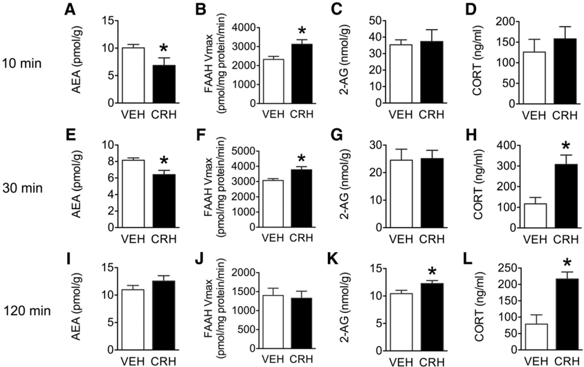
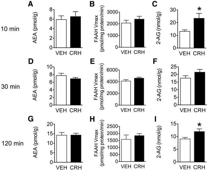
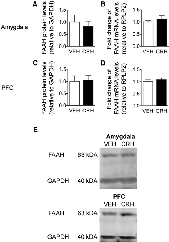
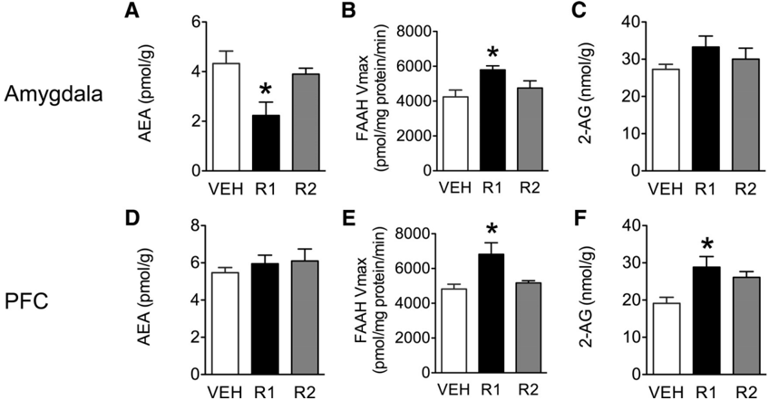
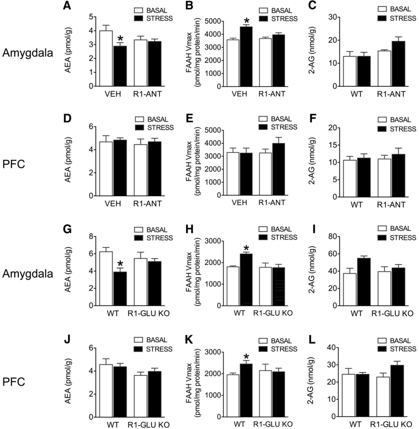
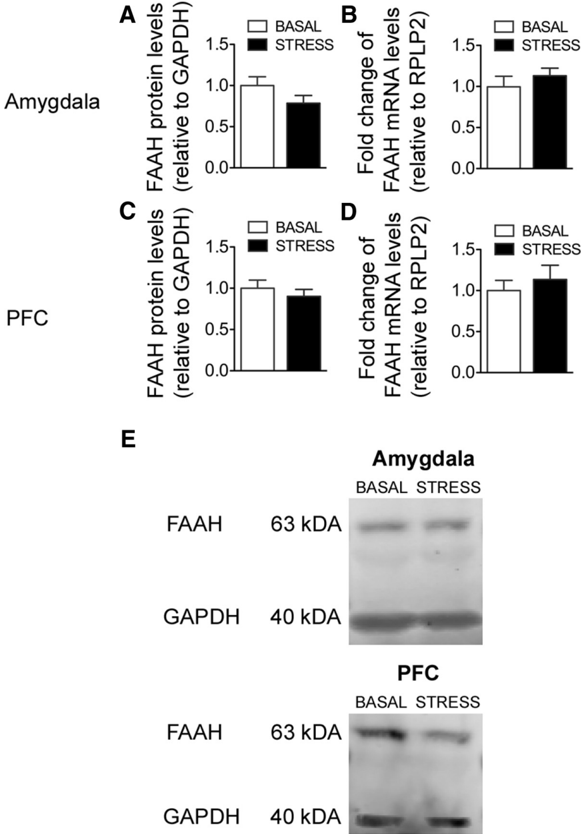
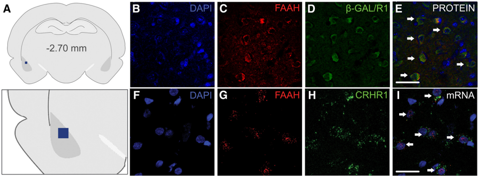
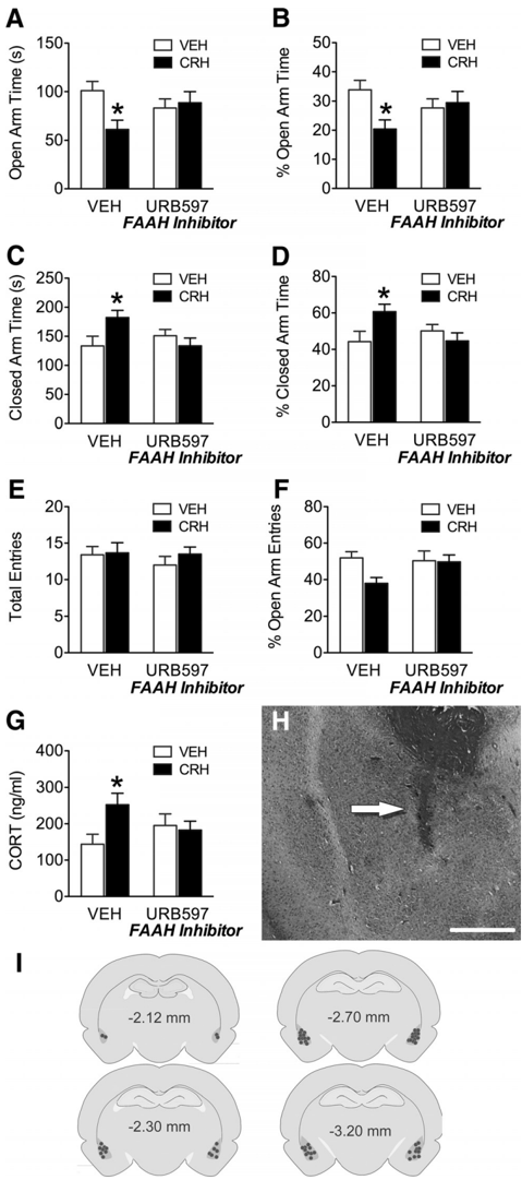
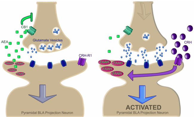
Corticotropin-releasing hormone (CRH) is a central integrator in the brain of endocrine and behavioral stress responses, whereas activation of the endocannabinoid CB1 receptor suppresses these responses. Although these systems regulate overlapping functions, few studies have investigated whether these systems interact. Here we demonstrate a novel mechanism of CRH-induced anxiety that relies on modulation of endocannabinoids. Specifically, we found that CRH, through activation of the CRH receptor type 1 (CRHR1), evokes a rapid induction of the enzyme fatty acid amide hydrolase (FAAH), which causes a reduction in the endocannabinoid anandamide (AEA), within the amygdala. Similarly, the ability of acute stress to modulate amygdala FAAH and AEA in both rats and mice is also mediated through CRHR1 activation. This interaction occurs specifically in amygdala pyramidal neurons and represents a novel mechanism of endocannabinoid-CRH interactions in regulating amygdala output. Functionally, we found that CRH signaling in the amygdala promotes an anxious phenotype that is prevented by FAAH inhibition. Together, this work suggests that rapid reductions in amygdala AEA signaling following stress may prime the amygdala and facilitate the generation of downstream stress-linked behaviors. Given that endocannabinoid signaling is thought to exert "tonic" regulation on stress and anxiety responses, these data suggest that CRH signaling coordinates a disruption of tonic AEA activity to promote a state of anxiety, which in turn may represent an endogenous mechanism by which stress enhances anxiety. These data suggest that FAAH inhibitors may represent a novel class of anxiolytics that specifically target stress-induced anxiety.
Overview
The claims and statistics of this paper haven’t been checked yet.
Run the check — free, no account needed. It reads the open-access text and checks the sources it cites.
Integrity signals
Registry
Cites
Statistics
Figures
Verification
Claims the paper makes
The claims and statistics of this paper haven’t been checked yet.
Run the check — free, no account needed. It reads the open-access text and checks the sources it cites.
Claims other papers make about this one
Each is checked against this paper’s own text: does it say what the citing paper says it does?
In the citing paper
“under conditions of stress, FAAH activity has been found to be rapidly increased in brain regions regulating anxiety, such as the amygdala, with a concomitant reduction in the signaling pool of AEA (Bluett et al., 2014; Gray et al., 2015; Hill et al., 2013b; Patel et al., 2005; Rademacher et al., 2008).”
From The endocannabinoid system as a target for novel anxiolytic drugs (2017) · cites this paper as Gray et al., 2015
In the citing paper
“gene deletion or selective pharmacological inhibition of FAAH, produced anxiolytic-like effects more reliably under conditions of high environmental aversiveness (Bluett et al., 2014; Carnevali et al., 2015; Gray et al., 2015; Haller et al., 2014; Haller et al., 2009; Hill et al., 2013b; Lomazzo et al., 2015; Naidu et al., 2007; Patel and Hillard, 2006; Rossi et al., 2010).”
From The endocannabinoid system as a target for novel anxiolytic drugs (2017) · cites this paper as Gray et al., 2015
Self-check
Percentages, sums and reported differences re-computed from the paper’s own numbers.
The self-check needs the paper’s full text.
Citations
Cites
(2012) · doi:10.1016/j.jmoldx.2011.08.002 · Cited by 2,567
The endogenous cannabinoid system controls extinction of aversive memories
Marsicano, G., Wotjak, C. T., Azad, S. C., Bisogno, T., Rammes, G., Cascio, M. G., Hermann, H., Tang, J., Hofmann, C., Zieglgänsberger, W., et al. (2002) · Nature · doi:10.1038/nature00839 · Cited by 1,728
Self-citationModulation of anxiety through blockade of anandamide hydrolysis
Kathuria, S., Gaetani, S., Fegley, D., Valiño, F., Duranti, A., Tontini, A., Mor, M., Tarzia, G., Rana, G. L., Calignano, A., et al. (2003) · Nature Medicine · doi:10.1038/nm803 · Cited by 1,293
Amygdala circuitry mediating reversible and bidirectional control of anxiety
Tye, K. M., Prakash, R., Kim, S.-Y., Fenno, L. E., Grosenick, L., Zarabi, H., Thompson, K. R., Gradinaru, V., Ramakrishnan, C., & Deisseroth, K. (2011) · Nature · doi:10.1038/nature09820 · Cited by 1,289
Timpl, P., Spanagel, R., Sillaber, I., Kresse, A., Reul, J. M. H. M., Stalla, G. K., Blanquet, V., Steckler, T., Holsboer, F., & Wurst, W. (1998) · Nature Genetics · doi:10.1038/520 · Cited by 919
Expression cloning of a human corticotropin-releasing-factor receptor.
Chen, R., Lewis, K. A., Perrin, M. H., & Vale, W. (1993) · Proceedings of the National Academy of Sciences · doi:10.1073/pnas.90.19.8967 · Cited by 919
Smith, G. W., Aubry, J.-M., Dellu, F., Contarino, A., Bilezikjian, L. M., Gold, L. H., Chen, R., Marchuk, Y., Hauser, C., Bentley, C. A., et al. (1998) · Neuron · doi:10.1016/s0896-6273(00)80491-2 · Cited by 892
Di, S., Malcher‐Lopes, R., Halmos, K. C., & Tasker, J. G. (2003) · Journal of Neuroscience · doi:10.1523/jneurosci.23-12-04850.2003 · Cited by 820
Genetic targeting of principal neurons in neocortex and hippocampus of NEX-Cre mice
Goebbels, S., Bormuth, I., Bode, U., Hermanson, O., Schwab, M. H., & Nave, K. (2006) · genesis · doi:10.1002/dvg.20256 · Cited by 579
Pich, E., Lorang, M., Yeganeh, M., Rodriguez de Fonseca, F., Raber, J., Koob, G., & Weiss, F. (1995) · The Journal of Neuroscience · doi:10.1523/jneurosci.15-08-05439.1995 · Cited by 565
Research works printed but not linked: 1
- 10.1210/en.140.1.79 · DOI · Paper page
Cited by
Sorted by citations
Mechanisms of stress in the brain
McEwen, B. S., Bowles, N. P., Gray, J. D., Hill, M. N., Hunter, R. G., Karatsoreos, I. N., & Nasca, C. (2015) · Nature Neuroscience · doi:10.1038/nn.4086 · Cited by 1,386
Self-citationCannabidiol as a Potential Treatment for Anxiety Disorders
Blessing, E., Steenkamp, M. M., Manzanares, J., & Marmar, C. R. (2015) · Neurotherapeutics · doi:10.1007/s13311-015-0387-1 · Cited by 752
Neurobiological Interactions Between Stress and the Endocannabinoid System
Morena, M., Patel, S., Bains, J. S., & Hill, M. N. (2015) · Neuropsychopharmacology · doi:10.1038/npp.2015.166 · Cited by 698
Self-citationThe endocannabinoid system in guarding against fear, anxiety and stress
Lutz, B., Marsicano, G., Maldonado, R., & Hillard, C. J. (2015) · Nature reviews. Neuroscience · doi:10.1038/nrn4036 · Cited by 506
Circulating Endocannabinoids: From Whence Do They Come and Where are They Going?
Hillard, C. J. (2017) · Neuropsychopharmacology · doi:10.1038/npp.2017.130 · Cited by 449
Metadata
- Authors
- Gray, J. M., Vecchiarelli, H. A., Morena, M., Lee, T. T. Y., Hermanson, D. J., Kim, A. B., McLaughlin, R. J., Hassan, K. I., Kühne, C., Wotjak, C. T., Deussing, J. M., Patel, S., & Hill, M. N.
- Affiliations
- Hotchkiss Brain Institute (CA)
- University of Calgary (CA)
- University of British Columbia (CA)
- Washington State University (US)
- Max Planck Institute of Psychiatry (DE)
- Vanderbilt University (US)
- ORCID
- Venue
- The Journal of Neuroscience
- Publisher
- Society for Neuroscience
- Published
- 2015-03-04
- Volume
- 35
- Issue
- 9
- Pages
- 3879-3892
- ISSN
- 0270-6474 (ISSN-L)
- 1529-2401
- PMC
- PMC4348185
- OpenAlex
- W2040268671
- Cited by
- 246
- References
- 82
- Citations by year
- 2026: 14 · 2025: 18 · 2024: 17 · 2023: 16 · 2022: 21 · 2021: 32 · 2020: 22 · 2019: 22 · 2018: 22 · 2017: 23 · 2016: 17 · 2015: 21
- FWCI
- 19.96
- Type
- journal article
- Language
- en
- Open access
- hybrid
- doi.org
- Licence
- Locations
- Journal of Neuroscience (publishedVersion, cc-by-nc-sa)
- PubMed Central (publishedVersion)
- PubMed (publishedVersion)
- IRIS Research product catalog (Sapienza University of Rome) (submittedVersion)
- MPG.PuRe (Max Planck Society) (submittedVersion)
- Funders
- Pfizer
- Vanderbilt University
- Alberta Innovates
- Branch Out Neurological Foundation
- National Institutes of Health (MH090412)
- Canadian Institutes of Health Research
- Natural Sciences and Engineering Research Council of Canada
- National Institute of Mental Health (R21MH103515, R01MH100096, K08MH090412, MH090412)
- National Institute on Drug Abuse (DA031572, F31DA031572)
- Eunice Kennedy Shriver National Institute of Child Health and Human Development (U54HD083211)
- Topics
- Health Sciences › Medicine › Pharmacology (medical) › Cannabis and Cannabinoid Research
- Life Sciences › Neuroscience › Behavioral Neuroscience › Stress Responses and Cortisol
- Life Sciences › Neuroscience › Cellular and Molecular Neuroscience › Neuropeptides and Animal Physiology
- Keywords
- corticotropin-releasing factor, anandamide, fatty acid amide hydrolase, amygdala, anxiety, CRHR1, endocannabinoid system, acute stress, FAAH inhibitors, anxiolytics, stress-induced anxiety
- MeSH terms
- CRF Receptor, Type 1, Fatty Acid Amide Hydrolases, Adrenocorticotropic Hormone, Amidohydrolases, Amygdala, Animals, Anxiety, Arachidonic Acids, Corticotropin-Releasing Hormone, Hydrolysis, Male, Stress, Psychological, Rats, Sprague-Dawley, Receptors, Corticotropin-Releasing Hormone, Mice, Knockout, Mice, Rats, Polyunsaturated Alkamides, Endocannabinoids
About these checks
Registry, retraction, Cites and statistics results are computed, not judged, with one exception: a printed reference that no index lists is read once by a model before it counts as a citation error. Claims are extracted by a model and kept only when their quote appears verbatim in the text; each cited source is then checked, with a model’s help, for whether it says what the claim relies on. Those verdicts can be wrong — read the claim’s quote and the cited source before relying on one.
Statistics and arithmetic are recomputed, not judged.
Confidence bands are shown only for fields calibrated on our annotated test set. None is calibrated yet, so no band is shown.
What counts as a claim ›How we measure confidence ›How we check this ›
Are you an author, or do you see a problem with this page? Request a review
