Cited by 173

The DOI matches its Crossref record; 2 of the 2 references it cites were found in a registry or an index.

Parkin Mediates Neuroprotection through Activation of IκB Kinase/Nuclear Factor-κB Signaling

Henn, Iris H., Bouman, Lena, Schlehe, Julia S., Schlierf, Anita, Schramm, Julia E., Wegener, Elmar, Nakaso, Kazuhiro, Culmsee, Carsten, Berninger, Benedikt, Krappmann, Daniel, Tatzelt, Jörg, Winklhofer, Konstanze F.·2007·The Journal of Neuroscience

Abstract

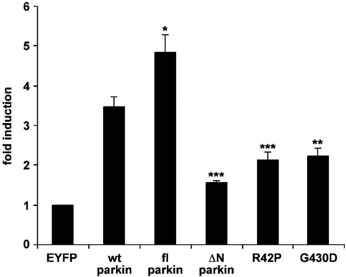
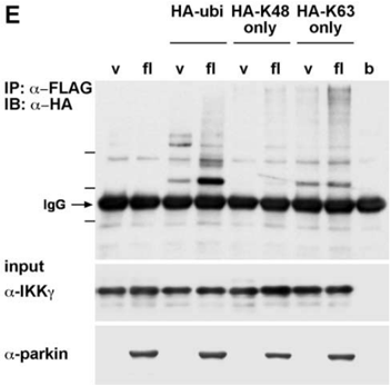
Mutations in the parkin gene are a major cause of autosomal recessive Parkinson's disease. Here we show that the E3 ubiquitin ligase parkin activates signaling through the IκB kinase (IKK)/nuclear factor κB (NF-κB) pathway. Our analysis revealed that activation of this signaling cascade is causally linked to the neuroprotective potential of parkin. Inhibition of NF-κB activation by an IκB super-repressor or a kinase-inactive IKKβ interferes with the neuroprotective activity of parkin. Furthermore, pathogenic parkin mutants with an impaired neuroprotective capacity show a reduced ability to stimulate NF-κB-dependent transcription. Finally, we present evidence that parkin interacts with and promotes degradation-independent ubiquitylation of IKKγ/NEMO (NF-κB essential modifier) and TRAF2 [TNF (tumor necrosis factor) receptor-associated factor 2], two critical components of the NF-κB pathway. Thus, our results support a direct link between the neuroprotective activity of parkin and ubiquitin signaling in the IKK/NF-κB pathway.

Overview

The claims and statistics of this paper haven’t been checked yet.

Run the check — free, no account needed. It reads the open-access text and checks the sources it cites.

A few free checks a day · papers already verified open instantly · results are public

Integrity signals

Registry

Crossref · 10.1523/jneurosci.5537-06.2007

Cites

2 of 2 references verified0 citation errors · 0 not checkable

Statistics

Figures

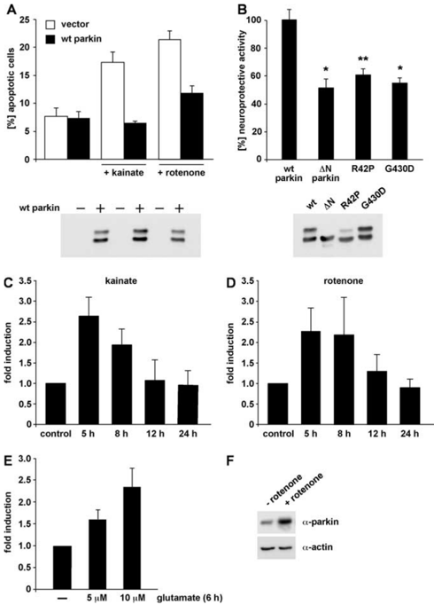
Figure 1 Parkin is a stress-inducible protein with neuroprotective capacity. A , B , Parkin has a neuroprotective capacity in cultured cells. A , SH-SY5Y cells were cotransfected with EYFP and wtparkin.At24haftertransfection,cellswereincubatedwithkainate(500 𝛍 M)orrotenone(10 𝛍 M) at 37°C for 3 h, fixed, permeabilized, and analyzed by indirect immunofluorescence. Activation of caspase-3 was detected using the anti-ACTIVE caspase-3 pAb. Shown is the percentage of apoptotic cells among the transfected cells. B , SH-SY5Y cells were cotransfected with EYFP and either wt parkin or the parkin mutants indicated. At 24 h after transfection, cells were incubated with kainate (500 𝛍 M) at 37°C for 3 h and analyzed by indirect immunofluorescence as described in A . * p < 0.01, ** p < 0.001 compared with cells expressing wt parkin. As a control for parkin expression, an aliquot of the cell lysates was immunoblotted with the antiparkin pAb hP1 (bottom). C-F , Endogenous parkin is upregulated after cellular stress. SH-SY5Y cells were incubated with 50 𝛍 M kainate ( C ) or 1 𝛍 M rotenone ( D ) for 3 h. Cells were harvested 5, 8, 12, and 24 h after the drug treatment, respectively. Total cellular RNA was isolated and subjected to quantitative RT-PCR using parkin-specific primers. The amount of RNA of each sample was normalized with respect to the endogenous housekeeping control gene 𝛃 -actin. Shownisthefoldincrease in the amount of parkin-specific mRNA compared with the untreated control. E , Primary cortical neurons derived from embryonic rat brain were incubated with glutamate (5 or 10 𝛍 M) for 6 h and then analyzed as described in C and D . F , SH-SY5Y cells were treated with rotenone (1 𝛍 M, 3 h) or mock treated, and expression of endogenous parkin was analyzed by Western blotting using the PRK8 mAb.
Figure 1 Parkin is a stress-inducible protein with neuroprotective capacity. A , B , Parkin has a neuroprotective capacity in cultured cells. A , SH-SY5Y cells were cotransfected with EYFP and wtparkin.At24haftertransfection,cellswereincubatedwithkainate(500 𝛍 M)orrotenone(10 𝛍 M) at 37°C for 3 h, fixed, permeabilized, and analyzed by indirect immunofluorescence. Activation of caspase-3 was detected using the anti-ACTIVE caspase-3 pAb. Shown is the percentage of apoptotic cells among the transfected cells. B , SH-SY5Y cells were cotransfected with EYFP and either wt parkin or the parkin mutants indicated. At 24 h after transfection, cells were incubated with kainate (500 𝛍 M) at 37°C for 3 h and analyzed by indirect immunofluorescence as described in A . * p < 0.01, ** p < 0.001 compared with cells expressing wt parkin. As a control for parkin expression, an aliquot of the cell lysates was immunoblotted with the antiparkin pAb hP1 (bottom). C-F , Endogenous parkin is upregulated after cellular stress. SH-SY5Y cells were incubated with 50 𝛍 M kainate ( C ) or 1 𝛍 M rotenone ( D ) for 3 h. Cells were harvested 5, 8, 12, and 24 h after the drug treatment, respectively. Total cellular RNA was isolated and subjected to quantitative RT-PCR using parkin-specific primers. The amount of RNA of each sample was normalized with respect to the endogenous housekeeping control gene 𝛃 -actin. Shownisthefoldincrease in the amount of parkin-specific mRNA compared with the untreated control. E , Primary cortical neurons derived from embryonic rat brain were incubated with glutamate (5 or 10 𝛍 M) for 6 h and then analyzed as described in C and D . F , SH-SY5Y cells were treated with rotenone (1 𝛍 M, 3 h) or mock treated, and expression of endogenous parkin was analyzed by Western blotting using the PRK8 mAb.CC-BY-NC-SA · From the PDF
Image 2
CC-BY-NC-SA · From the PDF
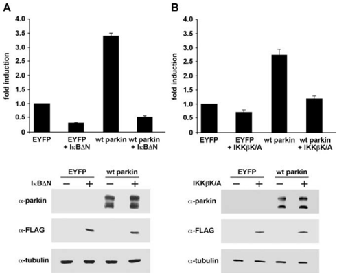
Figure 4 The effect of parkin on NF𝛋 B-dependent transcription can be antagonized by dominant-negative I 𝛋 B or kinase-inactive IKK 𝛃 . A , HEK293T cells were cotransfected with NF𝛋 Breporter plasmid, wt parkin, and the NF𝛋 Bsuper-repressor I 𝛋 B 𝚫 N. At 24 h after transfection, cell lysates were analyzed for luciferase activity. Shown is the fold induction of luciferase activity compared with the EYFP control. As a control for protein expression, aliquots of the cell lysates were immunoblotted with the anti-parkin pAb hP1 and anti-FLAG mAb (bottom panels). 𝛂 -Tubulin was used as a loading control. B , HEK293T cells were cotransfected with the NF𝛋 B reporter plasmid, wt parkin, and the kinase-inactive IKK 𝛃 mutant IKK 𝛃 K/A. Luciferase assay and immunoblotting were performed as described in A .
Figure 4 The effect of parkin on NF𝛋 B-dependent transcription can be antagonized by dominant-negative I 𝛋 B or kinase-inactive IKK 𝛃 . A , HEK293T cells were cotransfected with NF𝛋 Breporter plasmid, wt parkin, and the NF𝛋 Bsuper-repressor I 𝛋 B 𝚫 N. At 24 h after transfection, cell lysates were analyzed for luciferase activity. Shown is the fold induction of luciferase activity compared with the EYFP control. As a control for protein expression, aliquots of the cell lysates were immunoblotted with the anti-parkin pAb hP1 and anti-FLAG mAb (bottom panels). 𝛂 -Tubulin was used as a loading control. B , HEK293T cells were cotransfected with the NF𝛋 B reporter plasmid, wt parkin, and the kinase-inactive IKK 𝛃 mutant IKK 𝛃 K/A. Luciferase assay and immunoblotting were performed as described in A .CC-BY-NC-SA · From the PDF
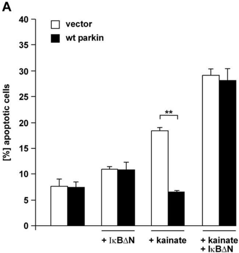
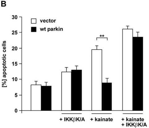
Figure 5 Activation of NF𝛋 B is essential for the neuroprotective effect of parkin. A , SHSY5Ycells were cotransfected with EYFP, wt parkin, and I 𝛋 B 𝚫 N. At 24 h after transfection, cells were incubated with kainate (500 𝛍 M) at 37°C for 3 h, fixed, permeabilized, and analyzed by indirect immunofluorescence. Activation of caspase-3 was detected by using the anti-ACTIVE caspase-3 pAb. Shown is the percentage of apoptotic cells among the transfected cells. ** p < 0.001 compared with EYFP-expressing cells. B , SH-SY5Y cells were cotransfected with EYFP, wt parkin, and the kinase-inactive IKK 𝛃 mutant IKK 𝛃 K/A and analyzed as described in A . ** p < 0.001 compared with EYFP-expressing cells.
Figure 5 Activation of NF𝛋 B is essential for the neuroprotective effect of parkin. A , SHSY5Ycells were cotransfected with EYFP, wt parkin, and I 𝛋 B 𝚫 N. At 24 h after transfection, cells were incubated with kainate (500 𝛍 M) at 37°C for 3 h, fixed, permeabilized, and analyzed by indirect immunofluorescence. Activation of caspase-3 was detected by using the anti-ACTIVE caspase-3 pAb. Shown is the percentage of apoptotic cells among the transfected cells. ** p < 0.001 compared with EYFP-expressing cells. B , SH-SY5Y cells were cotransfected with EYFP, wt parkin, and the kinase-inactive IKK 𝛃 mutant IKK 𝛃 K/A and analyzed as described in A . ** p < 0.001 compared with EYFP-expressing cells.CC-BY-NC-SA · From the PDF
Image 5
CC-BY-NC-SA · From the PDF
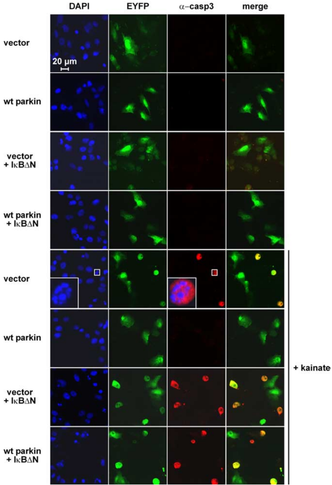
Figure 6 In the presence of the NF𝛋 B super-repressor I 𝛋 B 𝚫 N, parkin shows no neuroprotective activity. Immunofluorescence analysis of the experiment described in Figure 5 A . Activation of caspase-3 was detected by using the anti-ACTIVE caspase-3 pAb (red), and nuclei were stained with DAPI (blue). Scale bar, 20 𝛍 m
Figure 6 In the presence of the NF𝛋 B super-repressor I 𝛋 B 𝚫 N, parkin shows no neuroprotective activity. Immunofluorescence analysis of the experiment described in Figure 5 A . Activation of caspase-3 was detected by using the anti-ACTIVE caspase-3 pAb (red), and nuclei were stained with DAPI (blue). Scale bar, 20 𝛍 mCC-BY-NC-SA · From the PDF
Image 7
CC-BY-NC-SA · From the PDF
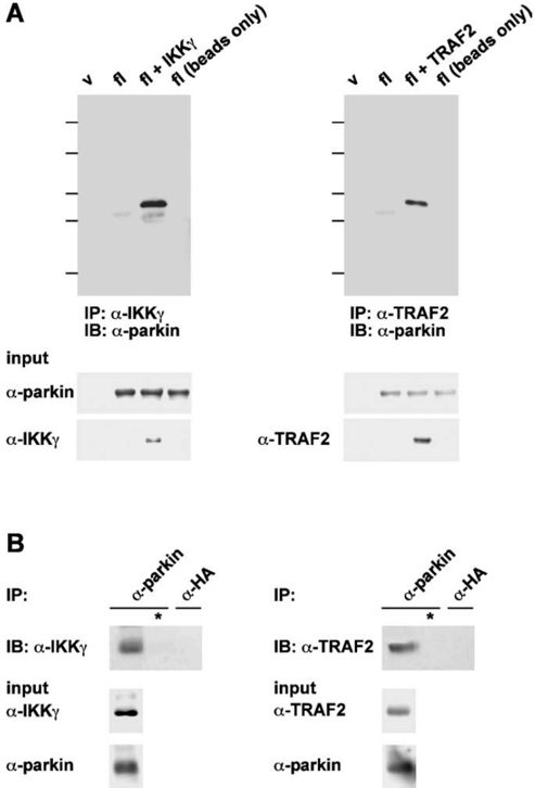
Figure8 Parkin is found in a complex together with IKK 𝛄 andTRAF2. A , HEK293T cells were cotransfected with full-length (fl) parkin and either IKK 𝛄 (top left panel) or TRAF2 (top right panel). One day after transfection, cells were harvested, lysed, and cleared by centrifugation. Equal protein amounts of the supernatant were incubated with a pAb against IKK 𝛄 or TRAF2 overnight at 4°C. Proteins present in the immunoprecipitates (IP) were resolved by SDS-PAGE and immunoblotted (IB) with the anti-parkin mAb PRK28. Aliquots of the supernatant were immunoblotted with antibodies against parkin (pAb hP1), IKK 𝛄 (mAb), and TRAF2 (mAb), respectively (input controls; bottom panels). B , Untransfected SH-SY5Y cells were lysed as described in A and incubated with the polyclonal anti-parkin antibody hP1 or with a polyclonal anti-HA antibody (both antibodies cross-linked to protein A-agarose) overnight at 4°C (* indicates buffer instead of cell lysate). Proteins present in the immunoprecipitates were resolved by SDS-PAGEandimmunoblottedwithmonoclonalantibodiesagainstIKK 𝛄 (left) or TRAF2 (right). For input controls, a parallel sample was analyzed by immunoprecipitation, followed by immunoblotting for the respective proteins. v, Vector control; b, buffer control (no lysate).
Figure8 Parkin is found in a complex together with IKK 𝛄 andTRAF2. A , HEK293T cells were cotransfected with full-length (fl) parkin and either IKK 𝛄 (top left panel) or TRAF2 (top right panel). One day after transfection, cells were harvested, lysed, and cleared by centrifugation. Equal protein amounts of the supernatant were incubated with a pAb against IKK 𝛄 or TRAF2 overnight at 4°C. Proteins present in the immunoprecipitates (IP) were resolved by SDS-PAGE and immunoblotted (IB) with the anti-parkin mAb PRK28. Aliquots of the supernatant were immunoblotted with antibodies against parkin (pAb hP1), IKK 𝛄 (mAb), and TRAF2 (mAb), respectively (input controls; bottom panels). B , Untransfected SH-SY5Y cells were lysed as described in A and incubated with the polyclonal anti-parkin antibody hP1 or with a polyclonal anti-HA antibody (both antibodies cross-linked to protein A-agarose) overnight at 4°C (* indicates buffer instead of cell lysate). Proteins present in the immunoprecipitates were resolved by SDS-PAGEandimmunoblottedwithmonoclonalantibodiesagainstIKK 𝛄 (left) or TRAF2 (right). For input controls, a parallel sample was analyzed by immunoprecipitation, followed by immunoblotting for the respective proteins. v, Vector control; b, buffer control (no lysate).CC-BY-NC-SA · From the PDF
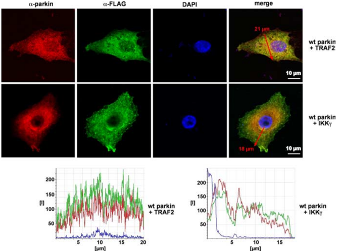
Figure 9 IKK 𝛄 and TRAF2 colocalize with parkin. SH-SY5Y cells attached to coverslips were transfected with wt parkin and FLAG-tagged IKK 𝛄 or TRAF2. One day after transfection, cells were fixed, permeabilized, and stained with the following antibodies: anti-parkin hP1 pAb (red) and anti-FLAG mAb (green). DAPI was added to the mounting medium to label nuclei. Red arrows indicate the regions in which intensity profiles (bottom panels) along a line were determined using Leica confocal software version 2.6.1. Scale bars, 10 𝛍 m
Figure 9 IKK 𝛄 and TRAF2 colocalize with parkin. SH-SY5Y cells attached to coverslips were transfected with wt parkin and FLAG-tagged IKK 𝛄 or TRAF2. One day after transfection, cells were fixed, permeabilized, and stained with the following antibodies: anti-parkin hP1 pAb (red) and anti-FLAG mAb (green). DAPI was added to the mounting medium to label nuclei. Red arrows indicate the regions in which intensity profiles (bottom panels) along a line were determined using Leica confocal software version 2.6.1. Scale bars, 10 𝛍 mCC-BY-NC-SA · From the PDF
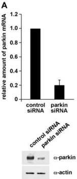
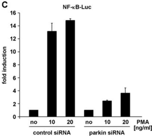
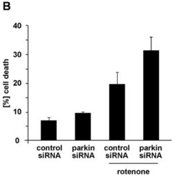
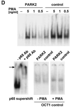
Figure 10 Loss of endogenous parkin increases cell death and compromises NF𝛋 B activation in response to stress. A , B , HEK293T cells were transfected with parkin-specific or control siRNA duplexes. A , At 48 h later, parkin mRNA levels were analyzed by RT-PCR (described in Fig. 1 C ), and endogenous parkin protein was analyzed by Western blotting (bottom) as described in Figure 1 F . B , Parallel cultures were exposed to rotenone (0.05 𝛍 M, 3 h), and, after additional 24h,cell viability was determined by trypan blue exclusion. C , HEK293T cells cotransfected with parkin-specific siRNA and the NF𝛋 B reporter plasmid were incubated with PMA (10 and 20 ng/ml, 3 h). At 5 h later, cells were harvested and analyzed for luciferase activity. D , Fibroblasts from a PARK2 patient or control fibroblasts were exposed to PMA for 30 min. Nuclear extracts were prepared, and NF𝛋 B binding activity was determined by electrophoretic mobility shift assays as described in Figure 2 E . Bottom left, Supershift induced by a pAb against p65 in nuclear extracts from PMA-treated control fibroblasts. Bottom right, As a control, OCT1 binding activity was determined in nuclear extracts from untreated and PMA-treated PARK2 and control fibroblasts.
Figure 10 Loss of endogenous parkin increases cell death and compromises NF𝛋 B activation in response to stress. A , B , HEK293T cells were transfected with parkin-specific or control siRNA duplexes. A , At 48 h later, parkin mRNA levels were analyzed by RT-PCR (described in Fig. 1 C ), and endogenous parkin protein was analyzed by Western blotting (bottom) as described in Figure 1 F . B , Parallel cultures were exposed to rotenone (0.05 𝛍 M, 3 h), and, after additional 24h,cell viability was determined by trypan blue exclusion. C , HEK293T cells cotransfected with parkin-specific siRNA and the NF𝛋 B reporter plasmid were incubated with PMA (10 and 20 ng/ml, 3 h). At 5 h later, cells were harvested and analyzed for luciferase activity. D , Fibroblasts from a PARK2 patient or control fibroblasts were exposed to PMA for 30 min. Nuclear extracts were prepared, and NF𝛋 B binding activity was determined by electrophoretic mobility shift assays as described in Figure 2 E . Bottom left, Supershift induced by a pAb against p65 in nuclear extracts from PMA-treated control fibroblasts. Bottom right, As a control, OCT1 binding activity was determined in nuclear extracts from untreated and PMA-treated PARK2 and control fibroblasts.CC-BY-NC-SA · From the PDF
Image 11
CC-BY-NC-SA · From the PDF
Image 12
CC-BY-NC-SA · From the PDF
Image 13
CC-BY-NC-SA · From the PDF
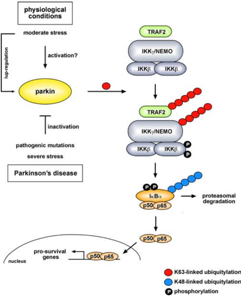
Figure 11 Model of the functional role of parkin. Under physiological conditions, parkin promotesregulatoryubiquitylationofTRAF2andIKK 𝛄 duringstress, leading to the activation of NF𝛋 B. As a consequence, transcription of prosurvival genes is upregulated. The activity of parkin can be modulated by stress. Under moderate stress, parkin is upregulated. For an immediate response, it is conceivable that parkin activity can be regulated by posttranslational modifications. In contrast, severe proteotoxic stress, for example induced by oxidized dopamine, causes misfolding and thus inactivation of parkin. Mutations in the parkin gene linked to familial PD also interfere with the capacity of parkin to stimulate the IKK/NF𝛋 B signaling pathway.
Figure 11 Model of the functional role of parkin. Under physiological conditions, parkin promotesregulatoryubiquitylationofTRAF2andIKK 𝛄 duringstress, leading to the activation of NF𝛋 B. As a consequence, transcription of prosurvival genes is upregulated. The activity of parkin can be modulated by stress. Under moderate stress, parkin is upregulated. For an immediate response, it is conceivable that parkin activity can be regulated by posttranslational modifications. In contrast, severe proteotoxic stress, for example induced by oxidized dopamine, causes misfolding and thus inactivation of parkin. Mutations in the parkin gene linked to familial PD also interfere with the capacity of parkin to stimulate the IKK/NF𝛋 B signaling pathway.CC-BY-NC-SA · From the PDF

Verification

Claims the paper makes

The claims and statistics of this paper haven’t been checked yet.

Run the check — free, no account needed. It reads the open-access text and checks the sources it cites.

A few free checks a day · papers already verified open instantly · results are public

Claims other papers make about this one

Each is checked against this paper’s own text: does it say what the citing paper says it does?

Self-check

Percentages, sums and reported differences re-computed from the paper’s own numbers.

The self-check needs the paper’s full text.

Citations

Cites

Show all 2 references

Research works printed but not linked: 47

  1. 10.1083/jcb.200502113 · DOI
  2. 10.1038/ncb0805-758 · DOI · Paper page
  3. 10.1093/hmg/ddg044 · DOI
  4. 10.1126/science.284.5412.309 · DOI
  5. 10.1016/S0092-8674(00)00126-4 · DOI · Paper page
  6. 10.1074/jbc.M413591200 · DOI
  7. 10.1042/BJ20031377 · DOI
  8. 10.1038/sj.emboj.7600808 · DOI
  9. 10.1101/gad.1228704 · DOI · Paper page
  10. 10.1186/1471-2202-5-14 · DOI
  11. 10.1111/j.1471-4159.2004.02854.x · DOI
  12. 10.1111/j.1471-4159.2004.02445.x · DOI
  13. 10.1074/jbc.C000447200 · DOI
  14. 10.1016/j.bbamcr.2005.05.009 · DOI
  15. 10.1038/ni0302-221 · DOI · Paper page
  16. 10.1038/33416 · DOI · Paper page
  17. Krappmann, 1996, Different mechanisms control signal-induced degradation and basal turnover of the NF-kappaB inhibitor IkappaB alpha in vivo, EMBO J, 15, 6716, 10.1002/j.1460-2075.1996.tb01061.x · DOI
  18. 10.1074/jbc.M003902200 · DOI
  19. 10.1128/MCB.21.19.6640-6650.2001 · DOI
  20. 10.1038/nm1314 · DOI
  21. 10.1073/pnas.0405313101 · DOI
  22. 10.1093/hmg/ddh089 · DOI
  23. 10.1074/jbc.M510393200 · DOI · Paper page
  24. 10.1016/0896-6273(93)90315-I · DOI · Paper page
  25. 10.1046/j.1471-4159.2000.740443.x · DOI
  26. 10.1093/hmg/ddh012 · DOI
  27. 10.1016/j.neulet.2006.02.035 · DOI
  28. 10.1074/jbc.M306889200 · DOI
  29. 10.1016/S0896-6273(02)01125-X · DOI
  30. 10.1016/j.cbpa.2004.09.009 · DOI
  31. 10.1016/j.cub.2004.09.074 · DOI
  32. Rawal, 2003, New parkin mutations and atypical phenotypes in families with autosomal recessive parkinsonism, Neurology, 60, 1378, 10.1212/01.WNL.0000056167.89221.BE · DOI
  33. 10.1074/jbc.M512649200 · DOI
  34. 10.1126/science.7544915 · DOI
  35. 10.1016/S0002-9440(10)61113-3 · DOI
  36. 10.1002/1531-8249(199905)45:5<668::AID-ANA19>3.0.CO;2-Z · DOI
  37. 10.1093/hmg/ddi292 · DOI
  38. 10.1016/S0896-6273(03)00084-9 · DOI
  39. 10.1007/s00439-005-0038-4 · DOI
  40. 10.1128/MCB.23.6.2029-2041.2003 · DOI
  41. 10.1093/hmg/ddi413 · DOI
  42. 10.1007/s00439-003-1074-6 · DOI
  43. Winklhofer KF Tatzelt J (2006) in Molecular chaperones in health and diseases, The role of chaperones in Parkinson's disease and prion diseases, ed Gaestel M (Springer, Berlin), pp 221–258. · DOI
  44. 10.1074/jbc.M104873200 · DOI
  45. 10.1074/jbc.M306769200 · DOI
  46. 10.1074/jbc.M407724200 · DOI
  47. 10.1016/S0896-6273(03)00143-0 · DOI

Cited by

Sorted by citations

Metadata

Authors
Henn, I. H., Bouman, L., Schlehe, J. S., Schlierf, A., Schramm, J. E., Wegener, E., Nakaso, K., Culmsee, C., Berninger, B., Krappmann, D., Tatzelt, J., & Winklhofer, K. F.
Venue
The Journal of Neuroscience
Published
2007-02-21
Volume
27
Issue
8
Pages
1868-1878
ISSN
  • 0270-6474
  • 1529-2401
Cited by
173
References
2
Language
en

About these checks

Registry, retraction, Cites and statistics results are computed, not judged, with one exception: a printed reference that no index lists is read once by a model before it counts as a citation error. Claims are extracted by a model and kept only when their quote appears verbatim in the text; each cited source is then checked, with a model’s help, for whether it says what the claim relies on. Those verdicts can be wrong — read the claim’s quote and the cited source before relying on one.

Statistics and arithmetic are recomputed, not judged.

Confidence bands are shown only for fields calibrated on our annotated test set. None is calibrated yet, so no band is shown.

What counts as a claim ›How we measure confidence ›How we check this ›

Are you an author, or do you see a problem with this page? Request a review