

The DOI matches its Crossref record; 455 of the 457 references it cites were found in a registry or an index.
Sarcoma treatment in the era of molecular medicine
Open accessAbstract
Sarcomas are heterogeneous and clinically challenging soft tissue and bone cancers. Although constituting only 1% of all human malignancies, sarcomas represent the second most common type of solid tumors in children and adolescents and comprise an important group of secondary malignancies. More than 100 histological subtypes have been characterized to date, and many more are being discovered due to molecular profiling. Owing to their mostly aggressive biological behavior, relative rarity, and occurrence at virtually every anatomical site, many sarcoma subtypes are in particular difficult‐to‐treat categories. Current multimodal treatment concepts combine surgery, polychemotherapy (with/without local hyperthermia), irradiation, immunotherapy, and/or targeted therapeutics. Recent scientific advancements have enabled a more precise molecular characterization of sarcoma subtypes and revealed novel therapeutic targets and prognostic/predictive biomarkers. This review aims at providing a comprehensive overview of the latest advances in the molecular biology of sarcomas and their effects on clinical oncology; it is meant for a broad readership ranging from novices to experts in the field of sarcoma.
Overview
The claims and statistics of this paper haven’t been checked yet.
Run the check — free, no account needed. It reads the open-access text and checks the sources it cites.
Integrity signals
Registry
Cites
Statistics
Figures
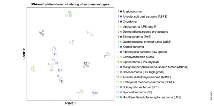
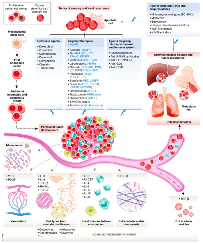
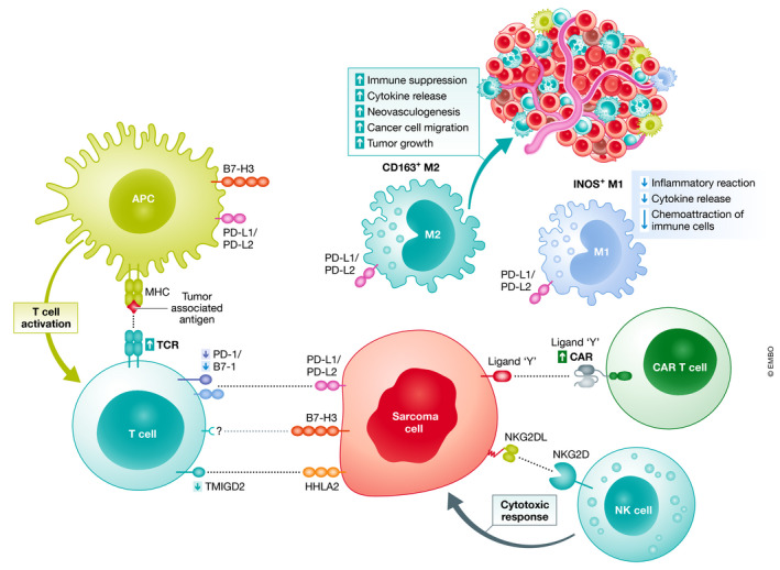
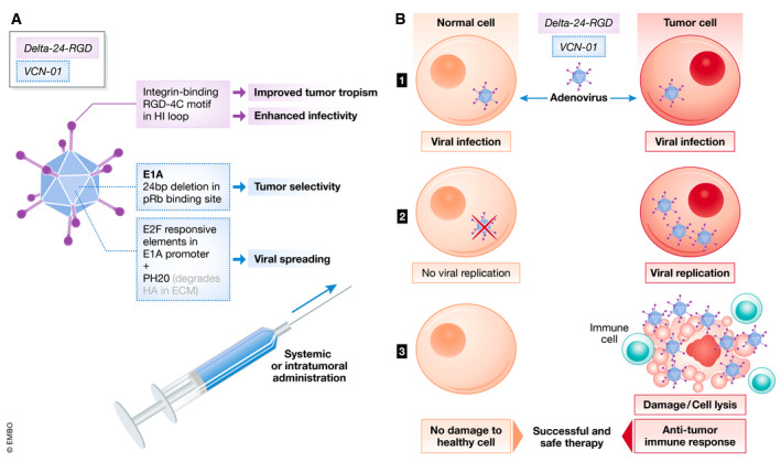
Verification
Claims the paper makes
The claims and statistics of this paper haven’t been checked yet.
Run the check — free, no account needed. It reads the open-access text and checks the sources it cites.
Claims other papers make about this one
Each is checked against this paper’s own text: does it say what the citing paper says it does?
In the citing paper
“This seed and soil phenomenon is driven by a microenvironment that modulates osteosarcoma cell behaviour and facilitates proliferation, quiescence, invasion, migration, and drug resistance 15,51,52,53 and contributes to their intrinsic heterogeneity 54,55 .”
From Osteosarcoma (2022) · cites this paper as [53]
Self-check
Percentages, sums and reported differences re-computed from the paper’s own numbers.
The self-check needs the paper’s full text.
Citations
Cites
1 retracted
Mu, J., Sun, P., Ma, Z., & Sun, P. (2019) · Cell Death and Disease · doi:10.1038/s41419-019-2170-4 · Cited by 36
RetractedThéry, C., Witwer, K. W., Aikawa, E., Alcaraz, M. J., Anderson, J. D., Andriantsitohaina, R., Antoniou, A., Arab, T., Archer, F., Atkin‐Smith, G. K., et al. (2018) · Journal of Extracellular Vesicles · doi:10.1080/20013078.2018.1535750 · Cited by 11,099
Self-citation3D bioprinting of tissues and organs
Murphy, S. V., & Atala, A. (2014) · Nature Biotechnology · doi:10.1038/nbt.2958 · Cited by 7,034
Master Transcription Factors and Mediator Establish Super-Enhancers at Key Cell Identity Genes
Whyte, W. A., Orlando, D. A., Hnisz, D., Abraham, B. J., Lin, C. Y., Kagey, M. H., Rahl, P. B., Lee, T. I., & Young, R. A. (2013) · Cell · doi:10.1016/j.cell.2013.03.035 · Cited by 4,143
Approaches to treat immune hot, altered and cold tumours with combination immunotherapies
Galon, J., & Bruni, D. (2019) · Nature Reviews Drug Discovery · doi:10.1038/s41573-018-0007-y · Cited by 3,520
DNA methylation-based classification of central nervous system tumours
Capper, D., Jones, D., Sill, M., Hovestadt, V., Schrimpf, D., Sturm, D., Koelsche, C., Sahm, F., Chávez, L., Reuß, D., et al. (2018) · Nature · doi:10.1038/nature26000 · Cited by 3,114
Self-citationA view on drug resistance in cancer
Vasan, N., Baselga, J., & Hyman, D. M. (2019) · Nature · doi:10.1038/s41586-019-1730-1 · Cited by 2,770
Talimogene Laherparepvec Improves Durable Response Rate in Patients With Advanced Melanoma
Andtbacka, R. H. I., Kaufman, H. L., Collichio, F. A., AMATRUDA, T. T., Senzer, N. N., Chesney, J. A., Delman, K. A., S̄pitler, L. E., Puzanov, I. J., Agarwala, S. S., et al. (2015) · Journal of Clinical Oncology · doi:10.1200/jco.2014.58.3377 · Cited by 2,728
The human tumor microbiome is composed of tumor type–specific intracellular bacteria
Nejman, D., Livyatan, I., Fuks, G., Gavert, N., Zwang, Y., Geller, L. T., Rotter-Maskowitz, A., Weiser, R., Mallel, G., Gigi, E., et al. (2020) · Science · doi:10.1126/science.aay9189 · Cited by 2,464
Self-citationPatient-derived organoids model treatment response of metastatic gastrointestinal cancers
Vlachogiannis, G., Hedayat, S., Vatsiou, A., Jamin, Y., Fernández-Mateos, J., Khan, K., Lampis, A., Eason, K., Huntingford, I., Burke, R., et al. (2018) · Science · doi:10.1126/science.aao2774 · Cited by 2,096
Research works printed but not linked: 1
- Heymann D (2014) Bone Cancer. Academic Press, San Diego, California, USA
Cited by
Sorted by citations
Items 201–300 of 316
Heater, N. K., Okuno, S. H., Robinson, S. I., Attia, S., Seetharam, M., Siontis, B. L., Yoon, J. M., Chawla, S. P., Milhem, M. M., Monga, V., et al. (2023) · Journal of Clinical Medicine · doi:10.3390/jcm12072561 · Cited by 2
Radak, M., Ghamari, N., & Fallahi, H. (2023) · Biosystems · doi:10.1016/j.biosystems.2023.105093 · Cited by 2
Chen, C., Packer, S., Hughes, G., Edeghere, O., Oliver, I., & Birney, E. (2021) · SSRN Electronic Journal · doi:10.2139/ssrn.3867682 · Cited by 2
Carreño-Gonzalez, M. J., Hanssen, K. M., Sadik, A., Henneberg, A., Knott, M. M. L., Ehlers, A. C., Ceranski, A. K., Kolodynska, Z., Zimmermann, M., Imle, R., et al. (2025) · bioRxiv (Cold Spring Harbor Laboratory) · doi:10.1101/2025.05.16.654502 · Cited by 2
Self-citationJulson, J. R., Hutchins, S. C., Quinn, C. H., Beierle, A. M., Bownes, L. V., Stewart, J. E., Aye, J. M., Yoon, K. J., & Beierle, E. A. (2023) · Journal of Pediatric Surgery · doi:10.1016/j.jpedsurg.2023.10.004 · Cited by 2
Lanzi, C., Pasquali, S., & Cassinelli, G. (2024) · Proteoglycan Research · doi:10.1002/pgr2.70005 · Cited by 3
Beyond the benign: A rare case report of myxoid pleomorphic liposarcoma
Ahmed, A., Ammar, M., Choudary, M. H. S., Sadiq, M. N., Ahmad, R. U., & Aziz, N. (2025) · Radiology Case Reports · doi:10.1016/j.radcr.2025.01.056 · Cited by 1
de Paula, F. E., Bonatelli, M., Reis, M. T., Rodrigues, K. E. D. S., van Kempen, L. C. L. T., Teixeira, G. R., & Reis, R. M. (2025) · Therapeutic Advances in Medical Oncology · doi:10.1177/17588359251318159 · Cited by 1
Lorimer, W. G., Lottering, M., Halkett, G. K. B., & O’Connor, M. (2025) · Cancer Survivorship Research & Care · doi:10.1080/28352610.2025.2473316 · Cited by 1
Ailts, I., Golafshar, M. A., Kunze, K. L., Klint, M. A., Barrus, K., Nussbaum, R. L., Esplin, E. D., Leach, B., Young, S., Samadder, N. J., et al. (2025) · International Journal of Molecular Sciences · doi:10.3390/ijms26072839 · Cited by 1
Dong, Y., Marin, T., Zhuo, Y., Najem, E., Beddok, A., Rozenblum, L., Moteabbed, M., Grogg, K. S., Xing, F., Woo, J., et al. (2025) · Medical Physics · doi:10.1002/mp.17865 · Cited by 1
Li, Z., Guo, J., Zhu, S., Zou, Y., Ma, W., Niu, J., Liu, R., & Zhao, K. (2025) · The FASEB Journal · doi:10.1096/fj.202501151rr · Cited by 1
Clinicopathologic Analysis of Sarcomas in the Oral and Maxillofacial Region: A Systematic Review
Ferreira, I. V., de Lima‐Souza, R. A., Kimura, T. D. C., Tincani, Á. J., Cattan, M. E. S., Antolini‐Tavares, A., ALTEMANI, A. M. D. A. M., & Mariano, F. V. (2025) · Oral Diseases · doi:10.1111/odi.70103 · Cited by 1
Functional Assays to Guide Personalized Oncological Treatment of Patients with Soft-Tissue Sarcomas
Yarar, S., & Tsagozis, P. (2025) · Cancers · doi:10.3390/cancers17213452 · Cited by 1
de Sousa, R. W. R., dos Santos, I. L., Rodrigues, D. C. D. N., do Nascimento, M. L. L. B., Conceição, M. L. P., de Freitas, M. M., Fernandes, G. D. S., Sousa, R. D. C. D. L., Souza, R. P., Pereira, I. C., et al. (2025) · Physiology & Behavior · doi:10.1016/j.physbeh.2025.115153 · Cited by 1
Xiao, J.-F., Ishaya, K., Ko, E. Y., Fowler, A., Broz, M. T., & Guarnerio, J. (2025) · Cancer Research Communications · doi:10.1158/2767-9764.crc-25-0301 · Cited by 1
Mitochondria in sarcoma and carcinoma: Achilles’ heel or Hercules’ strength?
Ashrafi, M. R., Cayrefourcq, L., Rajabi, S., & Alix‐Panabières, C. (2026) · Seminars in Cancer Biology · doi:10.1016/j.semcancer.2026.03.004 · Cited by 1
Rahimizadeh, P., MIALLOT, R., Bellis, C. D., Jolivet, P., & Przybył, J. (2026) · Journal of Cellular Physiology · doi:10.1002/jcp.70182 · Cited by 1
Onesti, C. E., Casini, B., Chiara, A. D., Fausti, V., Luksch, R., Milano, G. M., Palmerini, E., Vari, S., Ceddia, S., Riva, F., et al. (2026) · Frontiers in Oncology · doi:10.3389/fonc.2026.1641281 · Cited by 1
Jiménez‐Rodriguez, T. W., Ahmetaj, L. N., Alfaya, T., Boni, E., Bugadze, L., Chauhan, J., Vecillas, L. D. L., Ferastraoaru, D. E., Izquierdo, E., Karagiannis, S. N., et al. (2026) · Allergy · doi:10.1111/all.70421 · Cited by 1
Zhang, Z.-C., Guo, J., Zhang, N., Wang, Z.-Q., Lou, G., Cui, B., & Yang, C. Q. (2021) · Frontiers in Immunology · doi:10.3389/fimmu.2021.763791 · Cited by 1
Paediatric head and neck malignant neoplasms: A brazilian retrospective study
da CUNHA, W. A., Corazza, A., Rezende, K., Bönecker, M., & Gallottini, M. H. C. (2023) · Medicina oral, patología oral y cirugía bucal · doi:10.4317/medoral.25614 · Cited by 1
Single-Cell Sequencing in Cancer Research: Challenges and Opportunities
Prado‐Lòpez, S. (2023) · Springer eBooks · doi:10.1007/978-3-030-80962-1_143-1 · Cited by 1
Khalil, O. R. S., Omar, B. M. M., Khalil, L., Tarabieh, D., Al-Karaja, L., Alkhatib, H., & Asad, D. (2023) · Annals of Medicine and Surgery · doi:10.1097/ms9.0000000000000898 · Cited by 1
In-Silico Identification of Novel Pharmacological Synergisms: The Trabectedin Case
Mannarino, L., Ravasio, N., D’Incalci, M., Marchini, S., & Masseroli, M. (2024) · International Journal of Molecular Sciences · doi:10.3390/ijms25042059 · Cited by 1
Yadgarov, M. Y., Бондоевич, Б. Л., Ракова, Е., Kachanov, D. Y., & Likar, Y. N. (2024) · Clinical and Translational Imaging · doi:10.1007/s40336-024-00620-9 · Cited by 1
Goldsmith, R. M., Xing, J. L., Heal, C., de la Maza, M. C., & Stea, B. (2024) · Advances in Radiation Oncology · doi:10.1016/j.adro.2024.101517 · Cited by 1
Arid1a Loss Enhances Disease Progression in a Murine Model of Osteosarcoma
Fatema, K., Wang, Y., Pavek, A., Larson, Z., Nartker, C., Plyler, S., Jeppesen, A., Mehling, B., Capecchi, M. R., Jones, K. B., et al. (2024) · Cancers · doi:10.3390/cancers16152725 · Cited by 2
Undifferentiated sarcoma of thyroid gland: A case report and literature review
Qianlang, R., Hu, R., Fusheng, L., & Li, X. (2024) · Asian Journal of Surgery · doi:10.1016/j.asjsur.2024.07.289 · Cited by 1
Cipu, R. I., Stănişteanu, M.-L., Andrei, M.-A., Banciu, D. D., & Banciu, A. (2024) · Journal of Functional Biomaterials · doi:10.3390/jfb15100298 · Cited by 1
Assessing traditional Chinese medicine from a molecular perspective
Su, M., Wang, W., Xudong, S., Li, L., & Tan, W. (2024) · Journal of Traditional Chinese Medical Sciences · doi:10.1016/j.jtcms.2024.12.004 · Cited by 1
Ji, M., Fan, D., Yuan, Y., Dang, X., Feng, X., Yang, W., Dang, X., Xu, Y., & Wang, J. (2025) · Cancer Biotherapy and Radiopharmaceuticals · doi:10.1089/cbr.2025.0043 · Cited by 1
LAT1-Targeted Alpha Therapy Using 211At-AAMT for Bone and Soft Tissue Sarcomas
Takami, H., Imura, Y., Outani, H., Nakai, S., Inoue, A., Kotani, Y., Okada, S., & Kaneda‐Nakashima, K. (2025) · International Journal of Molecular Sciences · doi:10.3390/ijms26178599 · Cited by 1
Status: nosocomial transmission and prevention of SARS‐CoV‐2 in a Danish context
Calum, H., Sode, L. P., & Pedersen, M. (2021) · Apmis · doi:10.1111/apm.13160
Bone Tumours - A Comprehensive Review of Selected Topics
Amarasekera, H. (2022) · IntechOpen eBooks · doi:10.5772/intechopen.100923
Redox status of blood lymphocytes in patients with soft tissue sarcomas
Goroshinskaya, I. A., Kaplieva, I. V., Frantsiyants, E. M., Vashchenko, L. N., Kachesova, P. S., Lapteva, T. O., Ausheva, T. V., Nemashkalova, L. A., Trepitaki, L. K., & Pandova, O. V. (2023) · Bone and soft tissue sarcomas tumors of the skin · doi:10.17650/2782-3687-2023-15-3-21-33 · Cited by 1
Editorial: Extracellular Vesicles in Bone Oncology
Perut, F., Tasso, R., & Mannerström, B. (2022) · Frontiers in Oncology · doi:10.3389/fonc.2022.861335 · Cited by 1
Self-citationThiesen, A. H., Domanskyi, S., pour, A. F., Zhang, J., Sheridan, T., Neuhauser, S. B., Stetson, A. E., Dannheim, K., Cameron, D. B., Ahn, S. S., et al. (2025) · medRxiv · doi:10.1101/2025.06.10.25328700 · Cited by 1
Morice, S., Mullard, M., Brion, R., Dupuy, M., Renault, S., Tesfaye, R. A., Ory, B., Rédiní, F., & Verrecchia, F. (2020) · doi:10.21203/rs.3.rs-111013/v1
Self-citationSystems biology analysis identifies TCF7L1 as a key regulator of metastasis in Ewing sarcomaPreprint
Cidre‐Aranaz, F., Li, J., Hölting, T. L. B., Orth, M. F., Imle, R., Kutschmann, S., Ammirati, G. E. M., Ceranski, A. K., Carreño-Gonzalez, M. J., Kasan, M., et al. (2021) · bioRxiv (Cold Spring Harbor Laboratory) · doi:10.1101/2021.02.25.432862
Self-citationLiu, Y., Liu, C., & Yu, A. (2021) · Research Square · doi:10.21203/rs.3.rs-882267/v1
Fatehpur, S., Addicks, J. P., Tran, V. K., Verheggen, R., & Mirzaie, M. (2021) · Journal of Medical - Clinical Research & Reviews · doi:10.33425/2639-944x.1222
Shi, D., Mu, S., Pu, F., Zhong, B., Hu, B., Shao, Z., Zhang, Z., & Liu, J. (2022) · Research Square · doi:10.21203/rs.3.rs-1540777/v1
RETROPERITONEAL HIGH-GRADE SARCOMA IN A 76-YEARS-OLD MAN: Case Report.
Maruca, A. M., Sani, V., Troia, A. D., Traina, L., Oppici, D., & Carcoforo, P. (2022) · GLOBAL JOURNAL FOR RESEARCH ANALYSIS · doi:10.36106/gjra/0506124
Fuentes, T. B., Rivera-Saldívar, G., & Díaz, K. C. (2022) · Orthotips AMOT · doi:10.35366/105506
Circulating Tumor Cells and ctDNA in Sarcomas
Jubelin, C., Cochonneau, D., Moranton, E., Muñoz-García, J., & Heymann, D. (2022) · Springer eBooks · doi:10.1007/978-3-030-93084-4_12
Self-citationDrug Targeting of Chromosomal Translocations in Fusion-Positive Sarcoma
Richter, G. H. S. (2022) · IntechOpen eBooks · doi:10.5772/intechopen.106671
Undifferentiated Pleomorphic Sarcoma. From Supposition to Certainty: a Case Report
MEHEDINTU-IONESCU, M., ALEXA, G., TOADER, H., & COCIU, C. (2022) · Medicina Moderna - Modern Medicine · doi:10.31689/rmm.2022.29.4.339
Lokalisierte Weichgewebesarkome
Podleska, L. E., Bauer, S., Pöttgen, C., Streitbürger, A., Hardes, J., & Täger, G. (2023) · best practice onkologie · doi:10.1007/s11654-022-00458-5
Moyers, J. T., Pestana, R. C., Roszik, J., Hong, D. S., Naing, A., Fu, S., Piha-Paul, S. A., Yap, T. A., Karp, D. D., Rodon, J. A., et al. (2023) · doi:10.1158/1078-0432.c.6532743
Moyers, J. T., Pestana, R. C., Roszik, J., Hong, D. S., Naing, A., Fu, S., Piha-Paul, S. A., Yap, T. A., Karp, D. D., Rodon, J. A., et al. (2023) · doi:10.1158/1078-0432.c.6532743.v1
Morales‐Molina, Á., Rodríguez-Milla, M. Á., Gambera, S., Cejalvo, T., de Andrés, B., Gaspar, M.-L., & Garcı́a-Castro, J. (2023) · doi:10.1158/2767-9764.c.6551258
Self-citationMeet the Editorial Board Member
Heymann, D. (2023) · Current Medicinal Chemistry · doi:10.2174/092986733031230419122610
Self-citationPrinciples of Monoclonal and Small Molecular Targeting Agents for Pediatric Cancer Management
Wills, C. A., & Grant, C. N. (2023) · Springer eBooks · doi:10.1007/978-3-030-71113-9_38-1
Schöffski, P., Bahleda, R., Wagner, A. J., Burgess, M. A., Junker, N., Chisamore, M. J., Peterson, P. M., Szpurka, A. M., Ceccarelli, M., & Tap, W. D. (2023) · doi:10.1158/1078-0432.c.6749982.v1
Special Issue: “Pediatric Orthopedic Malignancy: Types, Symptoms, and Treatment”
Aiba, H., Miwa, S., Murakami, H., & Kimura, H. J. (2023) · Children · doi:10.3390/children10091545
Gopal, S., Imtiaz, Z., Ijishakin, O., Louise-Oluwasanmi, O., Berhe, H. S., Emmanuel, O. K., Oni, E., Agbo, G. J., Gabriel, K. L., Ezeamii, V. C., et al. (2023) · Journal of Advances in Medicine and Medical Research · doi:10.9734/jammr/2023/v35i225244
A 37-Year-Old Man with Myofibroblast Sarcoma Combined with Pleural Maculopathy: Case Report
Tu, D., Zhang, T., Wang, Y., Kang, D., & Li, H. (2023) · Case Reports in Oncology · doi:10.1159/000533554
STUDY THE GROWTH OF HUMAN SOFT TISSUE SARCOMA XENOGFAFT IN IMMUNODEFICIENCY MICE BALB/C NUDE
Владимировна, Г. А., Ausheva, T. V., Alikhanova, S. S., & Rostorguev, V. E. (2023) · doi:10.37539/231006.2023.80.54.003
Goroshinskaya, I. A., Kaplieva, I. V., Frantsiyants, E. M., Vashchenko, L. N., Lapteva, T. O., Ausheva, T. V., Nemashkalova, L. A., Kachesova, P. S., & Kozel, Y. Y. (2023) · Research and Practical Medicine Journal · doi:10.17709/2410-1893-2023-10-4-1
Predicting prognosis and immune status in sarcomas by identifying necroptosis-related lncRNAs
Wang, Z., He, A., Lu, Z., Xu, W., Wu, G., & Peng, T. (2024) · Aging · doi:10.18632/aging.205383
Lv, J., Qin, X., Wang, J., Li, J., Bai, J., & Lan, Y. (2024) · Research Square · doi:10.21203/rs.3.rs-3870105/v1
Role of Neuropeptides in Sarcomas
Galoian, K. A., Denny, C., Wagner, J. D., & Mosle, S. G. (2023) · Neurochemical Journal · doi:10.1134/s1819712423040104
A Comprehensive Review on the Role of Lurbinectedin in Soft Tissue Sarcomas
Khoury, R., Assi, T., Ibrahim, R., Ibrahim, T., Verret, B., Henon, C., Bahleda, R., & Cesne, A. L. (2024) · Current Treatment Options in Oncology · doi:10.1007/s11864-024-01178-4
Psychological Distress among Patients with Suspected Sarcoma Requiring Biopsy Confirmation
Ng, Y. H. (2023) · International Journal of Science and Research (IJSR) · doi:10.21275/sr23630155703
Mesenchymal Stem Cells and Sarcoma
Hatina, J., Kripnerová, M., Dejmek, J., Dolejšová, M., & Pešta, M. (2024) · Elsevier eBooks · doi:10.1016/b978-0-443-15717-2.00116-5
Li, X., Du, Y., Deng, Q., Cong, C., Zhang, Y., Li, Y., & Wang, D. (2024) · Research Square · doi:10.21203/rs.3.rs-4559224/v1
Ren, S., Pan, R., & Wang, Z. (2024) · Discover Oncology · doi:10.1007/s12672-024-01292-7
Ubiquitin specific protease 3,7 in bone cancer and squamous cell carcinoma
Velmurugan, S., Pauline, R., Arumugam, S., Muthuvel, R., Raja, S. A., Kulanthaivel, L., & Subbaraj, G. K. (2024) · Elsevier eBooks · doi:10.1016/b978-0-443-30098-1.00010-0
Lv, J., Qin, X., Wang, J., Li, J., Bai, J., & Lan, Y. (2024) · Medicine · doi:10.1097/md.0000000000040519
Coloma, R. N., Niler, S. P., Gustavo, F., Chavez-Velasquez, A., & Zavaleta-Corvera, C. (2024) · Pirogov Russian Journal of Surgery · doi:10.17116/hirurgia202412138
Hasnain, M., Bakhtawar, S., Khan, M., Ahmad, S., Raziq, M., Ashfaq, M., Khan, M. A., Shah, S. A., Halimi, S. M. A., & Farooq, I. (2025) · International Journal of Basic & Clinical Pharmacology · doi:10.18203/2319-2003.ijbcp20250475
Orientações Práticas sobre a Deteção de Sarcomas de Fusão NTRK: Estado Atual e Desafios Diagnósticos
Fernandes, I., Macedo, D., Gouveia, E. J., Ferreira, A. M., Lima, J., Lopez, D., Melo-Alvim, C., Carvalho, A., Freitas-Tavares, P., Rodrigues‐Santos, P., et al. (2025) · Acta Médica Portuguesa · doi:10.20344/amp.21925
Chen, M., Li, C., & Xu, Y. (2025) · Archives of Medical Science · doi:10.5114/aoms/199786
Peddio, A., Pizzolorusso, A., Clemente, O., Cannella, L., Balzamo, F., Scarpati, G. D. V., Papa, F., Picozzi, F., Garosi, C., Bartolo, D. D., et al. (2025) · Current Treatment Options in Oncology · doi:10.1007/s11864-025-01313-9
Chellini, L., Verme, A. D., Riccioni, V., & Paronetto, M. P. (2025) · Cellular & Molecular Biology Letters · doi:10.1186/s11658-025-00736-4
Saleem, W., Khan, S., Arif, M., Ali, N., Raza, A., & Khan, S. (2025) · SN Comprehensive Clinical Medicine · doi:10.1007/s42399-025-01970-9
Principles of Monoclonal and Small Molecular Targeting Agents for Pediatric Cancer Management
Wills, C. A., & Grant, C. N. (2025) · Springer eBooks · doi:10.1007/978-3-031-76882-8_38
Wu, M., Hua, H. J., Zha, H., Zhu, J., Ma, Q., Shu, H., Hu, Y. H., & Li, A. (2025) · Ultrasound in Medicine & Biology · doi:10.1016/j.ultrasmedbio.2025.08.004
Bulut, H., Kanay, E., Kara, A. N., Demiröz, S., Batıbay, S. G., & Özkan, K. (2025) · European Journal of Orthopaedic Surgery & Traumatology · doi:10.1007/s00590-025-04537-5
G Protein-Coupled Receptor 35 Holds Potential as a Beacon of Hope for Treating Chondrosarcoma
Tuncal, A., & Kalkan, R. (2025) · Balkan Journal of Medical Genetics · doi:10.2478/bjmg-2025-0013
Engin, M. N., Bural, G. G., Güler, E., & Boz, A. (2025) · Revista Española de Medicina Nuclear e Imagen Molecular (English Edition) · doi:10.1016/j.remnie.2025.500252
A tailored in vivo CRISPR screen identifies BAP1 as a potent tumor suppressor of soft tissue sarcomaPreprint
Huang, J., Liu, X., Floyd, W., Haugh, W., Sun, Z., Kasiewicz, M. J., Wu, Y., Piening, B., Welle, J. T., Rosales, W. K., et al. (2025) · bioRxiv (Cold Spring Harbor Laboratory) · doi:10.64898/2025.12.01.691734
Carroll, L. S., Smith-Fry, K., Kanmodi, R., Olsen, S., Larsen, A., Jolley, B., Morrison, L. C., Li, L., Li, J., & Jones, K. B. (2025) · medRxiv · doi:10.64898/2025.11.30.25341318
Molecular Alterations in Osteosarcomas of the Oral and Maxillofacial Region: A Scoping Review
Ferreira, I. V., de Lima‐Souza, R. A., Gonçalves, M. W. A., Chone, C. T., Antolini‐Tavares, A., ALTEMANI, A. M. D. A. M., & Mariano, F. V. (2025) · Journal of Oral Pathology and Medicine · doi:10.1111/jop.70103
Single-Cell Sequencing in Cancer Research: Challenges and Opportunities
Prado‐Lòpez, S. (2025) · Springer eBooks · doi:10.1007/978-3-032-00763-6_143
Genomic and Multi-Omic Reclassification of Sarcomas: Implications for Diagnosis and Targeted Therapy
Hariri, S., & Eilanlou, A. T. (2025) · The Cancer Review · doi:10.61882/tcr.202501.01.02
Murgas, K. A., Kim, T., Lozeau, D. F., Calabro, K. A., & Gow, K. W. (2026) · The American Journal of Surgery · doi:10.1016/j.amjsurg.2026.116858
Understanding Musculoskeletal Metastases
Kuthiala, R. S., Gonzalez, M. R., Méndez-Guerra, C., & Pretell-Mazzini, J. A. (2026) · Springer eBooks · doi:10.1007/978-3-032-09498-8_2
Liu, D., Liu, X., Li, M., & Li, X. (2026) · Frontiers in Immunology · doi:10.3389/fimmu.2026.1719285
Decoding Sarcoma Drug Resistance: From Molecular Mechanisms to Precision Interventions
Lyu, G., & Li, D. (2026) · Anti-Cancer Agents in Medicinal Chemistry · doi:10.2174/0118715206435280260122110804
Engin, M. N., Bural, G., Güler, E., & Boz, A. (2026) · Revista Española de Medicina Nuclear e Imagen Molecular · doi:10.1016/j.remn.2025.500252
Xia, J., Wu, J., Tao, Y., Wan, Z., Duan, W., & Weng, Y. (2026) · World Journal of Surgical Oncology · doi:10.1186/s12957-026-04308-4
Ali, R. H., Al-Otaibi, H. S., Abdulmoneim, S. A. K., Mohammed, E. M. A., Alsaber, A. R., Hassan, A., Khalifa, N., Ali, A. A., Ahmed, A. A., Bahzad, S., et al. (2026) · Frontiers in Oncology · doi:10.3389/fonc.2026.1752091
Kappa, S. S., Xu, S., Zobeck, M. C., Sartain, S. E., Rainusso, N. C., Wang, L., Montgomery, N. I., Wheaton, P., & Whittle, S. B. (2026) · Pediatric Blood & Cancer · doi:10.1002/1545-5017.70390
Zhang, L., Yan, W., Wang, D., & Ma, J. (2026) · Journal of Orthopaedic Translation · doi:10.1016/j.jot.2026.101118
Hu, X., Wang, Q.-F., & Lin, S.-S. (2026) · Molecular and Cellular Biochemistry · doi:10.1007/s11010-026-05578-y
Myers, S. P., Aldrich, A. M., Rush, L. J., Bitangacha, M., Hawthorn, R., McAlearney, A. S., & Alexander, J. H. (2026) · Journal of Adolescent and Young Adult Oncology · doi:10.1177/21565333261455257
Metadata
- Authors
- Grünewald, T. G., Alonso, M., Avnet, S., Banito, A., Burdach, S., Cidre‐Aranaz, F., Di Pompo, G., Distel, M., Dorado‐Garcia, H., Garcia‐Castro, J., González‐González, L., Grigoriadis, A. E., Kasan, M., Koelsche, C., Krumbholz, M., Lecanda, F., Lemma, S., Longo, D. L., Madrigal‐Esquivel, C., Morales‐Molina, Á., Musa, J., Ohmura, S., Ory, B., Pereira‐Silva, M., Perut, F., Rodriguez, R., Seeling, C., Al Shaaili, N., Shaabani, S., Shiavone, K., Sinha, S., Tomazou, E. M., Trautmann, M., Vela, M., Versleijen‐Jonkers, Y. M., Visgauss, J., Zalacain, M., Schober, S. J., Lissat, A., English, W. R., Baldini, N., & Heymann, D.
- Affiliations
- German Cancer Research Center (DE)
- Universidad de Navarra (ES)
- Istituto Ortopedico Rizzoli (IT)
- Technical University of Munich (DE)
- Pathologisches Institut (DE)
- Instituto de Salud Carlos III (ES)
- King's College London (GB)
- University Hospital Heidelberg (DE)
- Universitätsklinikum Erlangen (DE)
- Centro de Investigación Médica Aplicada (ES)
- Institute of Biostructure and Bioimaging (IT)
- University of Sheffield (GB)
- Heidelberg University (DE)
- Inserm (FR)
- University of Coimbra (PT)
- Universität Ulm (DE)
- University of Groningen (NL)
- University of Helsinki (FI)
- University of Münster (DE)
- Radboud University Medical Center (NL)
- Duke University (US)
- University Children's Hospital Zurich (CH)
- Institut de Cancérologie de l'Ouest (FR)
- Corresponding author
- Grünewald, Thomas GP
- Baldini, Nicola
- Heymann, Dominique
- ORCID
- Grünewald, Thomas GP
- Alonso, Marta
- Avnet, Sofia
- Banito, Ana
- Burdach, Stefan
- Cidre‐Aranaz, Florencia
- Distel, Martin
- González‐González, Laura
- Grigoriadis, Agamemnon E
- Kasan, Merve
- Koelsche, Christian
- Krumbholz, Manuela
- Lecanda, Fernando
- Lemma, Silvia
- Longo, Dario L
- Morales‐Molina, Álvaro
- Musa, Julian
- Ohmura, Shunya
- Ory, Benjamin
- Pereira‐Silva, Miguel
- Perut, Francesca
- Rodriguez, Rene
- Seeling, Carolin
- Shaabani, Shabnam
- Sinha, Snehadri
- Tomazou, Eleni M
- Trautmann, Marcel
- Versleijen‐Jonkers, Yvonne MH
- Visgauss, Julia
- Schober, Sebastian J
- Lissat, Andrej
- English, William R
- Baldini, Nicola
- Heymann, Dominique
- Venue
- EMBO Molecular Medicine
- Published
- 2020-10-13
- Volume
- 12
- Issue
- 11
- Pages
- e11131-e11131
- ISSN
- 1757-4676 (ISSN-L)
- 1757-4684
- PMC
- PMC7645378
- OpenAlex
- W3092176105
- Cited by
- 319
- References
- 457
- Citations by year
- 2026: 40 · 2025: 50 · 2024: 55 · 2023: 66 · 2022: 50 · 2021: 53 · 2020: 5
- FWCI
- 6.10
- Type
- journal article
- Language
- en
- Open access
- gold
- doi.org
- Locations
- EMBO Molecular Medicine (publishedVersion, cc-by)
- PubMed Central (publishedVersion, cc-by)
- PubMed (publishedVersion)
- MDC Repository (Max-Delbrueck-Center for Molecular Medicine) (acceptedVersion)
- White Rose Research Online (University of Leeds, The University of Sheffield, University of York) (acceptedVersion)
- Deposito Adademico Digital Universidad De Navarra (University of Navarra) (submittedVersion, cc-by)
- HAL (submittedVersion, cc-by)
- Työväentutkimus Vuosikirja (submittedVersion, other-oa)
- Archivio istituzionale della ricerca (Alma Mater Studiorum Università di Bologna) (submittedVersion, cc-by)
- DOAJ (DOAJ: Directory of Open Access Journals) (submittedVersion, cc-by-sa)
- Research Portal (King's College London) (submittedVersion, other-oa)
- mediaTUM – the media and publications repository of the Technical University Munich (Technical University Munich) (submittedVersion)
- University of Groningen research database (University of Groningen / Centre for Information Technology) (submittedVersion, cc-by)
- Radboud Repository (Radboud University) (submittedVersion)
- UEA Digital Repository (University of East Anglia) (submittedVersion)
- Radboud Repository (Radboud University) (submittedVersion, other-oa)
- Funders
- Agence Nationale de la Recherche (ANR-16-IDEX-0007)
- Topics
- Health Sciences › Medicine › Oncology › Sarcoma Diagnosis and Treatment
- Life Sciences › Biochemistry, Genetics and Molecular Biology › Molecular Biology › Chromatin Remodeling and Cancer
- Health Sciences › Medicine › Pathology and Forensic Medicine › Lymphoma Diagnosis and Treatment
- Keywords
- sarcoma, soft tissue sarcoma, bone sarcoma, sarcoma subtypes, multimodal therapy, combination chemotherapy, hyperthermia, immunotherapy, targeted therapy, prognostic factors, predictive biomarkers, pediatric sarcoma, secondary malignancies, therapeutic target
- MeSH terms
- Adolescent, Bone Neoplasms, Child, Humans, Sarcoma, Osteosarcoma, Soft Tissue Neoplasms, Molecular Medicine
About these checks
Registry, retraction, Cites and statistics results are computed, not judged, with one exception: a printed reference that no index lists is read once by a model before it counts as a citation error. Claims are extracted by a model and kept only when their quote appears verbatim in the text; each cited source is then checked, with a model’s help, for whether it says what the claim relies on. Those verdicts can be wrong — read the claim’s quote and the cited source before relying on one.
Statistics and arithmetic are recomputed, not judged.
Confidence bands are shown only for fields calibrated on our annotated test set. None is calibrated yet, so no band is shown.
What counts as a claim ›How we measure confidence ›How we check this ›
Are you an author, or do you see a problem with this page? Request a review
