Cited by 63

The DOI matches its Crossref record; 6 of the 6 references it cites were found in a registry or an index.

Increased uncoupling protein (UCP) activity in Drosophila insulin-producing neurons attenuates insulin signaling and extends lifespan

Fridell, Yih-Woei C., Hoh, Melissa, Kréneisz, Orsolya, Hosier, Suzanne, Chang, Chengyi, Scantling, Dane, Mulkey, Daniel K., Helfand, Stephen L.·2009·Aging

Overview

The claims and statistics of this paper haven’t been checked yet.

Run the check — free, no account needed. It reads the open-access text and checks the sources it cites.

A few free checks a day · papers already verified open instantly · results are public

Integrity signals

Registry

Crossref · 10.18632/aging.100067

Cites

6 of 6 references verified0 citation errors · 0 not checkable

Statistics

Figures

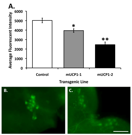
Figure 1. Constitutive UCP expression in adult IPCs results in decreased steady state intracellular Ca 2+ levels. (A) Under normal growth conditions, the Cg-2 protein in the adult IPCs produces fluorescent signals reflecting a steady state Ca2+ flux whereas a 21-51% decrease in average fluorescent intensity is detected in adult IPCs of two dilp2-Gal4/UAS-mucp1, UAS-cg-2 co-expressing fly lines (mUCP1-1 and mUCP1-2), consistent with attenuated insulin action. Each bar represents mean + S.E.M. *p=0.0004, **p<0.0001 (Student's t-test). Shown are average fluorescent signals from IPCs of 10 brains of each line in a representative experiment (see Materials and Methods). Reproducible results were obtained in three independent experiments. Representative images of IPCs from a control adult brain isolated from a dilp2-Gal4/UAS-cg-2 fly line (B) and an adult brain isolated from a dilp2-Gal4/UAS-mucp1, UAS-cg-2 fly line (C) are shown. Images were taken with a 40X objective. Scale bar, 100 μm.
Figure 1. Constitutive UCP expression in adult IPCs results in decreased steady state intracellular Ca 2+ levels. (A) Under normal growth conditions, the Cg-2 protein in the adult IPCs produces fluorescent signals reflecting a steady state Ca2+ flux whereas a 21-51% decrease in average fluorescent intensity is detected in adult IPCs of two dilp2-Gal4/UAS-mucp1, UAS-cg-2 co-expressing fly lines (mUCP1-1 and mUCP1-2), consistent with attenuated insulin action. Each bar represents mean + S.E.M. *p=0.0004, **p<0.0001 (Student's t-test). Shown are average fluorescent signals from IPCs of 10 brains of each line in a representative experiment (see Materials and Methods). Reproducible results were obtained in three independent experiments. Representative images of IPCs from a control adult brain isolated from a dilp2-Gal4/UAS-cg-2 fly line (B) and an adult brain isolated from a dilp2-Gal4/UAS-mucp1, UAS-cg-2 fly line (C) are shown. Images were taken with a 40X objective. Scale bar, 100 μm.CC BY · PubMed Central
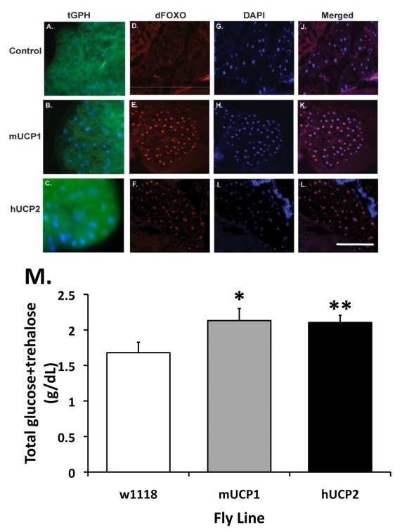
Figure 2. Normal and reduced systemic insulin signaling as reflected by the cellular localization of the PH-tagged GFP reporter protein (tGPH) and dFoxO in fat body cells. (A-C) Increased UCP activities in the adult IPCs attenuate systemic insulin signaling events. In control flies, under normal growth conditions and a full strength PI-3' kinase activity, tGPH is predominantly located at the plasma membrane of each fat body cell (A). (B-C) UCP expression in adult IPCs results in a diffused, cytoplasmic distribution of the tGPH protein. Control: dilp2-Gal4, tGPH; mUCP1: dilp2-Gal4/UAS-mucp1, tGPH; hUCP2: dilp2-Gal4/UAS-hucp2, tGPH. (D-F) Increased accumulation of dFoxO in the nucleus of pericerebral fat body cells in adult dilp2-Gal4/UAS-mucp1 and dilp2-Gal4/UAS-hucp2 flies indicates reduced insulin signaling. Cryosections of adult heads were stained with an α-dFoxO antibody followed by Alexa 568-conjugated secondary antibodies. A strong nuclear staining of the dFoxO protein was observed in the pericerebral fat body in both dilp2-Gal4/UAS-mucp1 (mUCP1, Panel E) and dilp2-Gal4/UAS-hucp2 (hUCP2, Panel F) flies but not in dilp2-Gal4/w1118 (Control, Panel D) flies. All sections were counter stained with DAPI (Panels G-I) to locate the nucleus of each cell. Merged images of anti-FoxO staining and DAPI are shown in Panels J-L. (M) Elevated levels of fasting circulating sugars are measured in adult dilp2-Gal4/UAS-mucp1 and dilp2-Gal4/UAS-hucp2 flies. An average of 29% increase in circulating sugars measured in 14-day-old dilp2-Gal4/UAS-mucp1 (mUCP1) and dilp2-Gal4/UAS-hucp2 (hUCP2) females as compared to control dilp2-Gal4/w1118 (w1118) females. Each bar represents mean + SEM. N=5-7, *p= 0.046, **p= 0.05 (Student's t test).
Figure 2. Normal and reduced systemic insulin signaling as reflected by the cellular localization of the PH-tagged GFP reporter protein (tGPH) and dFoxO in fat body cells. (A-C) Increased UCP activities in the adult IPCs attenuate systemic insulin signaling events. In control flies, under normal growth conditions and a full strength PI-3' kinase activity, tGPH is predominantly located at the plasma membrane of each fat body cell (A). (B-C) UCP expression in adult IPCs results in a diffused, cytoplasmic distribution of the tGPH protein. Control: dilp2-Gal4, tGPH; mUCP1: dilp2-Gal4/UAS-mucp1, tGPH; hUCP2: dilp2-Gal4/UAS-hucp2, tGPH. (D-F) Increased accumulation of dFoxO in the nucleus of pericerebral fat body cells in adult dilp2-Gal4/UAS-mucp1 and dilp2-Gal4/UAS-hucp2 flies indicates reduced insulin signaling. Cryosections of adult heads were stained with an α-dFoxO antibody followed by Alexa 568-conjugated secondary antibodies. A strong nuclear staining of the dFoxO protein was observed in the pericerebral fat body in both dilp2-Gal4/UAS-mucp1 (mUCP1, Panel E) and dilp2-Gal4/UAS-hucp2 (hUCP2, Panel F) flies but not in dilp2-Gal4/w1118 (Control, Panel D) flies. All sections were counter stained with DAPI (Panels G-I) to locate the nucleus of each cell. Merged images of anti-FoxO staining and DAPI are shown in Panels J-L. (M) Elevated levels of fasting circulating sugars are measured in adult dilp2-Gal4/UAS-mucp1 and dilp2-Gal4/UAS-hucp2 flies. An average of 29% increase in circulating sugars measured in 14-day-old dilp2-Gal4/UAS-mucp1 (mUCP1) and dilp2-Gal4/UAS-hucp2 (hUCP2) females as compared to control dilp2-Gal4/w1118 (w1118) females. Each bar represents mean + SEM. N=5-7, *p= 0.046, **p= 0.05 (Student's t test).CC BY · PubMed Central
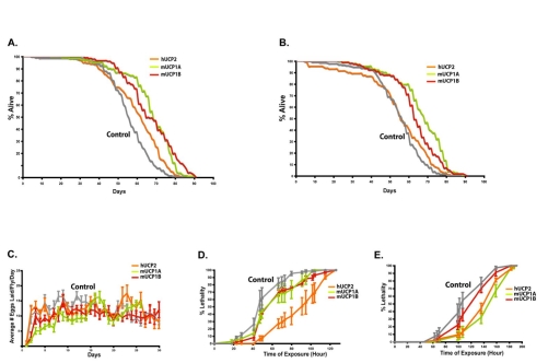
Figure 3. Targeted UCP expression in the adult IPCs extends life span and renders flies stress resistant with normal fecundity. Survivorship curves for female (A) and male (B) flies are shown. Two independent trials were performed and results from one trial are shown for one hUCP2 and two mUCP1 lines. Gray lines are control dilp2-Gal4/w1118 flies; two mUCP1 expressing dilp2-Gal4/UAS-mucp1 lines, mUCP1A and mUCP1B are green and red lines, respectively; and orange lines are dilp2-Gal4/UAS-hucp2. Panel A (females) median life spans are 55 for controls and 71, 63, and 65 for three UCP transgenic lines. Panel B (male) median life spans are 57 for controls and 70, 59, and 59 for three UCP transgenic lines. Log rank analysis shows an average of 19% increase in median life span in female with targeted UCP expression in the adult IPCs and an average of 10% increase in male (see SI Supplementary Table 1). (C) Female fecundity is similar for flies expressing mUCP1 or hUCP2 in the adult IPCs and control flies. Average number of eggs per day for 20 individual females was determined from daily counts of eggs produced from single mated pairs [6]. (D) Transgenic flies with targeted UCP expression in the adult IPCs are resistant to oxidative stress. Survival during administration of paraquat (20 mM paraquat in 5% sucrose) shows that 10-day-old flies expressing mUCP1 or hUCP2 in the IPCs are more resistant than controls. (E) mUCP1 or hUCP2-expressing flies are resistant to starvation. Ten-day-old dilp2-Gal4/UAS-mucp1 and dilp2-Gal4/UAS-hucp2 flies were placed in vials containing water soaked filters and the number of dead flies was counted at noted time intervals. Three independent assays were performed and a representative experiment is shown for females. All values are presented as mean ± S.E.M. Similar differences were seen for males and females. Each experiment included 8-10 vials with 20 flies in each vial, total of 160-200 flies for each condition.
Figure 3. Targeted UCP expression in the adult IPCs extends life span and renders flies stress resistant with normal fecundity. Survivorship curves for female (A) and male (B) flies are shown. Two independent trials were performed and results from one trial are shown for one hUCP2 and two mUCP1 lines. Gray lines are control dilp2-Gal4/w1118 flies; two mUCP1 expressing dilp2-Gal4/UAS-mucp1 lines, mUCP1A and mUCP1B are green and red lines, respectively; and orange lines are dilp2-Gal4/UAS-hucp2. Panel A (females) median life spans are 55 for controls and 71, 63, and 65 for three UCP transgenic lines. Panel B (male) median life spans are 57 for controls and 70, 59, and 59 for three UCP transgenic lines. Log rank analysis shows an average of 19% increase in median life span in female with targeted UCP expression in the adult IPCs and an average of 10% increase in male (see SI Supplementary Table 1). (C) Female fecundity is similar for flies expressing mUCP1 or hUCP2 in the adult IPCs and control flies. Average number of eggs per day for 20 individual females was determined from daily counts of eggs produced from single mated pairs [6]. (D) Transgenic flies with targeted UCP expression in the adult IPCs are resistant to oxidative stress. Survival during administration of paraquat (20 mM paraquat in 5% sucrose) shows that 10-day-old flies expressing mUCP1 or hUCP2 in the IPCs are more resistant than controls. (E) mUCP1 or hUCP2-expressing flies are resistant to starvation. Ten-day-old dilp2-Gal4/UAS-mucp1 and dilp2-Gal4/UAS-hucp2 flies were placed in vials containing water soaked filters and the number of dead flies was counted at noted time intervals. Three independent assays were performed and a representative experiment is shown for females. All values are presented as mean ± S.E.M. Similar differences were seen for males and females. Each experiment included 8-10 vials with 20 flies in each vial, total of 160-200 flies for each condition.CC BY · PubMed Central
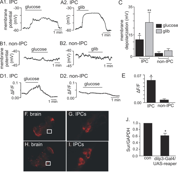
Figure 4. Electrophysiological, Ca 2+ influx, and expression evidence of functional KATP channels in adult IPCs. (A1-C) Membrane depolarization of adult IPCs in response to glucose and glibenclamide. (A1) Trace of membrane potential from an adult IPC in the whole brain preparation shows that exposure to high glucose (80 mM) evoked a reversible membrane depolarization. (A2) Trace of membrane potential from an adult IPC shows that exposure to a commonly known KATP channel blocker, glibenclamide (glib, 20 μM) also evoked a reversible membrane depolarization. (B1 and B2) traces of membrane potential from adult non-IPCs show that these cells do not respond to glucose or glibenclamide. (C) Average membrane potential response to glucose and glibenclamide of IPCs (N=5) and non-IPCs (N=3). Glucose (*) and glibenclamide (**) significantly increased membrane potential of IPCs as compared to non-IPCs, *p and **p <0.05 (Student's t test). Each bar represents mean + S.E.M. (D1-E) Glucose-dependent Ca2+ influx measured in adult IPCs. (D1) Normalized fluorescence trace (∆F/F) (see Materials and Methods) recorded from an acutely dissociated adult IPC shows that exposure to glucose (80 mM) increased fluorescence intensity, thus indicating an increase in intracellular Ca2+. (D2) Normalized fluorescence trace (∆F/F) recorded from an acutely dissociated non-IPC shows that glucose does not increase intracellular Ca2+ in these cells. (E). An average of normalized fluorescence intensity in response to glucose demonstrates a significant increase in Ca2+ influx recorded from IPCs (N=6) as compared to non-IPCs (N=3). *P= 0.007 (Student's t test). Each bar represents mean + S.E.M. (FI) In situ hybridization of whole mount adult brains demonstrates dSur expression in IPCs (F-G) when probed with anti-sense dSur probes and dilp2 expression (HI) when probed with anti-sense dilp2 probes. The IPCs marked in squares are shown in panel G for dSur signals and panel I for dilp2 signals. (J) Quantitative real-time PCR analysis reveals an average of 33% reduction in dSur transcripts when the IPCs are partially ablated using an IPC-specific driver, dilp3-Gal4 to drive the expression of a pro-apoptotic gene, reaper. The housekeeping gene GAPDH was used as the reference gene. Each bar represents mean + S.E.M. N=5, *p<0.001 (Student's t test). Control: dilp3-Gal4/w1118.
Figure 4. Electrophysiological, Ca 2+ influx, and expression evidence of functional KATP channels in adult IPCs. (A1-C) Membrane depolarization of adult IPCs in response to glucose and glibenclamide. (A1) Trace of membrane potential from an adult IPC in the whole brain preparation shows that exposure to high glucose (80 mM) evoked a reversible membrane depolarization. (A2) Trace of membrane potential from an adult IPC shows that exposure to a commonly known KATP channel blocker, glibenclamide (glib, 20 μM) also evoked a reversible membrane depolarization. (B1 and B2) traces of membrane potential from adult non-IPCs show that these cells do not respond to glucose or glibenclamide. (C) Average membrane potential response to glucose and glibenclamide of IPCs (N=5) and non-IPCs (N=3). Glucose (*) and glibenclamide (**) significantly increased membrane potential of IPCs as compared to non-IPCs, *p and **p <0.05 (Student's t test). Each bar represents mean + S.E.M. (D1-E) Glucose-dependent Ca2+ influx measured in adult IPCs. (D1) Normalized fluorescence trace (∆F/F) (see Materials and Methods) recorded from an acutely dissociated adult IPC shows that exposure to glucose (80 mM) increased fluorescence intensity, thus indicating an increase in intracellular Ca2+. (D2) Normalized fluorescence trace (∆F/F) recorded from an acutely dissociated non-IPC shows that glucose does not increase intracellular Ca2+ in these cells. (E). An average of normalized fluorescence intensity in response to glucose demonstrates a significant increase in Ca2+ influx recorded from IPCs (N=6) as compared to non-IPCs (N=3). *P= 0.007 (Student's t test). Each bar represents mean + S.E.M. (FI) In situ hybridization of whole mount adult brains demonstrates dSur expression in IPCs (F-G) when probed with anti-sense dSur probes and dilp2 expression (HI) when probed with anti-sense dilp2 probes. The IPCs marked in squares are shown in panel G for dSur signals and panel I for dilp2 signals. (J) Quantitative real-time PCR analysis reveals an average of 33% reduction in dSur transcripts when the IPCs are partially ablated using an IPC-specific driver, dilp3-Gal4 to drive the expression of a pro-apoptotic gene, reaper. The housekeeping gene GAPDH was used as the reference gene. Each bar represents mean + S.E.M. N=5, *p<0.001 (Student's t test). Control: dilp3-Gal4/w1118.CC BY · PubMed Central

Verification

Claims the paper makes

The claims and statistics of this paper haven’t been checked yet.

Run the check — free, no account needed. It reads the open-access text and checks the sources it cites.

A few free checks a day · papers already verified open instantly · results are public

Claims other papers make about this one

Each is checked against this paper’s own text: does it say what the citing paper says it does?

Self-check

Percentages, sums and reported differences re-computed from the paper’s own numbers.

The self-check needs the paper’s full text.

Citations

Cites

Research works printed but not linked: 28

  1. Kim, 2001, Uncoupling protein-2 negatively regulates insulin secretion and is a major link between obesity, beta cell dysfunction, and type 2 diabetes, Cell, 10, 745
  2. Brand, 2008, Uncoupling protein-2 contributes significantly to high mitochondrial proton leak in INS-1E insulinoma cells and attenuates glucose-stimulated insulin secretion, Biochem J, 40, 199
  3. Helfand, 2005, Targeted expression of the human uncoupling protein 2 (hUCP2) to adult neurons extends life span in the fly, Cell Metab, 1, 145, 10.1016/j.cmet.2005.01.005 · DOI
  4. Ashcroft, 2006, K(ATP) channels and insulin secretion: a key role in health and disease, Biochem Soc Trans, 3, 243, 10.1042/BST0340243 · DOI
  5. Lowell, 2005, The mitochondrial uncoupling-protein homologues, Nat Rev Mol Cell Biol, 6, 248, 10.1038/nrm1592 · DOI
  6. Rulifson, 2004, Conserved mechanisms of glucose sensing and regulation by Drosophila corpora cardiaca cells, Nature, 43, 316
  7. Nusse, 2002, Ablation of insulin-producing neurons in flies: growth and diabetic phenotypes, Science, 29, 1118
  8. Leevers, 2005, Longer lifespan, altered metabolism, and stress resistance in Drosophila from ablation of cells making insulin-like ligands, Proc Natl Acad Sci U S A, 10, 3105
  9. Park, 2004, Hemolymph sugar homeostasis and starvation-induced hyperactivity affected by genetic manipulations of the adipokinetic hormone-encoding gene in Drosophila melanogaster, Genetics, 16, 311
  10. Hafen, 2002, Nutrient-dependent expression of insulin-like peptides from neuroendocrine cells in the CNS contributes to growth regulation in Drosophila, Curr Biol, 1, 1293
  11. Gaskell, 1990, The fruitfly Drosophila melanogaster contains a novel charged adipokinetic-hormone-family peptide, Biochem J, 26, 315
  12. Schaffer, 1995, Identification and expression of the Drosophila adipokinetic hormone gene, Mol Cell Endocrinol, 10, 133
  13. Brand, 2008, On the role of uncoupling protein-2 in pancreatic beta cells, Biochim Biophys Acta, 177, 973
  14. Davis, 2003, Detection of calcium transients in Drosophila mushroom body neurons with camgaroo reporters, J Neurosci, 2, 64
  15. Cantley, 1999, The role of phosphoinositide 3-kinase lipid products in cell function, J Biol Chem, 27, 8347
  16. Edgar, 2002, Drosophila's insulin/PI3-kinase pathway coordinates cellular metabolism with nutritional conditions, Dev Cell, 2, 239, 10.1016/S1534-5807(02)00117-X · DOI
  17. Tjian, 2003, Control of cell number by Drosophila FOXO: downstream and feedback regulation of the insulin receptor pathway, Genes Dev, 1, 2006
  18. Helfand, 2007, Expression of dominant-negative Dmp53 in the adult fly brain inhibits insulin signaling, Proc Natl Acad Sci U S A, 10, 13355
  19. Antebi, 2003, The endocrine regulation of aging by insulin-like signals, Science, 29, 1346
  20. Nichols, 2005, Diabetes and insulin secretion: the ATP-sensitive K+ channel (K ATP) connection, Diabetes, 5, 3065
  21. Rorsman, 2004, Molecular defects in insulin secretion in type-2 diabetes, Rev Endocr Metab Disord, 5, 135, 10.1023/B:REMD.0000021435.87776.a7 · DOI
  22. Bodmer, 2006, The ATP-sensitive potassium (KATP) channel-encoded dSUR gene is required for Drosophila heart function and is regulated by tinman, Proc Natl Acad Sci U S A, 10, 11999
  23. Karschin, 2002, Inwardly rectifying K+ (Kir) channels in Drosophila. A crucial role of cellular milieu factors Kir channel function, J Biol Chem, 27, 25554
  24. Harshman, 2005, Insulin signaling is necessary for vitellogenesis in Drosophila melanogaster independent of the roles of juvenile hormone and ecdysteroids: female sterility of the chico1 insulin signaling mutation is autonomous to the ovary, J Insect Physiol, 5, 455
  25. Tatar, 2004, Drosophila dFOXO controls lifespan and regulates insulin signalling in brain and fat body, Nature, 42, 562
  26. Beutler, 2007, ATP-sensitive potassium channels mediate survival during infection in mammals and insects, Nat Genet, 3, 1453
  27. Wu, 2004, A modified minimal hemolymph-like solution, HL3.1, for physiological recordings at the neuromuscular junctions of normal and mutant Drosophila larvae, J Neurogenet, 1, 377
  28. Helfand, 2006, Involvement of Drosophila uncoupling protein 5 in metabolism and aging, Genetics, 17, 1699

Cited by

Sorted by citations

See all 63

Metadata

Authors
Fridell, Y.-W. C., Hoh, M., Kréneisz, O., Hosier, S., Chang, C., Scantling, D., Mulkey, D. K., & Helfand, S. L.
Affiliations
  • Department of Molecular Biology, Cell Biology, and Biochemistry, Brown University, Providence, RI 02912, USA
  • Department of Physiology and Neurobiology, University of Connecticut, Storrs, CT 06269, USA
Venue
Aging
Published
2009-07-21
Volume
1
Issue
8
Pages
699-713
ISSN
1945-4589
Cited by
63
References
6
Language
en

About these checks

Registry, retraction, Cites and statistics results are computed, not judged, with one exception: a printed reference that no index lists is read once by a model before it counts as a citation error. Claims are extracted by a model and kept only when their quote appears verbatim in the text; each cited source is then checked, with a model’s help, for whether it says what the claim relies on. Those verdicts can be wrong — read the claim’s quote and the cited source before relying on one.

Statistics and arithmetic are recomputed, not judged.

Confidence bands are shown only for fields calibrated on our annotated test set. None is calibrated yet, so no band is shown.

What counts as a claim ›How we measure confidence ›How we check this ›

Are you an author, or do you see a problem with this page? Request a review