

The DOI matches its Crossref record; 2 of the 2 references it cites were found in a registry or an index.
CRISPR/Cas9-mediated reversibly immortalized mouse bone marrow stromal stem cells (BMSCs) retain multipotent features of mesenchymal stem cells (MSCs)
Overview
The claims and statistics of this paper haven’t been checked yet.
Run the check — free, no account needed. It reads the open-access text and checks the sources it cites.
Integrity signals
Registry
Cites
Statistics
Figures
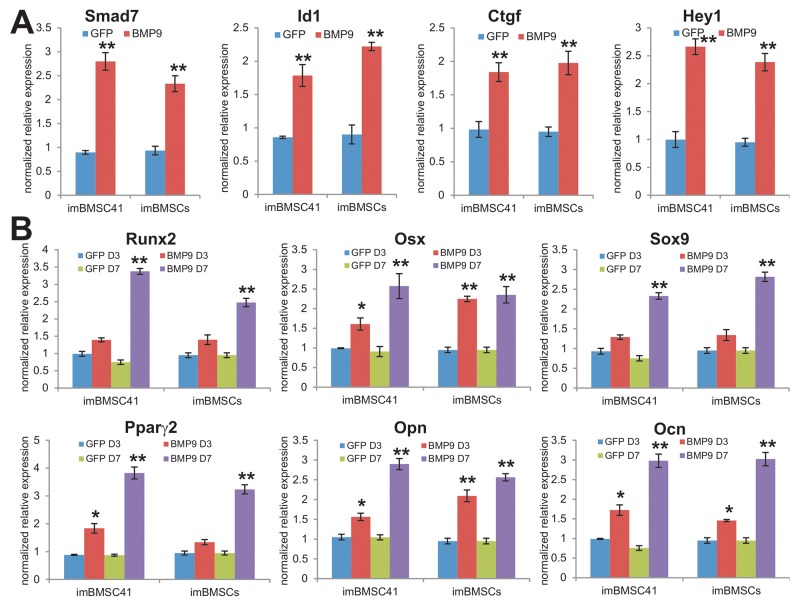
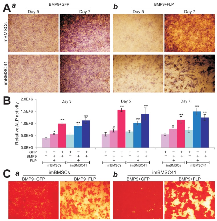
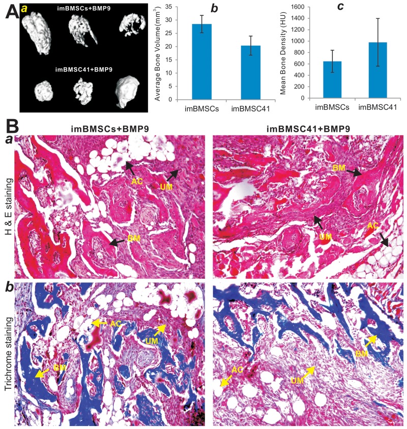
Verification
Claims the paper makes
The claims and statistics of this paper haven’t been checked yet.
Run the check — free, no account needed. It reads the open-access text and checks the sources it cites.
Claims other papers make about this one
Each is checked against this paper’s own text: does it say what the citing paper says it does?
In the citing paper
“The CRISPR/Cas9 system induces DNA double-strand breaks at specific sites of genomic DNA, which should allow a safer and more targeted gene delivery of the immortalizing genes [156].”
From Cell Immortality: In Vitro Effective Techniques to Achieve and Investigate Its Applications and Challenges (2024) · cites this paper as [156]
In the citing paper
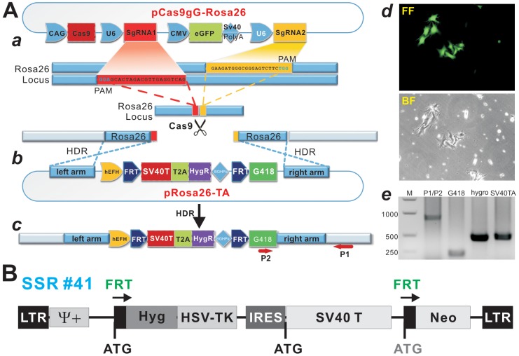
“In this study, by exploiting the CRISPR/Cas9-based homology-directed-repair (HDR) mechanism, they targeted SV40T to mouse Rosa26 locus and efficiently immortalized mouse BMSCs (i.e., imBMSCs) (Figure 4) [156].”
From Cell Immortality: In Vitro Effective Techniques to Achieve and Investigate Its Applications and Challenges (2024) · cites this paper as [156]
In the citing paper
“For example, in order to overcome the technical challenge of maintaining primary BMSCs in long-term culture, mouse bone marrow stromal stem cells (BMSCs), which are one of the most common mesenchymal stem cells, have been reversibly immortalized mediated CRISPR/Cas9, which can maintain the multipotent characteristics of mesenchymal stem cells (MSCs).”
From Cell Immortality: In Vitro Effective Techniques to Achieve and Investigate Its Applications and Challenges (2024) · cites this paper as [156]
Self-check
Percentages, sums and reported differences re-computed from the paper’s own numbers.
The self-check needs the paper’s full text.
Citations
Cites
Mali, P., Aach, J., Stranges, P. B., Esvelt, K. M., Moosburner, M., Kosuri, S., Yang, L., & Church, G. M. (2013) · Nature Biotechnology · doi:10.1038/nbt.2675 · Cited by 1,752 · cited 1×
“To reduce off-target effects of single gRNA-guided Cas9 nuclease, a previously reported “paired nicking” strategy was employed, in which two sgRNAs targeted to adjacent sites on opposite DNA strands [44, 50], as two sgRNA, driven by U6 promoter, were designed to target the first intron of the Rosa26 gene as reported (…”
MethodCas9 as a versatile tool for engineering biology
Mali, P., Esvelt, K. M., & Church, G. M. (2013) · Nature Methods · doi:10.1038/nmeth.2649 · Cited by 1,197
Research works printed but not linked: 86
- Marshak, 1999, Multilineage potential of adult human mesenchymal stem cells, Science, 284, 143, 10.1126/science.284.5411.143 · DOI · Paper page
- Gao, 2010, Mesenchymal progenitor cells and their orthopedic applications: forging a path towards clinical trials, Stem Cells Int, 2010, 519028
- Gao, 2010, Mesenchymal stem cells: molecular characteristics and clinical applications, World J Stem Cells, 2, 67, 10.4252/wjsc.v2.i4.67 · DOI
- Luu, 2008, Regulation of osteogenic differentiation during skeletal development, Front Biosci, 13, 2001, 10.2741/2819 · DOI
- Bruder, 2001, Mesenchymal stem cells: building blocks for molecular medicine in the 21st century, Trends Mol Med, 7, 259, 10.1016/S1471-4914(01)02016-0 · DOI
- Frolova, 1968, Heterotopic of bone marrow. Analysis of precursor cells for osteogenic and hematopoietic tissues, Transplantation, 6, 230
- Petrakova, 1966, Osteogenesis in transplants of bone marrow cells, J Embryol Exp Morphol, 16, 381
- Wang, 2000, Bone development, Annu Rev Cell Dev Biol, 16, 191, 10.1146/annurev.cellbio.16.1.191 · DOI
- Mansukhani, 2008, Osteoblast proliferation or differentiation is regulated by relative strengths of opposing signaling pathways, J Cell Physiol, 215, 442, 10.1002/jcp.21323 · DOI
- Lamplot, 2013, Wnt signaling in bone formation and its therapeutic potential for bone diseases, Ther Adv Musculoskelet Dis, 5, 13, 10.1177/1759720X12466608 · DOI
- He, 2016, The evolving roles of canonical WNT signaling in stem cells and tumorigenesis: implications in targeted cancer therapies, Lab Invest, 96, 116, 10.1038/labinvest.2015.144 · DOI
- Luu, 2015, Insulin-like growth factor (IGF) signaling in tumorigenesis and the development of cancer drug resistance, Genes Dis, 2, 13, 10.1016/j.gendis.2014.10.004 · DOI
- Reid, 2014, Fibroblast growth factor (FGF) signaling in development and skeletal diseases, Genes Dis, 1, 199, 10.1016/j.gendis.2014.09.005 · DOI · Paper page
- Haydon, 2014, The versatile functions of Sox9 in development, stem cells, and human diseases, Genes Dis, 1, 149, 10.1016/j.gendis.2014.09.004 · DOI
- Artavanis-Tsakonas, 2012, Notch and disease: a growing field, Semin Cell Dev Biol, 23, 473, 10.1016/j.semcdb.2012.02.005 · DOI
- Canalis, 2010, Notch and the skeleton, Mol Cell Biol, 30, 886, 10.1128/MCB.01285-09 · DOI
- Artavanis-Tsakonas, 2012, The notch signalling system: recent insights into the complexity of a conserved pathway, Nat Rev Genet, 13, 654, 10.1038/nrg3272 · DOI
- Wrana, 2005, The disparate role of BMP in stem cell biology, Oncogene, 24, 5713, 10.1038/sj.onc.1208919 · DOI
- He, 2007, Distinct roles of bone morphogenetic proteins in osteogenic differentiation of mesenchymal stem cells, J Orthop Res, 25, 665, 10.1002/jor.20359 · DOI
- Shen, 2014, Bone morphogenetic protein (BMP) signaling in development and human diseases, Genes Dis, 1, 87, 10.1016/j.gendis.2014.07.005 · DOI
- He, 2003, Osteogenic activity of the fourteen types of human bone morphogenetic proteins (BMPs), J Bone Joint Surg Am, 1544
- Li, 2004, Characterization of the distinct orthotopic bone-forming activity of 14 BMPs using recombinant adenovirus-mediated gene delivery, Gene Ther, 11, 1312, 10.1038/sj.gt.3302298 · DOI
- Gao, 2011, BMP-9 induced osteogenic differentiation of mesenchymal stem cells: molecular mechanism and therapeutic potential, Curr Gene Ther, 11, 229, 10.2174/156652311795684777 · DOI
- Zhang, 2013, BMP9 signaling in stem cell differentiation and osteogenesis, Am J Stem Cells, 2, 1
- Sensebe, 2013, Mesenchymal stromal cells: misconceptions and evolving concepts, Cytotherapy, 15, 140, 10.1016/j.jcyt.2012.11.005 · DOI
- Knippers, 1992, Structure and function of simian virus 40 large tumor antigen, Annu Rev Biochem, 61, 55, 10.1146/annurev.bi.61.070192.000415 · DOI
- McLean, 1993, Improved techniques for immortalizing animal cells, Trends Biotechnol, 11, 232, 10.1016/0167-7799(93)90134-U · DOI
- Hu, 2012, Conditionally immortalized mouse embryonic fibroblasts retain proliferative activity without compromising multipotent differentiation potential, PLoS One, 7, e32428, 10.1371/journal.pone.0032428 · DOI
- Zhang, 2012, Conditional immortalization establishes a repertoire of mouse melanocyte progenitors with distinct melanogenic differentiation potential, J Invest Dermatol, 132, 2479, 10.1038/jid.2012.145 · DOI
- Jiang, 2009, Wnt antagonist sfrp3 inhibits the differentiation of mouse hepatic progenitor cells, J Cell Biochem, 108, 295, 10.1002/jcb.22254 · DOI
- Kang, 2014, Functional characteristics of reversibly immortalized hepatic progenitor cells derived from mouse embryonic liver, Cell Physiol Biochem, 34, 1318, 10.1159/000366340 · DOI
- Zhang, 2013, Establishment and characterization of the reversibly immortalized mouse fetal heart progenitors, Int J Med Sci, 10, 1035, 10.7150/ijms.6639 · DOI
- Zhang, 2009, Retinoic acid signalling induces the differentiation of mouse fetal liver-derived hepatic progenitor cells, Liver Int, 29, 1569, 10.1111/j.1478-3231.2009.02111.x · DOI
- Mohammed, 2016, Bone morphogenetic protein 9 (BMP9) induces effective bone formation from reversibly immortalized multipotent adipose-derived (iMAD) mesenchymal stem cells, Am J Transl Res, 8, 3710
- Zhang, 2014, Bone morphogenetic protein-9 (BMP9) effectively induces osteo/odontoblastic differentiation of the reversibly immortalized stem cells of dental apical papilla, Stem Cells Dev, 23, 1405, 10.1089/scd.2013.0580 · DOI
- Deng, 2015, Reversibly immortalized mouse articular chondrocytes acquire long-term proliferative capability while retaining chondrogenic phenotype, Cell Transplant, 24, 1053, 10.3727/096368914X681054 · DOI
- He, 2016, Immortalized mouse achilles tenocytes demonstrate long-term proliferative capacity while retaining tenogenic properties, Tissue Eng Part C Methods, 22, 280, 10.1089/ten.tec.2015.0244 · DOI
- Li, 2017, BMP9 induces osteogenesis and adipogenesis in the immortalized human cranial suture progenitors from the patent sutures of craniosynostosis patients, J Cell Mol Med, 21, 2782, 10.1111/jcmm.13193 · DOI
- Liao, 2014, The piggybac transposon-mediated expression of SV40 t antigen efficiently immortalizes mouse embryonic fibroblasts (MEFs), PLoS One, 9, e97316, 10.1371/journal.pone.0097316 · DOI
- Charpentier, 2014, Genome editing. The new frontier of genome engineering with CRISPR-Cas9, Science, 346, 1258096, 10.1126/science.1258096 · DOI · Paper page
- Jacks, 2015, Applications of the CRISPR-Cas9 system in cancer biology, Nat Rev Cancer, 15, 387, 10.1038/nrc3950 · DOI · Paper page
- Qi, 2016, CRISPR/Cas9 in genome editing and beyond, Annu Rev Biochem, 85, 227, 10.1146/annurev-biochem-060815-014607 · DOI · Paper page
- He, 2014, CRISPR clear? dimeric Cas9-FoK1 nucleases improve genome-editing specificity, Genes Dis, 1, 6, 10.1016/j.gendis.2014.08.004 · DOI · Paper page
- Soriano, 1997, Disruption of overlapping transcripts in the rosa beta geo 26 gene trap strain leads to widespread expression of beta-galactosidase in mouse embryos and hematopoietic cells, Proc Natl Acad Sci U S A, 94, 3789, 10.1073/pnas.94.8.3789 · DOI
- Soriano, 1999, Generalized lacz expression with the ROSA26 cre reporter strain, Nat Genet, 21, 70, 10.1038/5007 · DOI
- Leboulch, 1996, Reversible immortalization of mammalian cells mediated by retroviral transfer and site-specific recombination, Proc Natl Acad Sci U S A, 93, 8971, 10.1073/pnas.93.17.8971 · DOI
- Haydon, 2015, Sustained high level transgene expression in mammalian cells mediated by the optimized piggybac transposon system, Genes Dis, 2, 96, 10.1016/j.gendis.2014.12.001 · DOI
- Ogawa, 2015, Genome editing in mouse spermatogonial stem cell lines using TALEN and double-nicking CRISPR/Cas9, Stem Cell Reports, 5, 75, 10.1016/j.stemcr.2015.05.011 · DOI
- Huang, 2009, A comprehensive analysis of the dual roles of BMPs in regulating adipogenic and osteogenic differentiation of mesenchymal progenitor cells, Stem Cells Dev, 18, 545, 10.1089/scd.2008.0130 · DOI
- He, 2003, Transcriptional characterization of bone morphogenetic proteins (BMPs)-mediated osteogenic signaling, J Cell Biochem, 90, 1149, 10.1002/jcb.10744 · DOI
- He, 2004, Inhibitor of DNA binding/differentiation helix-loop-helix proteins mediate bone morphogenetic protein-induced osteoblast differentiation of mesenchymal stem cells, J Biol Chem, 279, 32941, 10.1074/jbc.M403344200 · DOI
- He, 2004, Connective tissue growth factor (CTGF) is regulated by Wnt and bone morphogenetic proteins signaling in osteoblast differentiation of mesenchymal stem cells, J Biol Chem, 279, 55958, 10.1074/jbc.M407810200 · DOI
- Su, 2009, Hey1 basic helix-loop-helix protein plays an important role in mediating BMP9-induced osteogenic differentiation of mesenchymal progenitor cells, J Biol Chem, 284, 649, 10.1074/jbc.M806389200 · DOI
- MacDonald, 1990, Development of new cell lines for animal cell biotechnology, Crit Rev Biotechnol, 10, 155, 10.3109/07388559009068265 · DOI · Paper page
- Werbin, 1991, Defining the molecular mechanisms of human cell immortalization, Biochim Biophys Acta, 1072, 1
- Reddel, 2000, The role of senescence and immortalization in carcinogenesis, Carcinogenesis, 21, 477, 10.1093/carcin/21.3.477 · DOI
- Vinken, 2014, Strategies for immortalization of primary hepatocytes, J Hepatol, 61, 925, 10.1016/j.jhep.2014.05.046 · DOI
- Fanning, 1992, Simian virus 40 large T antigen: the puzzle, the pieces, and the emerging picture, J Virol, 66, 1289, 10.1128/JVI.66.3.1289-1293.1992 · DOI
- Wang, 2013, Noggin resistance contributes to the potent osteogenic capability of BMP9 in mesenchymal stem cells, J Orthop Res, 31, 1796, 10.1002/jor.22427 · DOI
- Luo, 2010, TGFbeta/BMP type i receptors ALK1 and ALK2 are essential for BMP9-induced osteogenic signaling in mesenchymal stem cells, J Biol Chem, 285, 29588, 10.1074/jbc.M110.130518 · DOI
- Wang, 2012, Growth hormone synergizes with BMP9 in osteogenic differentiation by activating the JAK/STAT/IGF1 pathway in murine multilineage cells, J Bone Miner Res, 27, 1566, 10.1002/jbmr.1622 · DOI
- Tang, 2009, BMP9-induced osteogenic differentiation of mesenchymal progenitors requires functional canonical Wnt/beta-catenin signaling, J Cell Mol Med, 13, 2448, 10.1111/j.1582-4934.2008.00569.x · DOI
- Wietholt, 2010, Insulin-like growth factor 2 (IGF-2) potentiates BMP-9-induced osteogenic differentiation and bone formation, J Bone Miner Res, 25, 2447, 10.1002/jbmr.133 · DOI
- Huang, 2010, Retinoic acids potentiate BMP9-induced osteogenic differentiation of mesenchymal progenitor cells, PLoS One, 5, e11917, 10.1371/journal.pone.0011917 · DOI
- Zhang, 2013, BMP9-regulated angiogenic signaling plays an important role in the osteogenic differentiation of mesenchymal progenitor cells, J Cell Sci, 126, 532, 10.1242/jcs.114231 · DOI
- Li, 2013, Cross-talk between egf and BMP9 signalling pathways regulates the osteogenic differentiation of mesenchymal stem cells, J Cell Mol Med, 17, 1160, 10.1111/jcmm.12097 · DOI
- Wang, 2015, Canonical Wnt signaling acts synergistically on BMP9-induced osteo/odontoblastic differentiation of stem cells of dental apical papilla (SCAPs), Biomaterials, 39, 145, 10.1016/j.biomaterials.2014.11.007 · DOI
- He, 2005, Gene therapy for bone regeneration, Curr Gene Ther, 5, 167, 10.2174/1566523053544218 · DOI
- Zhang, 2014, Overexpression of Ad5 precursor terminal protein accelerates recombinant adenovirus packaging and amplification in hek-293 packaging cells, Gene Ther, 21, 629, 10.1038/gt.2014.40 · DOI
- Qu, 2017, Engineering the rapid adenovirus production and amplification (RAPA) cell line to expedite the generation of recombinant adenoviruses, Cell Physiol Biochem, 41, 2383, 10.1159/000475909 · DOI
- Chen, 2017, Characterization of retroviral infectivity and superinfection resistance during retrovirus-mediated transduction of mammalian cells, Gene Ther, 24, 333, 10.1038/gt.2017.24 · DOI
- Yu, 2017, Noncanonical Wnt signaling plays an important role in modulating canonical Wnt-regulated stemness, proliferation and terminal differentiation of hepatic progenitors, Oncotarget, 8, 27105, 10.18632/oncotarget.15637 · DOI
- Wang, 2014, A simplified and versatile system for the simultaneous expression of multiple sirnas in mammalian cells using gibson DNA assembly, PLoS One, 9, e113064, 10.1371/journal.pone.0113064 · DOI
- Nadri, 2009, A protocol for isolation and culture of mesenchymal stem cells from mouse bone marrow, Nat Protoc, 4, 102, 10.1038/nprot.2008.221 · DOI
- Liu, 2017, Anthelmintic mebendazole enhances cisplatin’s effect on suppressing cell proliferation and promotes differentiation of head and neck squamous cell carcinoma (HNSCC), Oncotarget, 8, 12968, 10.18632/oncotarget.14673 · DOI
- Chen, 2016, A blockade of IGF signaling sensitizes human ovarian cancer cells to the anthelmintic niclosamide-induced anti-proliferative and anticancer activities, Cell Physiol Biochem, 39, 871, 10.1159/000447797 · DOI
- Vogelstein, 1998, A simplified system for generating recombinant adenoviruses, Proc Natl Acad Sci U S A, 95, 2509, 10.1073/pnas.95.5.2509 · DOI
- He, 2007, A protocol for rapid generation of recombinant adenoviruses using the AdEasy system, Nat Protoc, 2, 1236, 10.1038/nprot.2007.135 · DOI
- Zhang, 2014, Adenovirus-mediated gene transfer in mesenchymal stem cells can be significantly enhanced by the cationic polymer polybrene, PLoS One, 9, e92908, 10.1371/journal.pone.0092908 · DOI
- Wei, 2015, TqPCR: a touchdown qPCR assay with significantly improved detection sensitivity and amplification efficiency of sybr green qPCR, PLoS One, 10, e0132666, 10.1371/journal.pone.0132666 · DOI
- Luo, 2009, Activation of RXR and RAR signaling promotes myogenic differentiation of myoblastic C2C12 cells, Differentiation, 78, 195, 10.1016/j.diff.2009.06.001 · DOI
- Deng, 2014, Targeting BMP9-promoted human osteosarcoma growth by inactivation of notch signaling, Curr Cancer Drug Targets, 14, 274, 10.2174/1568009614666140305105805 · DOI
- Zhang, 2017, lncRNA H19 mediates BMP9-induced osteogenic differentiation of mesenchymal stem cells (MSCs) through notch signaling, Oncotarget, 8, 53851, 10.18632/oncotarget.18655 · DOI
- Huang, 2017, NEL-like molecule-1 (Nell1) is regulated by bone morphogenetic protein 9 (BMP9) and potentiates BMP9-induced osteogenic differentiation at the expense of adipogenesis in mesenchymal stem cells, Cell Physiol Biochem, 41, 484, 10.1159/000456885 · DOI
- Yu, 2017, Notch signaling augments BMP9-induced bone formation by promoting the osteogenesis-angiogenesis coupling process in mesenchymal stem cells (MSCs), Cell Physiol Biochem, 41, 1905, 10.1159/000471945 · DOI
- Zou, 2016, A thermoresponsive polydiolcitrate-gelatin scaffold and delivery system mediates effective bone formation from BMP9-transduced mesenchymal stem cells, Biomed Mater, 11, 025021, 10.1088/1748-6041/11/2/025021 · DOI
Cited by
Chalak, M., Hesaraki, M., Mirbahari, S. N., Yeganeh, M., Abdi, S., Rajabi, S., & Hemmatzadeh, F. (2024) · Life · doi:10.3390/life14030417 · Cited by 66
Metadata
- Authors
- Hu, X., Li, L., Yu, X., Zhang, R., Yan, S., Zeng, Z., Shu, Y., Zhao, C., Wu, X., Lei, J., Li, Y., Zhang, W., Yang, C., Wu, K., Wu, Y., An, L., Huang, S., Ji, X., Gong, C., Yuan, C., Zhang, L., Liu, W., Huang, B., Feng, Y., Zhang, B., Haydon, R. C., Luu, H. H., Reid, R. R., Lee, M. J., Wolf, J. M., Yu, Z., & He, T.-C.
- Affiliations
- Departments of Blood Transfusion, Nephrology, Orthopaedic Surgery, and General Surgery, The First Affiliated Hospital of Chongqing Medical University, Chongqing 400016, China
- Molecular Oncology Laboratory, Department of Orthopaedic Surgery and Rehabilitation Medicine, The University of Chicago Medical Center, Chicago, IL 60637, USA
- Venue
- Oncotarget
- Publisher
- Impact Journals, LLC
- Published
- 2017-12-05
- Volume
- 8
- Issue
- 67
- Pages
- 111847-111865
- ISSN
- 1949-2553
- PMC
- PMC5762364
- Cited by
- 62
- References
- 2
- Language
- en
About these checks
Registry, retraction, Cites and statistics results are computed, not judged, with one exception: a printed reference that no index lists is read once by a model before it counts as a citation error. Claims are extracted by a model and kept only when their quote appears verbatim in the text; each cited source is then checked, with a model’s help, for whether it says what the claim relies on. Those verdicts can be wrong — read the claim’s quote and the cited source before relying on one.
Statistics and arithmetic are recomputed, not judged.
Confidence bands are shown only for fields calibrated on our annotated test set. None is calibrated yet, so no band is shown.
What counts as a claim ›How we measure confidence ›How we check this ›
Are you an author, or do you see a problem with this page? Request a review
How to cite
Cite CoChat for the checks on this page, and the paper for its own findings.
- This page
- CoChat. (2026). Citation check: CRISPR/Cas9-mediated reversibly immortalized mouse bone marrow stromal stem cells (BMSCs) retain multipotent features of mesenchymal stem cells (MSCs). https://app.cochat.ai/papers/10.18632/oncotarget.22915
- The paper
- Hu, X., Li, L., Yu, X., Zhang, R., Yan, S., Zeng, Z., Shu, Y., Zhao, C., Wu, X., Lei, J., Li, Y., Zhang, W., Yang, C., Wu, K., Wu, Y., An, L., Huang, S., Ji, X., Gong, C., . . . He, T.-C. (2017). CRISPR/Cas9-mediated reversibly immortalized mouse bone marrow stromal stem cells (BMSCs) retain multipotent features of mesenchymal stem cells (MSCs). Oncotarget. https://doi.org/10.18632/oncotarget.22915
