

The DOI matches its Crossref record; 1 of the 1 references it cites were found in a registry or an index.
Activation-induced cytidine deaminase, an antibody diversification enzyme, interacts with chromatin modifier UBN1 in B-cells
Open accessPreprint — published as 10.1038/s41598-023-46448-7 · Paper page
Abstract
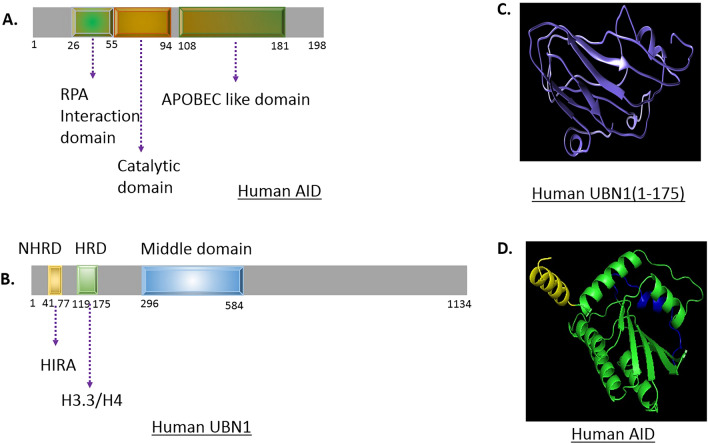
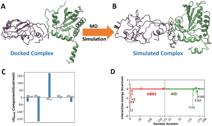
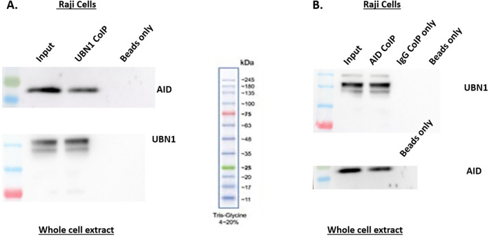
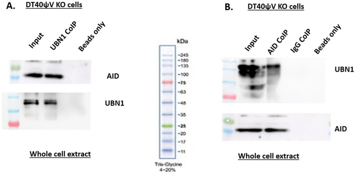
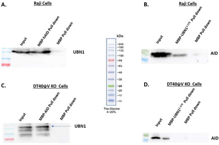
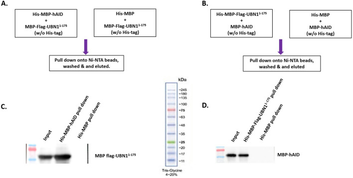
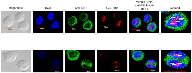
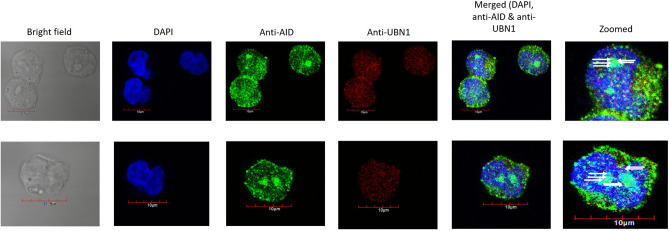
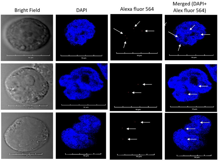
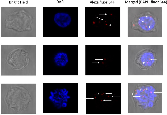
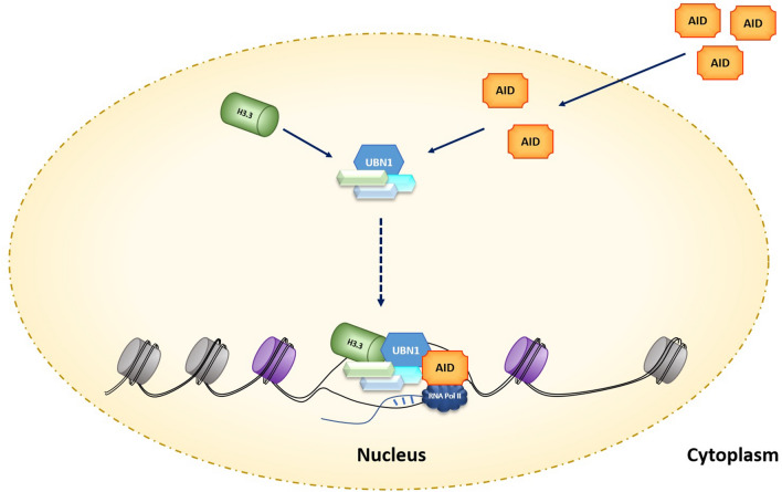
Activation-induced cytidine deaminase (AID) is the key mediator of antibody diversification in activated B-cells by the process of somatic hypermutation (SHM) and class switch recombination (CSR). Targeting of AID to the Ig genes requires transcription (initiation and elongation), and enhancers as well as its interaction with numerous factors. Furthermore, the HIRA chaperone complex, a regulator of chromatin architecture, is indispensable for SHM. The HIRA chaperone complex consists of UBN1, ASF1a, HIRA, and CABIN1 that deposit H3.3 onto the DNA, which is a hallmark of SHM. We explored whether UBN1 interacts with AID using computational as well as in-vitro experiments. Interestingly, our in-silico studies, such as molecular docking and molecular dynamics simulation results, predict that AID interacts with UBN1. Subsequent co-immunoprecipitation experiments established interactions between UBN1 and AID inside B-cells. Additionally, a double immunofluorescence assay confirmed that AID and UBN1 were co-localized in the human as well as chicken B-cell lines. Moreover, proximity ligation assay studies validated that AID interacts with UBN1. Ours is the first report on the interaction of genome mutator enzyme AID with UBN1. Nevertheless, the fate of interaction between UBN1 and AID is yet to be explored in the context of SHM or CSR.
Overview
The claims and statistics of this paper haven’t been checked yet.
Run the check — free, no account needed. It reads the open-access text and checks the sources it cites.
Integrity signals
Registry
Cites
Statistics
Figures
Verification
Claims the paper makes
The claims and statistics of this paper haven’t been checked yet.
Run the check — free, no account needed. It reads the open-access text and checks the sources it cites.
Self-check
Percentages, sums and reported differences re-computed from the paper’s own numbers.
The self-check needs the paper’s full text.
Citations
Cites
Highly accurate protein structure prediction with AlphaFold
Jumper, J., Evans, R., Pritzel, A., Green, T., Figurnov, M., Ronneberger, O., Tunyasuvunakool, K., Bates, R., Žídek, A., Potapenko, A., et al. (2021) · Nature · doi:10.1038/s41586-021-03819-2 · Cited by 43,764
Research works printed but not linked: 51
- Di Noia J, Neuberger MS. Altering the pathway of immunoglobulin hypermutation by inhibiting uracil-DNA glycosylase. Nature. 2002 Sep;419(6902):43–8. · DOI · Paper page
- Nagaoka H, Tran TH, Kobayashi M, Aida M, Honjo T. Preventing AID, a physiological mutator, from deleterious activation: regulation of the genomic instability that is associated with antibody diversity. Int Immunol. 2010 Apr;22(4):227–35. · DOI
- Muramatsu M, Kinoshita K, Fagarasan S, Yamada S, Shinkai Y, Honjo T. Class switch recombination and hypermutation require activation-induced cytidine deaminase (AID), a potential RNA editing enzyme. Cell. 2000 Sep;102(5):553–63. · DOI · Paper page
- Shinkura R, Ito S, Begum NA, Nagaoka H, Muramatsu M, Kinoshita K, et al. Separate domains of AID are required for somatic hypermutation and class-switch recombination. Nat Immunol. 2004 Jul;5(7):707–12. · DOI
- Ito S, Nagaoka H, Shinkura R, Begum N, Muramatsu M, Nakata M, et al. Activation-induced cytidine deaminase shuttles between nucleus and cytoplasm like apolipoprotein B mRNA editing catalytic polypeptide 1. Proc Natl Acad Sci U S A [Internet]. 2004/02/09. 2004 Feb 17;101(7):1975–80. Available from: https://www.ncbi.nlm.nih.gov/pubmed/14769937
- Rogozin IB, Diaz M. Cutting edge: DGYW/WRCH is a better predictor of mutability at G:C bases in Ig hypermutation than the widely accepted RGYW/WRCY motif and probably reflects a two-step activation-induced cytidine deaminase-triggered process. J Immunol. 2004 Mar;172(6):3382–4. · DOI
- Pham P, Afif SA, Shimoda M, Maeda K, Sakaguchi N, Pedersen LC, et al. Activation-induced deoxycytidine deaminase: Structural basis for favoring WRC hot motif specificities unique among APOBEC family members. Vol. 54, DNA repair. 2017. p. 8–12.
- Quinlan EM, King JJ, Amemiya CT, Hsu E, Larijani M. Biochemical Regulatory Features of Activation-Induced Cytidine Deaminase Remain Conserved from Lampreys to Humans. Mol Cell Biol. 2017 Oct;37(20). · DOI
- Betz AG, Milstein C, Gonzalez-Fernandez A, Pannell R, Larson T, Neuberger MS. Elements regulating somatic hypermutation of an immunoglobulin kappa gene: critical role for the intron enhancer/matrix attachment region. Cell. 1994 Apr;77(2):239–48. · DOI · Paper page
- Peters A, Storb U. Somatic hypermutation of immunoglobulin genes is linked to transcription initiation. Immunity [Internet]. 1996 Jan;4(1):57–65. Available from: https://www.ncbi.nlm.nih.gov/pubmed/8574852
- Kodgire P, Mukkawar P, Ratnam S, Martin TE, Storb U. Changes in RNA polymerase II progression influence somatic hypermutation of Ig-related genes by AID. J Exp Med. 2013 Jul;210(7):1481–92. · DOI
- Aida M, Hamad N, Stanlie A, Begum NA, Honjo T. Accumulation of the FACT complex, as well as histone H3.3, serves as a target marker for somatic hypermutation. Proc Natl Acad Sci U S A. 2013 May;110(19):7784–9. · DOI
- Yu G, Zhang Y, Gupta V, Zhang J, MacCarthy T, Duan Z, et al. The role of HIRA-dependent H3.3 deposition and its modifications in the somatic hypermutation of immunoglobulin variable regions. Proc Natl Acad Sci U S A. 2021 Dec;118(50).
- Pavri R, Gazumyan A, Jankovic M, Di Virgilio M, Klein I, Ansarah-Sobrinho C, et al. Activation-induced cytidine deaminase targets DNA at sites of RNA polymerase II stalling by interaction with Spt5. Cell. 2010 Oct;143(1):122–33. · DOI · Paper page
- Chaudhuri J, Khuong C, Alt FW. Replication protein A interacts with AID to promote deamination of somatic hypermutation targets. Nature. 2004 Aug;430(7003):992–8. · DOI · Paper page
- Kumar Singh A, Tamrakar A, Jaiswal A, Kanayama N, Agarwal A, Tripathi P, et al. Splicing regulator SRSF1-3 that controls somatic hypermutation of IgV genes interacts with topoisomerase 1 and AID. Mol Immunol. 2019 Dec;116:63–72. · DOI
- Conticello SG, Ganesh K, Xue K, Lu M, Rada C, Neuberger MS. Interaction between antibody-diversification enzyme AID and spliceosome-associated factor CTNNBL1. Mol Cell. 2008 Aug;31(4):474–84. · DOI
- Nowak U, Matthews AJ, Zheng S, Chaudhuri J. The splicing regulator PTBP2 interacts with the cytidine deaminase AID and promotes binding of AID to switch-region DNA. Nat Immunol. 2011 Feb;12(2):160–6. · DOI
- Ahmad K, Henikoff S. The histone variant H3.3 marks active chromatin by replication-independent nucleosome assembly. Mol Cell. 2002 Jun;9(6):1191–200. · DOI · Paper page
- Sherwood PW, Tsang S V, Osley MA. Characterization of HIR1 and HIR2, two genes required for regulation of histone gene transcription in Saccharomyces cerevisiae. Mol Cell Biol. 1993 Jan;13(1):28–38.
- Xiong C, Wen Z, Yu J, Chen J, Liu C-P, Zhang X, et al. UBN1/2 of HIRA complex is responsible for recognition and deposition of H3.3 at cis-regulatory elements of genes in mouse ES cells. BMC Biol. 2018 Oct;16(1):110. · DOI
- Kanehiro Y, Todo K, Negishi M, Fukuoka J, Gan W, Hikasa T, et al. Activation-induced cytidine deaminase (AID)-dependent somatic hypermutation requires a splice isoform of the serine/arginine-rich (SR) protein SRSF1. Proc Natl Acad Sci U S A. 2012 Jan;109(4):1216–21. · DOI
- Singh AK, Tamrakar A, Jaiswal A, Kanayama N, Kodgire P. SRSF1-3, a splicing and somatic hypermutation regulator, controls transcription of IgV genes via chromatin regulators SATB2, UBN1 and histone variant H3.3. Mol Immunol. 2020 Jan;119:69–82. · DOI
- Zhang H, Gan H, Wang Z, Lee J-H, Zhou H, Ordog T, et al. RPA Interacts with HIRA and Regulates H3.3 Deposition at Gene Regulatory Elements in Mammalian Cells. Mol Cell. 2017 Jan;65(2):272–84. · DOI
- Roy A, Kucukural A, Zhang Y. I-TASSER: a unified platform for automated protein structure and function prediction. Nat Protoc. 2010 Apr;5(4):725–38. · DOI · Paper page
- Yang J, Yan R, Roy A, Xu D, Poisson J, Zhang Y. The I-TASSER Suite: protein structure and function prediction. Vol. 12, Nature methods. 2015. p. 7–8.
- Lyskov S, Gray JJ. The RosettaDock server for local protein-protein docking. Nucleic Acids Res. 2008 Jul;36(Web Server issue):W233-8.
- Case DA, Cheatham TE 3rd, Darden T, Gohlke H, Luo R, Merz KMJ, et al. The Amber biomolecular simulation programs. J Comput Chem. 2005 Dec;26(16):1668–88. · DOI · Paper page
- Maier JA, Martinez C, Kasavajhala K, Wickstrom L, Hauser KE, Simmerling C. ff14SB: Improving the Accuracy of Protein Side Chain and Backbone Parameters from ff99SB. J Chem Theory Comput [Internet]. 2015 Aug 11;11(8):3696–713. Available from: https://doi.org/10.1021/acs.jctc.5b00255 · DOI · Paper page
- Price DJ, Brooks CL 3rd. A modified TIP3P water potential for simulation with Ewald summation. J Chem Phys. 2004 Nov;121(20):10096–103. · DOI · Paper page
- Kräutler V, van Gunsteren WF, Hünenberger PH. A fast SHAKE algorithm to solve distance constraint equations for small molecules in molecular dynamics simulations. J Comput Chem [Internet]. 2001 Apr 15;22(5):501–8. Available from: https://doi.org/10.1002/1096-987X(20010415)22:5%3C501::AID-JCC1021%3E3.0.CO · doi:10.1002/1096-987x(20010415)22
- Darden TA, York DM, Pedersen LG. Particle mesh Ewald: An p-log(N) method for Ewald sums in large systems. J Chem Phys. 1993;98:10089–92. · DOI · Paper page
- Pastor RW, Brooks BR, Szabo A. An analysis of the accuracy of Langevin and molecular dynamics algorithms. Mol Phys [Internet]. 1988 Dec 20;65(6):1409–19. Available from: https://doi.org/10.1080/00268978800101881 · DOI · Paper page
- Roy R, Mishra A, Poddar S, Nayak D, Kar P. Investigating the mechanism of recognition and structural dynamics of nucleoprotein-RNA complex from Peste des petits ruminants virus via Gaussian accelerated molecular dynamics simulations. J Biomol Struct Dyn. 2022 Mar;40(5):2302–15. · DOI
- Roy R, Jonniya NA, Poddar S, Sk MF, Kar P. Unraveling the Molecular Mechanism of Recognition of Human Interferon-Stimulated Gene Product 15 by Coronavirus Papain-Like Proteases: A Multiscale Simulation Study. J Chem Inf Model. 2021 Dec;61(12):6038–52. · DOI
- Roe DR, Cheatham TE. PTRAJ and CPPTRAJ: Software for Processing and Analysis of Molecular Dynamics Trajectory Data. J Chem Theory Comput [Internet]. 2013 Jul 9;9(7):3084–95. Available from: https://doi.org/10.1021/ct400341p · DOI · Paper page
- Kollman P. Free energy calculations: Applications to chemical and biochemical phenomena. Chem Rev [Internet]. 1993 Nov 1;93(7):2395–417. Available from: https://doi.org/10.1021/cr00023a004 · DOI · Paper page
- Gohlke H, Kiel C, Case DA. Insights into protein-protein binding by binding free energy calculation and free energy decomposition for the Ras-Raf and Ras-RalGDS complexes. J Mol Biol. 2003 Jul;330(4):891–913. · DOI · Paper page
- Lyskov S, Chou F-C, Conchúir SÓ, Der BS, Drew K, Kuroda D, et al. Serverification of Molecular Modeling Applications: The Rosetta Online Server That Includes Everyone (ROSIE). PLoS One [Internet]. 2013 May 22;8(5):e63906. Available from: https://doi.org/10.1371/journal.pone.0063906 · DOI · Paper page
- Chelico L, Pham P, Petruska J, Goodman MF. Biochemical basis of immunological and retroviral responses to DNA-targeted cytosine deamination by activation-induced cytidine deaminase and APOBEC3G. J Biol Chem. 2009 Oct;284(41):27761–5. · DOI
- Bransteitter R, Pham P, Calabrese P, Goodman MF. Biochemical analysis of hypermutational targeting by wild type and mutant activation-induced cytidine deaminase. J Biol Chem. 2004 Dec;279(49):51612–21. · DOI
- Revy P, Muto T, Levy Y, Geissmann F, Plebani A, Sanal O, et al. Activation-induced cytidine deaminase (AID) deficiency causes the autosomal recessive form of the Hyper-IgM syndrome (HIGM2). Cell. 2000 Sep;102(5):565–75. · DOI · Paper page
- Kodgire P, Mukkawar P, North JA, Poirier MG, Storb U. Nucleosome stability dramatically impacts the targeting of somatic hypermutation. Mol Cell Biol. 2012 May;32(10):2030–40. · DOI
- Singh AK, Jaiswal A, Kodgire P. AID preferentially targets the top strand in nucleosome sequences. Mol Immunol. 2019 Aug;112:198–205. · DOI
- Romanello M, Schiavone D, Frey A, Sale JE. Histone H3.3 promotes IgV gene diversification by enhancing formation of AID-accessible single-stranded DNA. EMBO J. 2016 Jul;35(13):1452–64. · DOI
- Goldberg AD, Banaszynski LA, Noh K-M, Lewis PW, Elsaesser SJ, Stadler S, et al. Distinct factors control histone variant H3.3 localization at specific genomic regions. Cell. 2010 Mar;140(5):678–91. · DOI · Paper page
- Ray-Gallet D, Quivy J-P, Scamps C, Martini EM-D, Lipinski M, Almouzni G. HIRA is critical for a nucleosome assembly pathway independent of DNA synthesis. Mol Cell. 2002 May;9(5):1091–100. · DOI · Paper page
- Banumathy G, Somaiah N, Zhang R, Tang Y, Hoffmann J, Andrake M, et al. Human UBN1 is an ortholog of yeast Hpc2p and has an essential role in the HIRA/ASF1a chromatin-remodeling pathway in senescent cells. Mol Cell Biol. 2009 Feb;29(3):758–70. · DOI
- Ray-Gallet D, Woolfe A, Vassias I, Pellentz C, Lacoste N, Puri A, et al. Dynamics of histone H3 deposition in vivo reveal a nucleosome gap-filling mechanism for H3.3 to maintain chromatin integrity. Mol Cell. 2011 Dec;44(6):928–41. · DOI · Paper page
- Dominguez PM, Teater M, Chambwe N, Kormaksson M, Redmond D, Ishii J, et al. DNA Methylation Dynamics of Germinal Center B Cells Are Mediated by AID. Cell Rep. 2015 Sep;12(12):2086–98. · DOI
- Béguelin W, Popovic R, Teater M, Jiang Y, Bunting KL, Rosen M, et al. EZH2 is required for germinal center formation and somatic EZH2 mutations promote lymphoid transformation. Cancer Cell. 2013 May;23(5):677–92. · DOI · Paper page
Metadata
- Authors
- Jaiswal, A., Roy, R., Tamarkar, A., Singh, A. K., Kar, P., & Kodgire, P.
- Affiliations
- Indian Institute of Technology Indore (IN)
- Indian Institute of Technology Indore
- Corresponding author
- Kodgire, Prashant
- Venue
- Scientific Reports
- Publisher
- Research Square Platform LLC
- Published
- 2022-12-21
- Volume
- 13
- Issue
- 1
- Pages
- 19615-19615
- ISSN
- 2045-2322 (ISSN-L)
- PMC
- PMC10638239
- OpenAlex
- W4388573268
- Cited by
- 3
- References
- 1
- Citations by year
- 2026: 2 · 2025: 1
- FWCI
- 0.27
- Type
- journal article
- Language
- en
- Open access
- gold
- doi.org
- Locations
- Scientific Reports (publishedVersion, cc-by)
- Research Square (acceptedVersion, cc-by)
- PubMed Central (publishedVersion, cc-by)
- PubMed (publishedVersion)
- DOAJ (DOAJ: Directory of Open Access Journals) (submittedVersion, cc-by-sa)
- Funders
- Indian Council of Medical Research (OMI/17/2020-ECD-1)
- Science and Engineering Research Board (CRG/2020/358)
- Topics
- Life Sciences › Biochemistry, Genetics and Molecular Biology › Molecular Biology › Genomics and Chromatin Dynamics
- Life Sciences › Biochemistry, Genetics and Molecular Biology › Molecular Biology › DNA Repair Mechanisms
- Life Sciences › Immunology and Microbiology › Immunology › T-cell and B-cell Immunology
- Keywords
- activation-induced cytidine deaminase, antibody diversification, somatic hypermutation, class switch recombination, B cells, protein-protein interaction, chromatin modification, immunoglobulin genes
- MeSH terms
- AICDA (Activation-Induced Cytidine Deaminase), Chromatin, Cytidine Deaminase, Humans, Immunoglobulins, Nuclear Proteins, Transcription Factors, Immunoglobulin Class Switching, Molecular Chaperones, Somatic Hypermutation, Immunoglobulin, Molecular Docking Simulation
About these checks
Registry, retraction, Cites and statistics results are computed, not judged, with one exception: a printed reference that no index lists is read once by a model before it counts as a citation error. Claims are extracted by a model and kept only when their quote appears verbatim in the text; each cited source is then checked, with a model’s help, for whether it says what the claim relies on. Those verdicts can be wrong — read the claim’s quote and the cited source before relying on one.
Statistics and arithmetic are recomputed, not judged.
Confidence bands are shown only for fields calibrated on our annotated test set. None is calibrated yet, so no band is shown.
What counts as a claim ›How we measure confidence ›How we check this ›
Are you an author, or do you see a problem with this page? Request a review
How to cite
Cite CoChat for the checks on this page, and the paper for its own findings.
- This page
- CoChat. (2026). Citation check: Activation-induced cytidine deaminase, an antibody diversification enzyme, interacts with chromatin modifier UBN1 in B-cells. https://app.cochat.ai/papers/10.21203/rs.3.rs-2379831/v1
- The paper
- Jaiswal, A., Roy, R., Tamarkar, A., Singh, A. K., Kar, P., & Kodgire, P. (2022). Activation-induced cytidine deaminase, an antibody diversification enzyme, interacts with chromatin modifier UBN1 in B-cells. Scientific Reports. https://doi.org/10.21203/rs.3.rs-2379831/v1
Data sources
Bibliographic data: Semantic Scholar, OpenAlex, Crossref.
Contains information from the Semantic Scholar Academic Graph Datasets, made available under the ODC Attribution License (ODC-BY 1.0).
Semantic Scholar’s data is described in: Kinney, R., et al. (2023). The Semantic Scholar Open Data Platform. arXiv:2301.10140.
