

The DOI matches its Crossref record; 1 of the 1 references it cites were found in a registry or an index.
A role for the ATP-dependent DNA ligase Lig E of Neisseria gonorrhoeae in biofilm formation
Open accessPreprint — published as 10.1186/s12866-024-03193-9 · Paper page
Abstract
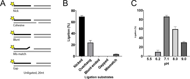
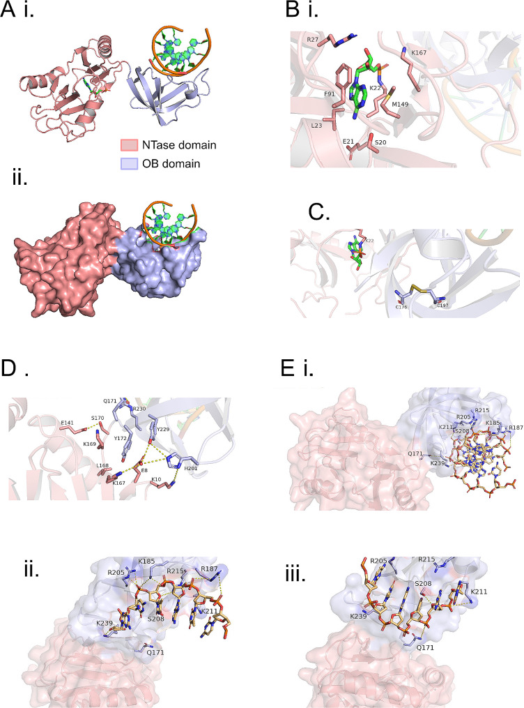
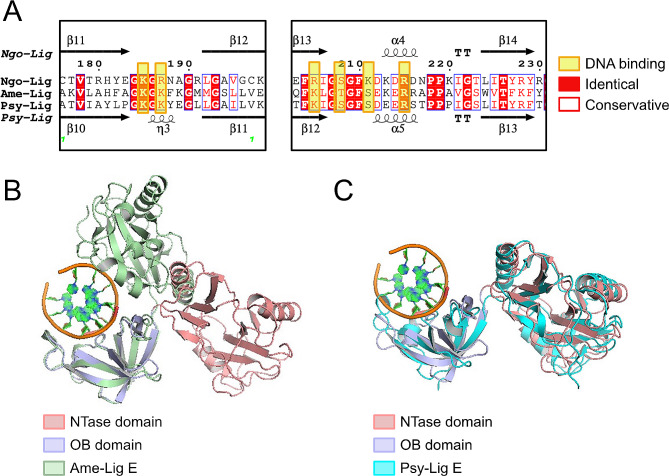
The ATP-dependent DNA ligase Lig E is present as an accessory DNA ligase in numerous proteobacterial genomes, including many disease-causing species. Here we have constructed a genomic Lig E knock-out in the obligate human pathogen Neisseria gonorrhoeae and characterised its growth and infection characteristics. This demonstrates that N. gonorrhoeae Lig E is a non-essential gene and its deletion does not cause defects in replication or survival of DNA-damaging stressors. Knock-out strains were partially defective in biofilm formation on an artificial surface as well as adhesion to epithelial cells which coupled with the predicted extracellular/ periplasmic location of Lig E indicates a role in extracellular DNA joining. In addition to in vivo characterisation, we have recombinantly expressed and assayed N. gonorrhoeae Lig E and determined the crystal structure of the enzyme-adenylate engaged with DNA substrate in an open non-catalytic conformation, providing insight into the binding dynamics of these minimal DNA ligases.
Overview
The claims and statistics of this paper haven’t been checked yet.
Run the check — free, no account needed. It reads the open-access text and checks the sources it cites.
Integrity signals
Registry
Cites
Statistics
Figures
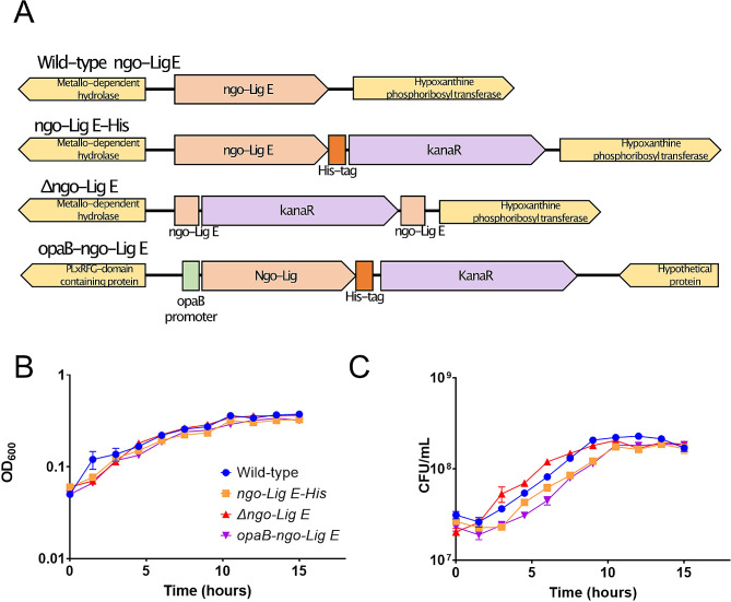
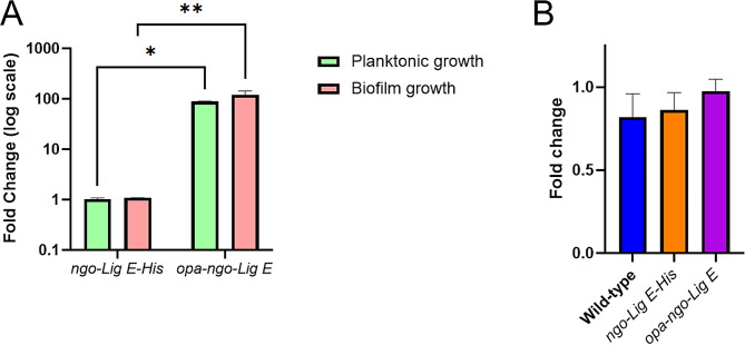
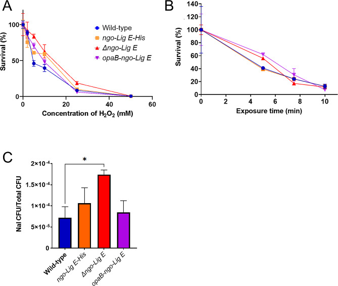
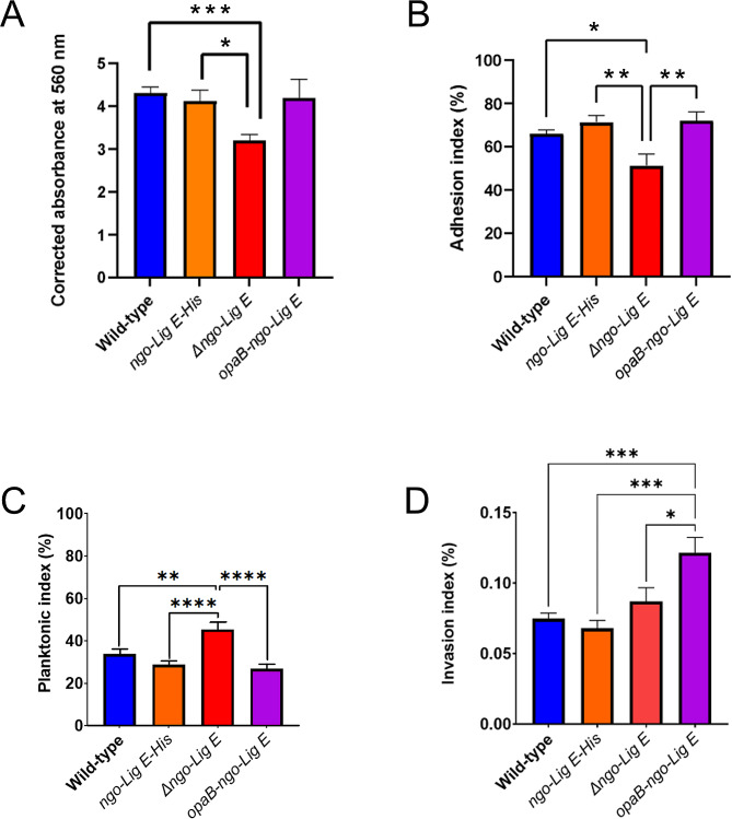
Verification
Claims the paper makes
The claims and statistics of this paper haven’t been checked yet.
Run the check — free, no account needed. It reads the open-access text and checks the sources it cites.
Self-check
Percentages, sums and reported differences re-computed from the paper’s own numbers.
The self-check needs the paper’s full text.
Citations
Cites
Highly accurate protein structure prediction with AlphaFold
Jumper, J., Evans, R., Pritzel, A., Green, T., Figurnov, M., Ronneberger, O., Tunyasuvunakool, K., Bates, R., Žídek, A., Potapenko, A., et al. (2021) · Nature · doi:10.1038/s41586-021-03819-2 · Cited by 43,764
Research works printed but not linked: 48
- Płociński P, et al. DNA Ligase C and Prim-PolC participate in base excision repair in mycobacteria. Nat Commun. 2017;8:1251. https://doi.org:10.1038/s41467-017-01365-y. · DOI
- Zhu H, Shuman S. Gap filling activities of Pseudomonas DNA ligase D (LigD) polymerase and functional interactions of LigD with the DNA end-binding Ku protein. J Biol Chem. 2010;285:4815–25. https://doi.org:10.1074/jbc.M109.073874. · DOI
- Shuman S, Glickman MS. Bacterial DNA repair by non-homologous end joining. Nat Rev Microbiol. 2007;5:852–61. https://doi.org:10.1038/nrmicro1768. · DOI
- Ejaz A, Goldgur Y, Shuman S. Activity and structure of Pseudomonas putida MPE, a manganese-dependent single-strand DNA endonuclease encoded in a nucleic acid repair gene cluster. J Biol Chem. 2019;294:7931–41. https://doi.org:10.1074/jbc.RA119.008049. · DOI
- Ejaz A, Shuman S. Characterization of Lhr-Core DNA helicase and manganese- dependent DNA nuclease components of a bacterial gene cluster encoding nucleic acid repair enzymes. J Biol Chem. 2018;293:17491–504. https://doi.org:10.1074/jbc.RA118.005296. · DOI
- Pan J, Lian K, Sarre A, Leiros HS, Williamson A. Bacteriophage origin of some minimal ATP-dependent DNA ligases: a new structure from Burkholderia pseudomallei with striking similarity to Chlorella virus ligase. Sci Rep. 2021;11:18693. https://doi.org:10.1038/s41598-021-98155-w. · DOI
- Williamson A, Hjerde E, Kahlke T. Analysis of the distribution and evolution of the ATP-dependent DNA ligases of bacteria delineates a distinct phylogenetic group ‘Lig E’. Mol Microbiol. 2016;99:274–90. https://doi.org:10.1111/mmi.13229. · DOI
- Berg K, Leiros I, Williamson A. Temperature adaptation of DNA ligases from psychrophilic organisms. Extremophiles. 2019;23:305–17. https://doi.org:10.1007/s00792-019-01082-y. · DOI
- Williamson A, Grgic M, Leiros HS. DNA binding with a minimal scaffold: structure-function analysis of Lig E DNA ligases. Nucleic Acids Res. 2018;46:8616–29. https://doi.org:10.1093/nar/gky622. · DOI
- Williamson A, Rothweiler U, Schroder Leiros H-K. Enzyme-adenylate structure of a bacterial ATP-dependent DNA ligase with a minimized DNA-binding surface. Acta Crystallogr Sect D. 2014;70:3043–56. https://doi.org:doi:10.1107/S1399004714021099. · DOI
- Williamson A, Pedersen H. Recombinant expression and purification of an ATP-dependent DNA ligase from Aliivibrio salmonicida. Protein Expres Purif. 2014;97:29–36. //dx.doi.org/10.1016/j.pep.2014.02.008. https://doi.org:http:. · DOI
- Cehovin A, Lewis SB. Mobile genetic elements in Neisseria gonorrhoeae: movement for change. Pathogens and disease. 2017;75. https://doi.org:10.1093/femspd/ftx071. · DOI
- Unemo M, Shafer WM. Antimicrobial resistance in Neisseria gonorrhoeae in the 21st century: past, evolution, and future. Clin Microbiol Rev. 2014;27:587–613. https://doi.org:10.1128/cmr.00010-14. · DOI
- Martín-Sánchez M, et al. Clinical presentation of asymptomatic and symptomatic heterosexual men who tested positive for urethral gonorrhoea at a sexual health clinic in Melbourne, Australia. BMC Infect Dis. 2020;20:486. https://doi.org:10.1186/s12879-020-05197-y. · DOI
- Unemo M, et al. Gonorrhoea. Nat Reviews Disease Primers. 2019;5:79. https://doi.org:10.1038/s41572-019-0128-6. · DOI
- Falsetta ML, et al. The Composition and Metabolic Phenotype of Neisseria gonorrhoeae Biofilms. Front Microbiol. 2011;2:75. https://doi.org:10.3389/fmicb.2011.00075. · DOI
- Kouzel N, Oldewurtel ER, Maier B. Gene Transfer Efficiency in Gonococcal Biofilms: Role of Biofilm Age, Architecture, and Pilin Antigenic Variation. J Bacteriol. 2015;197:2422–31. https://doi.org:10.1128/jb.00171-15. · DOI
- Steichen CT, Shao JQ, Ketterer MR, Apicella MA. Gonococcal cervicitis: a role for biofilm in pathogenesis. J Infect Dis. 2008;198:1856–61. https://doi.org:10.1086/593336. · DOI
- Greiner LL, et al. Biofilm Formation by Neisseria gonorrhoeae. Infect Immun. 2005;73:1964–70. https://doi.org:10.1128/iai.73.4.1964-1970.2005. · DOI
- Zweig M, et al. Secreted single-stranded DNA is involved in the initial phase of biofilm formation by Neisseria gonorrhoeae. Environ Microbiol. 2014;16:1040–52. https://doi.org:10.1111/1462-2920.12291. · DOI · Paper page
- Phillips NJ, et al. Proteomic analysis of Neisseria gonorrhoeae biofilms shows shift to anaerobic respiration and changes in nutrient transport and outermembrane proteins. PLoS ONE. 2012;7:e38303. https://doi.org:10.1371/journal.pone.0038303. · DOI
- Steichen CT, Cho C, Shao JQ, Apicella MA. The Neisseria gonorrhoeae biofilm matrix contains DNA, and an endogenous nuclease controls its incorporation. Infect Immun. 2011;79:1504–11. https://doi.org:10.1128/iai.01162-10. · DOI
- Dillard JP, Unit4A.2. Genetic Manipulation of Neisseria gonorrhoeae. Curr protocols Microbiol Chap. 2011;4. https://doi.org:10.1002/9780471729259.mc04a02s23. · DOI
- Callaghan MM, Dillard JP. Transformation in Neisseria gonorrhoeae. Methods in molecular biology (Clifton, N.J.) 1997, 143–162 (2019). https://doi.org:10.1007/978-1-4939-9496-0_10. · DOI
- Schmittgen TD, Livak KJ. Analyzing real-time PCR data by the comparative CT method. Nat Protoc. 2008;3:1101–8. https://doi.org:10.1038/nprot.2008.73. · DOI · Paper page
- Petersen TN, Brunak S, von Heijne G, Nielsen H. SignalP 4.0: discriminating signal peptides from transmembrane regions. Nat Methods. 2011;8:785–6. https://doi.org:10.1038/nmeth.1701. · DOI · Paper page
- Sharma JK, et al. Methods for competitive enrichment and evaluation of superior DNA ligases. Methods Enzymol. 2020;644:209–25. https://doi.org:10.1016/bs.mie.2020.04.061. · DOI
- Schneider CA, Rasband WS, Eliceiri K. W. NIH Image to ImageJ: 25 years of image analysis. Nat Meth. 2012;9:671–5. · DOI · Paper page
- Aragão D, et al. MX2: a high-flux undulator microfocus beamline serving both the chemical and macromolecular crystallography communities at the Australian Synchrotron. J Synchrotron Radiat. 2018;25:885–91. https://doi.org:10.1107/s1600577518003120. · DOI
- Winn MD, et al. Overview of the CCP4 suite and current developments. Acta Crystallogr D. 2011;67:235–42. https://doi.org:10.1107/s0907444910045749. · DOI · Paper page
- Kabsch WXDS. Acta Crystallogr Sect D: Biol Crystallogr. 2010;66:125–32. https://doi.org:10.1107/S0907444909047337. · DOI · Paper page
- Adams PD, et al. PHENIX: a comprehensive Python-based system for macromolecular structure solution. Acta Crystallogr D. 2010;66:213–21. https://doi.org:10.1107/s0907444909052925. · DOI · Paper page
- McCoy AJ, et al. Phaser crystallographic software. J Appl Crystallogr. 2007;40:658–74. https://doi.org:10.1107/s0021889807021206. · DOI · Paper page
- Afonine PV, et al. Towards automated crystallographic structure refinement with phenix.refine. Acta Crystallogr D. 2012;68:352–67. https://doi.org:10.1107/s0907444912001308. · DOI · Paper page
- Emsley P, Lohkamp B, Scott WG, Cowtan K. Features and development of Coot. Acta Crystallogr D. 2010;66:486–501. https://doi.org:10.1107/S0907444910007493. · DOI · Paper page
- Cheng CH, Shuman S. Characterization of an ATP-dependent DNA ligase encoded by Haemophilus influenzae. Nucleic Acids Res. 1997;25:1369–74. · DOI
- Magnet S, Blanchard JS. Mechanistic and kinetic study of the ATP-dependent DNA ligase of Neisseria meningitidis. Biochemistry-Us. 2004;43:710–7. https://doi.org:10.1021/bi0355387. · DOI
- Seib KL, et al. Defenses against oxidative stress in Neisseria gonorrhoeae: a system tailored for a challenging environment. Microbiol Mol biology reviews: MMBR. 2006;70:344–61. https://doi.org:10.1128/mmbr.00044-05. · DOI
- Cahoon LA, Stohl EA, Seifert HS. The Neisseria gonorrhoeae photolyase orthologue phrB is required for proper DNA supercoiling but does not function in photo-reactivation. Mol Microbiol. 2011;79:729–42. https://doi.org:10.1111/j.1365-2958.2010.07481.x. · DOI
- Cadet J, Wagner JR. DNA base damage by reactive oxygen species, oxidizing agents, and UV radiation. Cold Spring Harb Perspect Biol. 2013;5. https://doi.org:10.1101/cshperspect.a012559. · DOI · Paper page
- Juneau RA, Stevens JS, Apicella MA, Criss AK. A thermonuclease of Neisseria gonorrhoeae enhances bacterial escape from killing by neutrophil extracellular traps. J Infect Dis. 2015;212:316–24. https://doi.org:10.1093/infdis/jiv031. · DOI
- Higashi DL, et al. Dynamics of Neisseria gonorrhoeae attachment: microcolony development, cortical plaque formation, and cytoprotection. Infect Immun. 2007;75:4743–53. https://doi.org:10.1128/iai.00687-07. · DOI
- Winther-Larsen HC, et al. Neisseria gonorrhoeae PilV, a type IV pilus-associated protein essential to human epithelial cell adherence. Proc Natl Acad Sci U S A. 2001;98:15276–81. https://doi.org:10.1073/pnas.261574998. · DOI
- Quillin SJ, Seifert HS. Neisseria gonorrhoeae host adaptation and pathogenesis. Nat Rev Microbiol. 2018;16:226–40. https://doi.org:10.1038/nrmicro.2017.169. · DOI
- LeVan A, et al. Construction and characterization of a derivative of Neisseria gonorrhoeae strain MS11 devoid of all opa genes. J Bacteriol. 2012;194:6468–78. https://doi.org:10.1128/jb.00969-12. · DOI
- Walker E, van Niekerk S, Hanning K, Kelton W, Hicks J. Mechanisms of host manipulation by Neisseria gonorrhoeae. Front Microbiol. 2023;14. https://doi.org:10.3389/fmicb.2023.1119834. · DOI
- Bender N, Hennes M, Maier B. Mobility of extracellular DNA within gonococcal colonies. Biofilm. 2022;4:100078. https://doi.org:10.1016/j.bioflm.2022.100078. · DOI
- Bauer RJ, Jurkiw TJ, Evans TC Jr., Lohman GJ. Rapid Time Scale Analysis of T4 DNA Ligase-DNA Binding. Biochemistry-Us. 2017;56:1117–29. https://doi.org:10.1021/acs.biochem.6b01261. · DOI
Metadata
- Authors
- Pan, J., Hicks, J., & Williamson, A.
- Affiliations
- University of Waikato (NZ)
- Corresponding author
- Williamson, Adele
- Venue
- BMC Microbiology
- Publisher
- Research Square Platform LLC
- Published
- 2023-10-06
- Volume
- 24
- Issue
- 1
- Pages
- 29-29
- ISSN
- 1471-2180 (ISSN-L)
- PMC
- PMC10799422
- OpenAlex
- W4391052468
- Cited by
- 9
- References
- 1
- Citations by year
- 2026: 4 · 2025: 3 · 2024: 2
- FWCI
- 2.39
- Type
- journal article
- Language
- en
- Open access
- gold
- doi.org
- Locations
- BMC Microbiology (publishedVersion, cc-by)
- Research Square (acceptedVersion, cc-by)
- PubMed Central (publishedVersion, cc-by)
- PubMed (publishedVersion)
- DOAJ (DOAJ: Directory of Open Access Journals) (submittedVersion, cc-by-sa)
- Funders
- Australian Nuclear Science and Technology Organisation
- University of Waikato
- Australian Cancer Research Foundation
- Australian Synchrotron
- Health Research Council of New Zealand (21/754, 23/534)
- Topics
- Life Sciences › Biochemistry, Genetics and Molecular Biology › Genetics › Bacterial Genetics and Biotechnology
- Life Sciences › Biochemistry, Genetics and Molecular Biology › Molecular Biology › Biochemical and Structural Characterization
- Life Sciences › Biochemistry, Genetics and Molecular Biology › Molecular Biology › Bacterial biofilms and quorum sensing
- Keywords
- ATP-dependent DNA ligase, Neisseria gonorrhoeae, biofilm formation, periplasmic enzymes, crystal structure
- MeSH terms
- DNA Ligase ATP, DNA, Humans, Neisseria gonorrhoeae, DNA Ligases, Biofilms
About these checks
Registry, retraction, Cites and statistics results are computed, not judged, with one exception: a printed reference that no index lists is read once by a model before it counts as a citation error. Claims are extracted by a model and kept only when their quote appears verbatim in the text; each cited source is then checked, with a model’s help, for whether it says what the claim relies on. Those verdicts can be wrong — read the claim’s quote and the cited source before relying on one.
Statistics and arithmetic are recomputed, not judged.
Confidence bands are shown only for fields calibrated on our annotated test set. None is calibrated yet, so no band is shown.
What counts as a claim ›How we measure confidence ›How we check this ›
Are you an author, or do you see a problem with this page? Request a review
