

The DOI matches its Crossref record; 1 of the 1 references it cites were found in a registry or an index.
Deciphering the Allosteric Regulation of Mycobacterial Inosine-5′-Monophosphate Dehydrogenase
Open accessPreprint — published as 10.1038/s41467-024-50933-6 · Paper page
Abstract
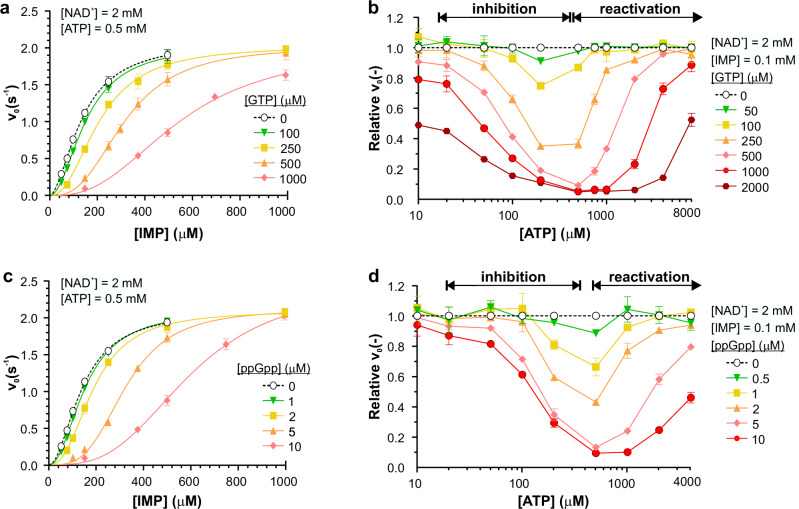
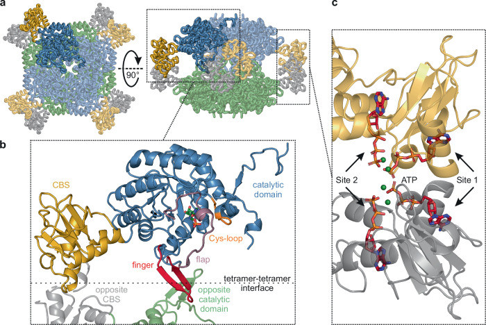
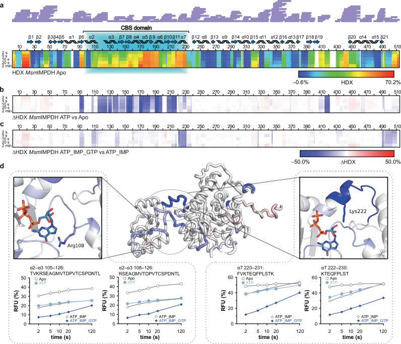
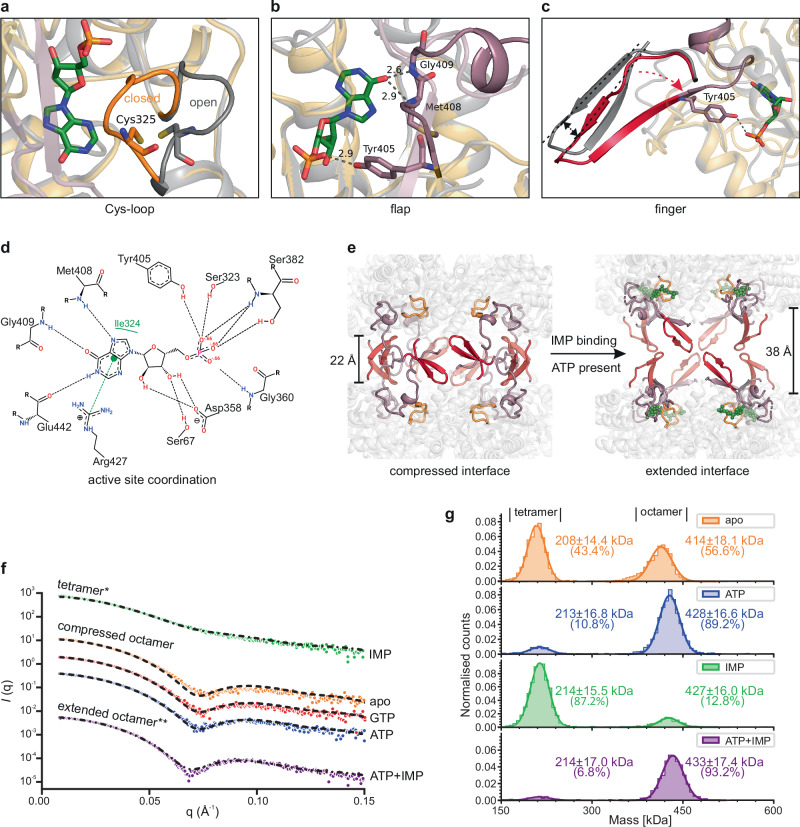
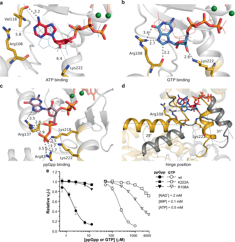
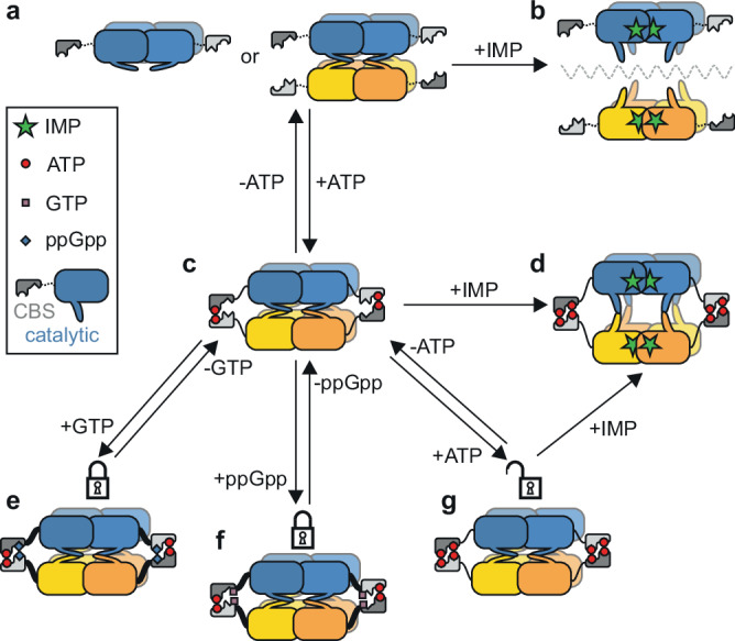
Allosteric regulation of inosine 5′-monophosphate dehydrogenase (IMPDH), an essential enzyme of purine metabolism, contributes to the homeostasis of adenine and guanine nucleotides. However, the precise molecular mechanism of IMPDH regulation in bacteria remains unclear. Using biochemical, and cryo-EM approaches, we reveal the intricate molecular mechanism of the IMPDH allosteric regulation in mycobacteria. The enzyme is inhibited by both GTP and (p)ppGpp which bind to the regulatory CBS domains and, via interactions with basic residues in hinge regions, lock the catalytic core domains in a compressed conformation. This results in occlusion of inosine monophosphate (IMP) substrate binding to the active site and, ultimately, inhibition of the enzyme. The GTP and (p)ppGpp allosteric effectors bind to their dedicated sites but stabilize the compressed octamer by a common mechanism. Inhibition is relieved by the competitive displacement of GTP or (p)ppGpp by ATP allowing IMP-induced enzyme expansion. The structural knowledge and mechanistic understanding presented here open up new possibilities for the development of allosteric inhibitors with antibacterial potential.
Overview
The claims and statistics of this paper haven’t been checked yet.
Run the check — free, no account needed. It reads the open-access text and checks the sources it cites.
Integrity signals
Registry
Cites
Statistics
Figures
Verification
Claims the paper makes
The claims and statistics of this paper haven’t been checked yet.
Run the check — free, no account needed. It reads the open-access text and checks the sources it cites.
Self-check
Percentages, sums and reported differences re-computed from the paper’s own numbers.
The self-check needs the paper’s full text.
Citations
Cites
Highly accurate protein structure prediction with AlphaFold
Jumper, J., Evans, R., Pritzel, A., Green, T., Figurnov, M., Ronneberger, O., Tunyasuvunakool, K., Bates, R., Žídek, A., Potapenko, A., et al. (2021) · Nature · doi:10.1038/s41586-021-03819-2 · Cited by 43,764
Research works printed but not linked: 48
- Hedstrom LIMP, Dehydrogenase (2009) Structure, Mechanism, and Inhibition. Chem Rev 109 7:2903–2928. https://doi.org/10.1021/cr900021w · DOI
- Buey RM, Fernández-Justel D, Jiménez A, Revuelta JL (2022) The Gateway to Guanine Nucleotides: Allosteric Regulation of IMP Dehydrogenases. Protein Sci 31(9):e4399. https://doi.org/10.1002/pro.4399 · DOI
- Burrell AL, Nie C, Said M, Simonet JC, Fernández-Justel D, Johnson MC, Quispe J, Buey RM, Peterson JR, Kollman JM (2021) IMPDH1 Retinal Variants Control Filament Architecture to Tune Allosteric Regulation; ; p 2021.08.03.454821. https://doi.org/10.1101/2021.08.03.454821 · DOI
- Hedstrom L, Liechti G, Goldberg JB, Gollapalli DR (2011) The Antibiotic Potential of Prokaryotic IMP Dehydrogenase Inhibitors. Curr Med Chem 18(13):1909–1918 · DOI
- Singh V, Donini S, Pacitto A, Sala C, Hartkoorn RC, Dhar N, Keri G, Ascher DB, Mondésert G, Vocat A, Lupien A, Sommer R, Vermet H, Lagrange S, Buechler J, Warner DF, McKinney JD, Pato J, Cole ST, Blundell TL, Rizzi M, Mizrahi V (2017) The Inosine Monophosphate Dehydrogenase, GuaB2, Is a Vulnerable New Bactericidal Drug Target for Tuberculosis. ACS Infect Dis 3(1):5–17. https://doi.org/10.1021/acsinfecdis.6b00102 · DOI
- Alexandre T, Rayna B, Munier-Lehmann H (2015) Two Classes of Bacterial IMPDHs According to Their Quaternary Structures and Catalytic Properties. PLoS ONE 10(2):e0116578. https://doi.org/10.1371/journal.pone.0116578 · DOI
- Ereño-Orbea J, Oyenarte I, Martínez-Cruz LACBS, Domains (2013) Ligand Binding Sites and Conformational Variability. Arch Biochem Biophys 540(1–2):70–81. https://doi.org/10.1016/j.abb.2013.10.008 · DOI
- Buey RM, Ledesma-Amaro R, Velázquez-Campoy A, Balsera M, Chagoyen M, de Pereda JM, Revuelta JL (2015) Guanine Nucleotide Binding to the Bateman Domain Mediates the Allosteric Inhibition of Eukaryotic IMP Dehydrogenases. Nat Commun 6:8923. https://doi.org/10.1038/ncomms9923 · DOI
- Fernández-Justel D, Marcos-Alcalde Í, Abascal F, Vidaña N, Gómez-Puertas P, Jiménez A, Revuelta JL, Buey RM (2022) Diversity of Mechanisms to Control Bacterial GTP Homeostasis by the Mutually Exclusive Binding of Adenine and Guanine Nucleotides to IMP Dehydrogenase. Protein Sci 31(5):e4314. https://doi.org/10.1002/pro.4314 · DOI
- Rostirolla DC, Assunção TM (2014) Biochemical Characterization of Mycobacterium Tuberculosis IMP Dehydrogenase: Kinetic Mechanism, Metal Activation and Evidence of a Cooperative System. RSC Adv 4(50):26271–26287. https://doi.org/10.1039/C4RA02142H. BizarroC. V.Basso, L. A.; Santos, D. S. · DOI
- Usha V, Gurcha SS, Lovering AL, Lloyd AJ, Papaemmanouil A, Reynolds RC, Besra GS (2011) Identification of Novel Diphenyl Urea Inhibitors of Mt-GuaB2 Active against Mycobacterium Tuberculosis. Microbiology 157(1):290–299. https://doi.org/10.1099/mic.0.042549-0 · DOI
- Makowska-Grzyska M, Kim Y, Maltseva N, Osipiuk J, Gu M, Zhang M, Mandapati K, Gollapalli DR, Gorla SK, Hedstrom L, Joachimiak AA (2015) Novel Cofactor-Binding Mode in Bacterial IMP Dehydrogenases Explains Inhibitor Selectivity. J Biol Chem 290(9):5893–5911. https://doi.org/10.1074/jbc.M114.619767 · DOI
- Labesse G, Alexandre T, Vaupré L, Salard-Arnaud I, Him JLK, Raynal B, Bron P, Munier-Lehmann H (2013) MgATP Regulates Allostery and Fiber Formation in IMPDHs. Structure 21 6, 975–985. https://doi.org/10.1016/j.str.2013.03.011 · DOI
- Labesse G, Alexandre T, Gelin M, Haouz A, Munier-Lehmann H (2015) Crystallographic Studies of Two Variants of Pseudomonas Aeruginosa IMPDH with Impaired Allosteric Regulation. Acta Crystallogr D Biol Crystallogr 71(Pt 9):1890–1899. https://doi.org/10.1107/S1399004715013115 · DOI
- Pimkin M, Markham GD (2008) The CBS Subdomain of Inosine 5′-Monophosphate Dehydrogenase Regulates Purine Nucleotide Turnover. Mol Microbiol 68(2):342–359. https://doi.org/10.1111/j.1365-2958.2008.06153.x · DOI
- Pimkin M, Pimkina J, Markham GD (2009) A Regulatory Role of the Bateman Domain of IMP Dehydrogenase in Adenylate Nucleotide Biosynthesis. J Biol Chem 284(12):7960–7969. https://doi.org/10.1074/jbc.M808541200 · DOI
- Giammarinaro PI, Young MKM, Steinchen W, Mais C-N, Hochberg G, Yang J, Stevenson DM, Amador-Noguez D, Paulus A, Wang JD, Bange G (2022) Diadenosine Tetraphosphate Regulates Biosynthesis of GTP in Bacillus Subtilis. Nat Microbiol 1–11. https://doi.org/10.1038/s41564-022-01193-x · DOI
- Makowska-Grzyska M, Kim Y, Wu R, Wilton R, Gollapalli DR, Wang XK, Zhang R, Jedrzejczak R, Mack JC, Maltseva N, Mulligan R, Binkowski TA, Gornicki P, Kuhn ML, Anderson WF, Hedstrom L, Joachimiak A (2012) Bacillus Anthracis Inosine 5′-Monophosphate Dehydrogenase in Action: The First Bacterial Series of Structures of Phosphate Ion-, Substrate-, and Product-Bound Complexes. Biochemistry 51(31). 10.1021/bi300511w · DOI
- Knejzlík Z, Doležal M, Herkommerová K, Clarova K, Klíma M, Dedola M, Zborníková E, Rejman D, Pichová I (2022) The Mycobacterial guaB1 Gene Encodes a Guanosine 5′-Monophosphate Reductase with a Cystathionine-β-Synthase Domain. FEBS J 289(18):5571–5598. https://doi.org/10.1111/febs.16448 · DOI
- Imholz NCE, Noga MJ, van den Broek NJF, Bokinsky G (2020) Calibrating the Bacterial Growth Rate Speedometer: A Re-Evaluation of the Relationship Between Basal ppGpp, Growth, and RNA Synthesis in Escherichia Coli. Frontiers in Microbiology 11. https://doi.org/10.3389/fmicb.2020.574872 · DOI
- Steinchen W, Zegarra V, Bange G (2020) (P)ppGpp: Magic Modulators of Bacterial Physiology and Metabolism. Front. Microbiol. 11 (2072). https://doi.org/10.3389/fmicb.2020.02072 · DOI · Paper page
- Fernández-Justel D, Peláez R, Revuelta JL, Buey RM (2019) The Bateman Domain of IMP Dehydrogenase Is a Binding Target for Dinucleoside Polyphosphates. J Biol Chem 294(40):14768–14775. https://doi.org/10.1074/jbc.AC119.010055 · DOI
- Despotović D, Brandis A, Savidor A, Levin Y, Fumagalli L, Tawfik DS (2017) Diadenosine Tetraphosphate (Ap4A) – an E. Coli Alarmone or a Damage Metabolite? FEBS J 284(14):2194–2215. https://doi.org/10.1111/febs.14113 · DOI
- Cox JAG, Mugumbate G, Del Peral LV-G, Jankute M, Abrahams KA, Jervis P, Jackenkroll S, Perez A, Alemparte C, Esquivias J, Lelièvre J, Ramon F, Barros D, Ballell L, Besra GS (2016) Novel Inhibitors of Mycobacterium Tuberculosis GuaB2 Identified by a Target Based High-Throughput Phenotypic Screen. Sci Rep 6(1):38986. https://doi.org/10.1038/srep38986 · DOI
- Makowska-Grzyska M, Kim Y, Gorla SK, Wei Y, Mandapati K, Zhang M, Maltseva N, Modi G, Boshoff HI, Gu M, Aldrich C, Cuny GD, Hedstrom L, Joachimiak A (2015) Mycobacterium Tuberculosis IMPDH in Complexes with Substrates, Products and Antitubercular Compounds. PLoS ONE 10(10):e0138976. https://doi.org/10.1371/journal.pone.0138976 · DOI
- Chen L, Wilson DJ, Xu Y, Aldrich CC, Felczak K, Sham YY, Pankiewicz KW (2010) Triazole-Linked Inhibitors of Inosine Monophosphate Dehydrogenase from Human and Mycobacterium Tuberculosis. J Med Chem 53(12):4768–4778. https://doi.org/10.1021/jm100424m · DOI
- Trapero A, Pacitto A, Singh V, Sabbah M, Coyne AG, Mizrahi V, Blundell TL, Ascher DB, Abell C (2018) Fragment-Based Approach to Targeting Inosine-5′-Monophosphate Dehydrogenase (IMPDH) from Mycobacterium Tuberculosis. J Med Chem 61(7):2806–2822. https://doi.org/10.1021/acs.jmedchem.7b01622 · DOI
- Gollapalli DR, MacPherson IS, Liechti G, Gorla SK, Goldberg JB, Hedstrom L (2010) Structural Determinants of Inhibitor Selectivity in Prokaryotic IMP Dehydrogenases. Chem Biol 17(10):1084–1091. https://doi.org/10.1016/j.chembiol.2010.07.014 · DOI
- Alexandre T, Lupan A, Helynck O, Vichier-Guerre S, Dugué L, Gelin M, Haouz A, Labesse G, Munier-Lehmann H (2019) First-in-Class Allosteric Inhibitors of Bacterial IMPDHs. Eur J Med Chem 167:124–132. https://doi.org/10.1016/j.ejmech.2019.01.064 · DOI
- Andersen KR, Leksa NC, Schwartz TU, Optimized E (2013) Coli Expression Strain LOBSTR Eliminates Common Contaminants from His-Tag Purification. Proteins 81(11):1857–1861. https://doi.org/10.1002/prot.24364 · DOI
- Studier FW (2005) Protein Production by Auto-Induction in High-Density Shaking Cultures. Protein Expression Purif 41(1):207–234. https://doi.org/10.1016/j.pep.2005.01.016 · DOI
- Zheng SQ, Palovcak E, Armache J-P, Verba KA, Cheng Y, Agard DA (2017) MotionCor2: Anisotropic Correction of Beam-Induced Motion for Improved Cryo-Electron Microscopy. Nat Methods 14(4):331–332. https://doi.org/10.1038/nmeth.4193 · DOI
- Rohou A, Grigorieff N (2015) CTFFIND4: Fast and Accurate Defocus Estimation from Electron Micrographs. J Struct Biol 192(2):216–221. https://doi.org/10.1016/j.jsb.2015.08.008 · DOI · Paper page
- Kimanius D, Dong L, Sharov G, Nakane T, Scheres SH (2021) W. New Tools for Automated Cryo-EM Single-Particle Analysis in RELION-4.0. Biochem J 478(24):4169–4185. https://doi.org/10.1042/BCJ20210708 · DOI
- Burnley T, Palmer CM, Winn M (2017) Recent Developments in the CCP-EM Software Suite. Acta Crystallogr D Struct Biol 73(6):469–477. https://doi.org/10.1107/S2059798317007859 · DOI
- Jakobi AJ, Wilmanns M, Sachse C (2017) Model-Based Local Density Sharpening of Cryo-EM Maps. eLife 6, e27131. https://doi.org/10.7554/eLife.27131 · DOI
- Pettersen EF, Goddard TD, Huang CC, Meng EC, Couch GS, Croll TI, Morris JH, Ferrin TE (2021) UCSF ChimeraX: Structure Visualization for Researchers, Educators, and Developers. Protein Sci 30(1):70–82. https://doi.org/10.1002/pro.3943 · DOI
- Emsley P, Lohkamp B, Scott WG, Cowtan K (2010) Features and Development of Coot. Acta Cryst D 66(4):486–501. https://doi.org/10.1107/S0907444910007493 · DOI · Paper page
- Croll TI, ISOLDE: (2018) A Physically Realistic Environment for Model Building into Low-Resolution Electron-Density Maps. Acta Cryst D 74(6):519–530. https://doi.org/10.1107/S2059798318002425 · DOI
- Afonine PV, Poon BK, Read RJ, Sobolev OV, Terwilliger TC, Urzhumtsev A, Adams PD (2018) Real-Space Refinement in PHENIX for Cryo-EM and Crystallography. Acta Cryst D 74(6):531–544. https://doi.org/10.1107/S2059798318006551 · DOI
- Hoh SW, Burnley T, Cowtan K (2020) Current Approaches for Automated Model Building into Cryo-EM Maps Using Buccaneer with CCP-EM. Acta Cryst D 76(6):531–541. https://doi.org/10.1107/S2059798320005513 · DOI
- Stierand K, Rarey M (2010) Drawing the PDB: Protein – Ligand Complexes in Two Dimensions. ACS Med Chem Lett 1(9):540–545. https://doi.org/10.1021/ml100164p · DOI
- Sayers E (2022) A General Introduction to the E-Utilities. Entrez Programming Utilities Help [Internet]. National Center for Biotechnology Information (US)
- Altschul SF, Gish W, Miller W, Myers EW, Lipman DJ (1990) Basic Local Alignment Search Tool. J Mol Biol 215(3):403–410. https://doi.org/10.1016/S0022-2836(05)80360-2 · DOI · Paper page
- Chapman B, Chang J, Biopython (2000) Python Tools for Computational Biology. SIGBIO Newsl 20(2):15–19. https://doi.org/10.1145/360262.360268 · DOI
- Neuwald AF, Poleksic A, PSI-BLAST Searches Using Hidden Markov Models of Structural Repeats (2000) Prediction of an Unusual Sliding DNA Clamp and of β-Propellers in UV-Damaged DNA-Binding Protein. Nucleic Acids Res 28(18):3570–3580. https://doi.org/10.1093/nar/28.18.3570 · DOI
- Camacho C, Coulouris G, Avagyan V, Ma N, Papadopoulos J, Bealer K, Madden TL, BLAST+ (2009) Architecture and Applications. BMC Bioinformatics 10(1):421. https://doi.org/10.1186/1471-2105-10-421 · DOI · Paper page
- Schneider TD, Stephens RM, Sequence Logos (1990) A New Way to Display Consensus Sequences. Nucleic Acids Res 18(20):6097–6100. https://doi.org/10.1093/nar/18.20.6097 · DOI · Paper page
Metadata
- Authors
- Pichova, I., Bulvas, O., Knejzlík, Z., Sýs, J., Filimoněnko, A., Čížková, M., Clarová, K., Rejman, D., & Kouba, T.
- Affiliations
- Institute of Organic Chemistry and Biochemistry of the CAS
- Czech Academy of Sciences, Institute of Organic Chemistry and Biochemistry (CZ)
- IOCB Prague
- ORCID
- Venue
- Nature Communications
- Published
- 2024-01-10
- Volume
- 15
- Issue
- 1
- Pages
- 6673-6673
- ISSN
- 2041-1723 (ISSN-L)
- PMC
- PMC11303537
- OpenAlex
- W4401358016
- Cited by
- 13
- References
- 1
- Citations by year
- 2026: 4 · 2025: 8 · 2024: 1
- FWCI
- 1.76
- Type
- journal article
- Language
- en
- Open access
- gold
- doi.org
- Locations
- Nature Communications (publishedVersion, cc-by)
- Research Square (acceptedVersion, cc-by)
- PubMed Central (publishedVersion, cc-by)
- PubMed (publishedVersion)
- RePEc: Research Papers in Economics (submittedVersion)
- DOAJ (DOAJ: Directory of Open Access Journals) (submittedVersion)
- Funders
- European Commission (LX22NPO5103, CZ.02.1.01)
- Ministerstvo Školství, Mládeže a Tělovýchovy (LM2023050, CZ.02.1.01/0.0/0.0/18_046/0015974, CZ.02.1.01/0.0/0.0/18_046/0016045, CZ.02.1.01, LX22NPO5103, LM2023042)
- Central European Institute of Technology
- Masarykova Univerzita
- Vysoká Škola Chemicko-technologická v Praze
- European Regional Development Fund (02.1.01/0.0/0.0, CZ.02.1.01/0.0/0.0/18_046/0015974, CZ.02, CZ.02.1.01, CZ.02.01, CZ.02.1.01/0.0/0.0/18_046/0016045, CZ.02.1.01/0.0/0.0, CZ.02.01.01/00/23_015/0008205)
- NextGenerationEU
- Topics
- Life Sciences › Biochemistry, Genetics and Molecular Biology › Molecular Biology › Biochemical and Molecular Research
- Life Sciences › Biochemistry, Genetics and Molecular Biology › Structural Biology › Enzyme Structure and Function
- Life Sciences › Biochemistry, Genetics and Molecular Biology › Molecular Biology › Protein Structure and Dynamics
- Keywords
- inosine 5'-monophosphate dehydrogenase, allosteric regulation, mycobacteria, purine metabolism, CBS domain, GTP inhibition, (p)ppGpp, nucleotide signaling, enzyme conformational change, active site occlusion, ATP-competitive inhibition, allosteric inhibitors, antibiotic targets
- MeSH terms
- Adenosine Triphosphate, Allosteric Regulation, Bacterial Proteins, Guanosine Pentaphosphate, Guanosine Triphosphate, IMP Dehydrogenase, Inosine Monophosphate, Models, Molecular, Protein Binding, Mycobacterium smegmatis, Catalytic Domain, Cryoelectron Microscopy
About these checks
Registry, retraction, Cites and statistics results are computed, not judged, with one exception: a printed reference that no index lists is read once by a model before it counts as a citation error. Claims are extracted by a model and kept only when their quote appears verbatim in the text; each cited source is then checked, with a model’s help, for whether it says what the claim relies on. Those verdicts can be wrong — read the claim’s quote and the cited source before relying on one.
Statistics and arithmetic are recomputed, not judged.
Confidence bands are shown only for fields calibrated on our annotated test set. None is calibrated yet, so no band is shown.
What counts as a claim ›How we measure confidence ›How we check this ›
Are you an author, or do you see a problem with this page? Request a review
Data sources
Bibliographic data: Semantic Scholar, OpenAlex, Crossref.
Contains information from the Semantic Scholar Academic Graph Datasets, made available under the ODC Attribution License (ODC-BY 1.0).
Semantic Scholar’s data is described in: Kinney, R., et al. (2023). The Semantic Scholar Open Data Platform. arXiv:2301.10140.
