PreprintCited by 0T-cell and B-cell ImmunologyProtein Tyrosine PhosphatasesProtein Kinase Regulation and GTPase Signaling

The DOI matches its Crossref record; 1 of the 1 references it cites were found in a registry or an index.

Themis and Grb2 form a constitutive structural hub in T cell receptor signalling

Open access

Preprint — published as 10.1038/s41467-026-73359-8 · Paper page

Abstract

Positive selection of thymocytes is essential for laying the foundations of the mammalian immune system that include the T cell repertoire, self-tolerance, and prevention of autoimmunity. Themis, the archetypal member of a metazoan protein family featuring distinctive CABIT domains, crucially regulates thymocyte positive selection by linking signalling by the T cell receptor (TCR) to the linker of activation of TCR (LAT). Intriguingly, Themis has been proposed to function via a constitutive complex with the multifunctional adaptor Grb2. Although poised to represent a paradigm shift in our understanding of TCR signalling, the structural and mechanistic basis of such an assembly has remained enigmatic. Here, we present the cryo-EM structure of Themis in complex with Grb2, which reveals how the tandem CABIT domains of Themis engulf the C-terminal SH3 domain of Grb2 (Grb2 SH3C ) to enable its latching onto the proline rich sequence of Themis. The remaining two domains of Grb2 adopt at least three conformational poses set to interact with other binding partners such as Sos1. Structural insights from unbound Themis unmask the pronounced flexibility of the CABIT domains of Themis, which becomes ordered upon binding to Grb2 to create a binding hotspot for their constitutive complex. Indeed, Themis variants that abrogate interactions with Grb2 also fail to activate the tyrosine phosphatase SHP-1 after TCR stimulation, analogous to the functional phenotype of Themis-deficient cells. Collectively, our study draws the blueprint of the Themis-Grb2 complex as a dynamic structural hub in T cell development.

Overview

The claims and statistics of this paper haven’t been checked yet.

Run the check — free, no account needed. It reads the open-access text and checks the sources it cites.

A few free checks a day · papers already verified open instantly · results are public

Integrity signals

Registry

Crossref · 10.21203/rs.3.rs-6042814/v1

Cites

1 of 1 references verified0 citation errors · 0 not checkable

Statistics

Figures

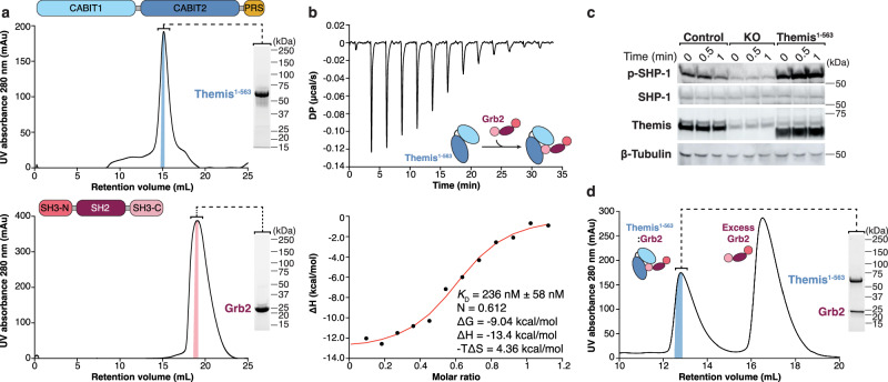
Fig. 1 Biochemical and biophysical characterisation of the Themis1–563-Grb2 complex. a Schematic domain delineations, representative chromatograms and SDS-PAGE gels for the purification of Themis1-–563 (top) and Grb2FL (bottom). Chromatograms and SDS-PAGE analysis are representative of different protein batches of at least five independent experiments each. b Representative isothermal titration calorimetry (ITC) thermograms for the titration of Themis1–563 (7 μM) with Grb2FL (41.6 μM). DP, differential electrical power. Fitted values are provided with their fitting errors as reported by the MicroCal PEAQ-ITC Analysis software version 1.1.0.1262. Similar results were obtained from four independent experiments. c Western blot analysis of SHP-1 phosphorylation (pY564) after TCR stimulation. Jurkat cells reconstituted with an empty lentiviral vector (control), Themis-deficient Jurkat cells (KO) generated by CRISPR/Cas9 gene editing and Jurkat KO cells reconstituted with Themis1–563 were stimulated with plate-bound anti-CD3 (2 μg/mL) and soluble anti-CD28 (1 μg/mL) for the indicated time period. The indicated proteins were analyzed by Western blot. Data are representative of three independent experiments. d Representative chromatogram for the co-expression and purification of the Themis1–563-Grb2FL complex from HEK293 suspension cells. Data are representative of at least six independent experiments.
Fig. 1 Biochemical and biophysical characterisation of the Themis1–563-Grb2 complex. a Schematic domain delineations, representative chromatograms and SDS-PAGE gels for the purification of Themis1-–563 (top) and Grb2FL (bottom). Chromatograms and SDS-PAGE analysis are representative of different protein batches of at least five independent experiments each. b Representative isothermal titration calorimetry (ITC) thermograms for the titration of Themis1–563 (7 μM) with Grb2FL (41.6 μM). DP, differential electrical power. Fitted values are provided with their fitting errors as reported by the MicroCal PEAQ-ITC Analysis software version 1.1.0.1262. Similar results were obtained from four independent experiments. c Western blot analysis of SHP-1 phosphorylation (pY564) after TCR stimulation. Jurkat cells reconstituted with an empty lentiviral vector (control), Themis-deficient Jurkat cells (KO) generated by CRISPR/Cas9 gene editing and Jurkat KO cells reconstituted with Themis1–563 were stimulated with plate-bound anti-CD3 (2 μg/mL) and soluble anti-CD28 (1 μg/mL) for the indicated time period. The indicated proteins were analyzed by Western blot. Data are representative of three independent experiments. d Representative chromatogram for the co-expression and purification of the Themis1–563-Grb2FL complex from HEK293 suspension cells. Data are representative of at least six independent experiments.CC BY-NC-ND · PubMed Central
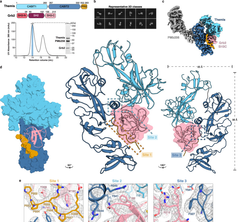
Fig. 2 Structure of the Themis-Grb2 complex. a Schematic representation of the domain organisation of Themis1–563 and Grb2 and sample preparation of the Themis1–563-Grb2-PMb256 complex for cryo-EM. Representative SEC chromatogram and SDS-PAGE analysis of the Themis1–563-Grb2 complex incubated with a 2 molar excess of PMb256. The second SEC peak corresponds to excess PMb256. The data are representative of two independent experiments. Newly delineated domain boundaries based on structural models are indicated. b Representative cryo-EM 2D class averages of the Themis1–563-Grb2-PMb256 complex. Scale bars represent 100 Å. c Cryo-EM map of the Themis1–563-Grb2-PMb256 complex. The map was sharpened using DeepEMhancer, contoured at 0.12 and coloured per domain with ThemisCABIT1 domain in cyan, ThemisCABIT2 in dark blue, ThemisPRS in orange, Grb2SH3C in pink and PMb256 in grey. d Real-space refined atomic model of the Themis1–563-Grb2SH3C complex. Cartoon representations of front and back views are shown, with Grb2SH3C depicted in surface view and the PMb256 model removed for clarity. Side view of the complex shown with a surface representation of Themis, illustrating how Grb2SH3C is engulfed by the CABIT domains. The model is coloured per domain, as in (c). e Zoom-in views of the three interaction sites in the Themis1–563-Grb2SH3C interface fitted in a local refinement 3D map displayed as a grey mesh. Grb2SH3C, ThemisPRS, ThemisCABIT1, ThemisCABIT2 are shown in pink, orange, cyan and dark blue, respectively.
Fig. 2 Structure of the Themis-Grb2 complex. a Schematic representation of the domain organisation of Themis1–563 and Grb2 and sample preparation of the Themis1–563-Grb2-PMb256 complex for cryo-EM. Representative SEC chromatogram and SDS-PAGE analysis of the Themis1–563-Grb2 complex incubated with a 2 molar excess of PMb256. The second SEC peak corresponds to excess PMb256. The data are representative of two independent experiments. Newly delineated domain boundaries based on structural models are indicated. b Representative cryo-EM 2D class averages of the Themis1–563-Grb2-PMb256 complex. Scale bars represent 100 Å. c Cryo-EM map of the Themis1–563-Grb2-PMb256 complex. The map was sharpened using DeepEMhancer, contoured at 0.12 and coloured per domain with ThemisCABIT1 domain in cyan, ThemisCABIT2 in dark blue, ThemisPRS in orange, Grb2SH3C in pink and PMb256 in grey. d Real-space refined atomic model of the Themis1–563-Grb2SH3C complex. Cartoon representations of front and back views are shown, with Grb2SH3C depicted in surface view and the PMb256 model removed for clarity. Side view of the complex shown with a surface representation of Themis, illustrating how Grb2SH3C is engulfed by the CABIT domains. The model is coloured per domain, as in (c). e Zoom-in views of the three interaction sites in the Themis1–563-Grb2SH3C interface fitted in a local refinement 3D map displayed as a grey mesh. Grb2SH3C, ThemisPRS, ThemisCABIT1, ThemisCABIT2 are shown in pink, orange, cyan and dark blue, respectively.CC BY-NC-ND · PubMed Central
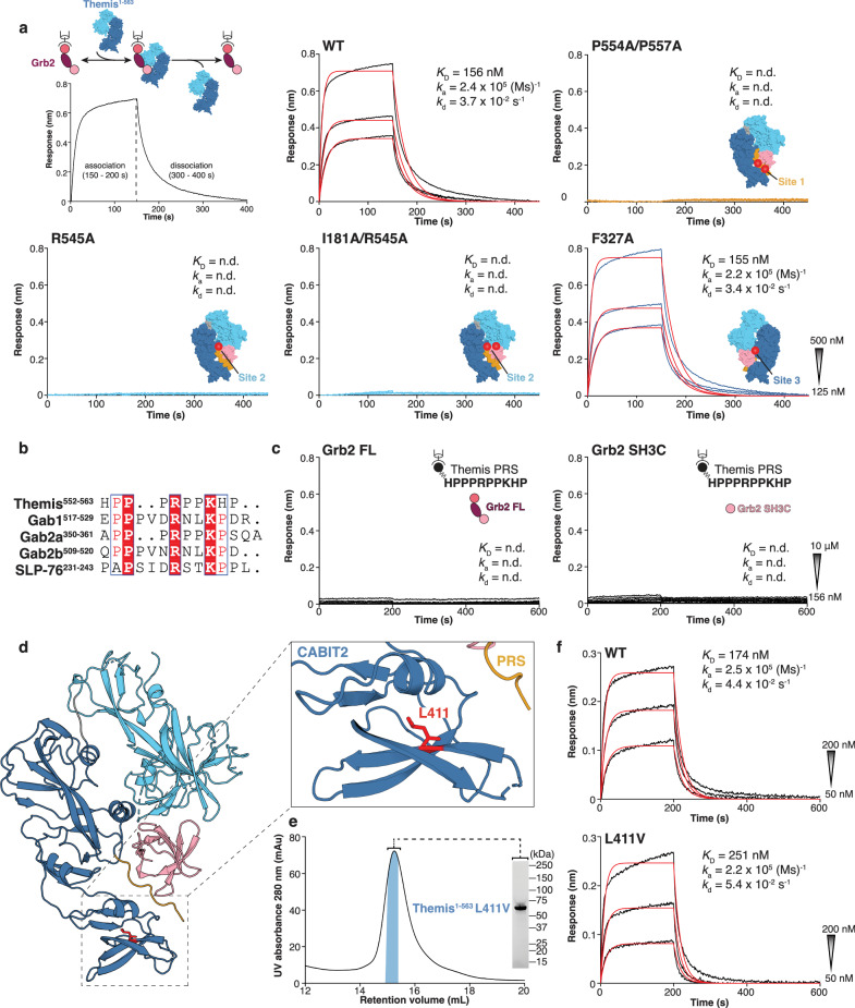
Fig. 3 Structural and functional interrogation of Themis1–563 mutants. a Experimental BLI setup and representative response curves fitted with a 1:1 binding model (red) to quantify the kinetics (ka, kd) and binding affinity (KD) of wild-type and mutant Themis1–563 to Grb2FL. The location of the mutations in the Themis1–563-Grb2SH3C interface is indicated on a surface representation of the complex. b Sequence alignment of ThemisPRS with proline-rich motifs from other Grb2SH3C binding partners, Gab1, Gab2 and SLP-76. c Representative BLI response curves to quantify the interaction of Grb2FL and Grb2SH3C to an immobilised biotinylated ThemisPRS peptide. The peptide sequence is shown in an inset. d Zoom-in view of the ThemisCABIT2 domain showing residue L411 coloured in red. e SEC chromatogram and SDS-PAGE analysis for the purification of the ThemisL411V mutant variant. The data are representative of two independent experiments. f Representative BLI response curves fitted with a 1:1 binding model (red) to compare the kinetics (ka, kd) and binding affinity (KD) of ThemisWT and ThemisL411V to immobilised Grb2. For all BLI experiments, start and end concentrations of the two-fold dilution series are shown as an inset.
Fig. 3 Structural and functional interrogation of Themis1–563 mutants. a Experimental BLI setup and representative response curves fitted with a 1:1 binding model (red) to quantify the kinetics (ka, kd) and binding affinity (KD) of wild-type and mutant Themis1–563 to Grb2FL. The location of the mutations in the Themis1–563-Grb2SH3C interface is indicated on a surface representation of the complex. b Sequence alignment of ThemisPRS with proline-rich motifs from other Grb2SH3C binding partners, Gab1, Gab2 and SLP-76. c Representative BLI response curves to quantify the interaction of Grb2FL and Grb2SH3C to an immobilised biotinylated ThemisPRS peptide. The peptide sequence is shown in an inset. d Zoom-in view of the ThemisCABIT2 domain showing residue L411 coloured in red. e SEC chromatogram and SDS-PAGE analysis for the purification of the ThemisL411V mutant variant. The data are representative of two independent experiments. f Representative BLI response curves fitted with a 1:1 binding model (red) to compare the kinetics (ka, kd) and binding affinity (KD) of ThemisWT and ThemisL411V to immobilised Grb2. For all BLI experiments, start and end concentrations of the two-fold dilution series are shown as an inset.CC BY-NC-ND · PubMed Central
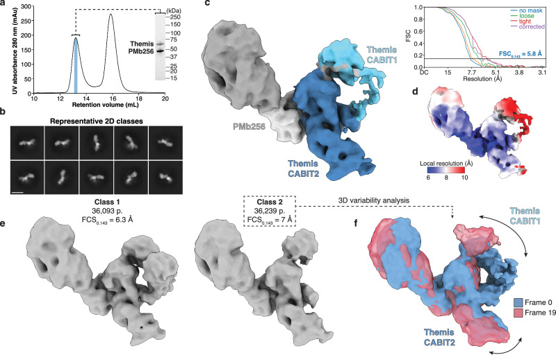
Fig. 4 Themis CABIT domains are highly flexible and are stabilised by Grb2 binding. a Sample preparation of the Themis1–563-PMb256 complex for cryo-EM. Representative SEC chromatogram and SDS-PAGE analysis of Themis1–563 incubated with a 2 molar excess of PMb256. The second SEC peak contains excess PMb256. Data are representative of one independent experiment. b Representative cryo-EM 2D class averages of the Themis1–563-PMb256 complex. Scale bar represents 100 Å. c Cryo-EM map of the Themis1–563-PMb256 complex. The displayed 3D map is contoured at 0.12 and coloured with Themis CABIT domains in blue, and PMb256 in grey. Gold-standard Fourier Shell Correlation (FSC) curves are shown, with the estimated resolution at FSC = 0.143 (dark blue line) shown for the corrected FSC curve (purple line). d 3D cryo-EM map shown in c coloured according to local resolution. e 3D classification without alignment of the particles contributing to the map shown in (c) results in two classes in which the CABIT1 domain is present (class 1) or absent (class 2). Sharpened maps from subsequent non-uniform refinement are shown contoured at 0.12. f 3D variability analysis of particles from class 2 shows movement of the unbound ThemisCABIT1 domain. 3D volumes from the start (Frame 0, blue) and end (Frame 19, red) of the component analysis are shown.
Fig. 4 Themis CABIT domains are highly flexible and are stabilised by Grb2 binding. a Sample preparation of the Themis1–563-PMb256 complex for cryo-EM. Representative SEC chromatogram and SDS-PAGE analysis of Themis1–563 incubated with a 2 molar excess of PMb256. The second SEC peak contains excess PMb256. Data are representative of one independent experiment. b Representative cryo-EM 2D class averages of the Themis1–563-PMb256 complex. Scale bar represents 100 Å. c Cryo-EM map of the Themis1–563-PMb256 complex. The displayed 3D map is contoured at 0.12 and coloured with Themis CABIT domains in blue, and PMb256 in grey. Gold-standard Fourier Shell Correlation (FSC) curves are shown, with the estimated resolution at FSC = 0.143 (dark blue line) shown for the corrected FSC curve (purple line). d 3D cryo-EM map shown in c coloured according to local resolution. e 3D classification without alignment of the particles contributing to the map shown in (c) results in two classes in which the CABIT1 domain is present (class 1) or absent (class 2). Sharpened maps from subsequent non-uniform refinement are shown contoured at 0.12. f 3D variability analysis of particles from class 2 shows movement of the unbound ThemisCABIT1 domain. 3D volumes from the start (Frame 0, blue) and end (Frame 19, red) of the component analysis are shown.CC BY-NC-ND · PubMed Central
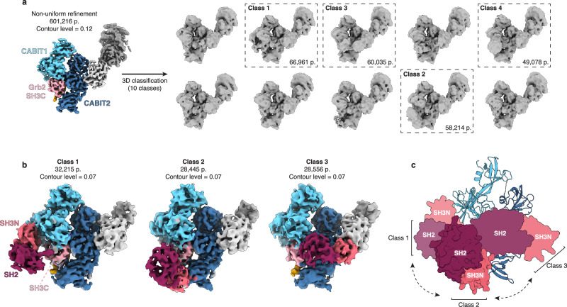
Fig. 5 Grb2 SH3N and SH2 domains are highly flexible and sample multiple conformational states. a The particle stack contributing to the consensus map (Fig. 2d) was further processed using 3D classification without alignment in cryoSPARC using 10 classes as input. The resulting classes are shown, with four classes showing volume corresponding to Grb2SH3N and Grb2SH2 (dashed boxes). b Maps corresponding to class 1–3 from 3D classification were further processed by particle subtraction to remove the PMb moiety, followed by local refinement. The displayed 3D maps are contoured at 0.07 and coloured as before, with the additional Grb2SH3N and Grb2SH2 domains in dark pink and wine red, respectively. Particle numbers (p.) contributing to the map for each class are shown. For all maps, the back view of the complex is shown. c Schematic showing the distinct conformational states of Grb2 captured by cryo-EM analysis. Grb2SH3C domain is locked in one position through its interaction with Themis, while Grb2SH2 and Grb2SH3N exhibit 180-degree movement at the back of the Themis-Grb2 molecule. Surface representations of Grb2SH2 and Grb2SH3N are shown based on rigid body fitting of individual domains in the cryo-EM maps from (b) in ChimeraX.
Fig. 5 Grb2 SH3N and SH2 domains are highly flexible and sample multiple conformational states. a The particle stack contributing to the consensus map (Fig. 2d) was further processed using 3D classification without alignment in cryoSPARC using 10 classes as input. The resulting classes are shown, with four classes showing volume corresponding to Grb2SH3N and Grb2SH2 (dashed boxes). b Maps corresponding to class 1–3 from 3D classification were further processed by particle subtraction to remove the PMb moiety, followed by local refinement. The displayed 3D maps are contoured at 0.07 and coloured as before, with the additional Grb2SH3N and Grb2SH2 domains in dark pink and wine red, respectively. Particle numbers (p.) contributing to the map for each class are shown. For all maps, the back view of the complex is shown. c Schematic showing the distinct conformational states of Grb2 captured by cryo-EM analysis. Grb2SH3C domain is locked in one position through its interaction with Themis, while Grb2SH2 and Grb2SH3N exhibit 180-degree movement at the back of the Themis-Grb2 molecule. Surface representations of Grb2SH2 and Grb2SH3N are shown based on rigid body fitting of individual domains in the cryo-EM maps from (b) in ChimeraX.CC BY-NC-ND · PubMed Central
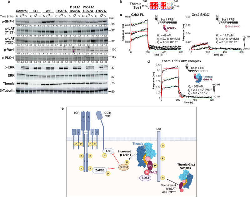
Fig. 6 Themis-Grb2 complex acts as a signalling hub downstream of TCR activation. a Western blot analysis of SHP-1 phosphorylation (pY564) after TCR activation. Themis-deficient Jurkat cells (KO) were transduced with a lentiviral vector expressing full-length wild-type Themis (WT) or Themis mutants, as indicated. Jurkat cells reconstituted with an empty lentiviral vector were used as a control. Cells were stimulated with plate-bound anti-CD3 (2 μg/mL) and soluble anti-CD28 (1 μg/mL). The indicated proteins were analysed by Western blot. Band intensities for p-SHP-1, p-LAT, p-Vav1 and p-PLCγ1 were quantified and normalised to total ERK. Values below the blots indicate quantified fold changes in phosphorylation relative to untreated control cells. Data are representative of three independent experiments. b Sequence alignment of the ThemisPRS and Sos1PRS peptide. Residue numbers for each peptide are indicated. c Representative response curves fitted with a 1:1 binding model (red) to quantify the kinetics (ka, kd) and binding affinity (KD) of Grb2FL, Grb2SH3C and d Themis1–563-Grb2FL complex to Sos1 PRS peptide. Schematics show the BLI setup for each experiment. Start and end concentrations of the two-fold dilution series are shown as an inset. e Schematic depicting the role of the Themis-Grb2 complex as a proximal signalling hub downstream of the TCR. Upon TCR stimulation, the tyrosine kinase Lck is recruited to the membrane via the CD4 and CD8 co-receptors. Lck phosphorylates immunoreceptor tyrosine-based activation motifs (ITAM) in the TCR CD3 chains, facilitating the recruitment of the protein tyrosine kinase ZAP70. ZAP70 phosphorylates the transmembrane adaptor protein LAT, promoting the formation of the LAT signalosome. The Themis-Grb2 complex is recruited to the membrane via Grb2SH2 binding to phosphorylated LAT. Once there, Themis is positioned to interact with the protein tyrosine phosphatase SHP-1 and regulate its activity to fine-tune TCR signalling. Simultaneously, the Grb2SH3N domain is available for binding to other target proteins, such as Sos1, contributing to the regulation of other downstream signalling pathways. Tyrosine phosphorylation (P) is depicted in yellow.
Fig. 6 Themis-Grb2 complex acts as a signalling hub downstream of TCR activation. a Western blot analysis of SHP-1 phosphorylation (pY564) after TCR activation. Themis-deficient Jurkat cells (KO) were transduced with a lentiviral vector expressing full-length wild-type Themis (WT) or Themis mutants, as indicated. Jurkat cells reconstituted with an empty lentiviral vector were used as a control. Cells were stimulated with plate-bound anti-CD3 (2 μg/mL) and soluble anti-CD28 (1 μg/mL). The indicated proteins were analysed by Western blot. Band intensities for p-SHP-1, p-LAT, p-Vav1 and p-PLCγ1 were quantified and normalised to total ERK. Values below the blots indicate quantified fold changes in phosphorylation relative to untreated control cells. Data are representative of three independent experiments. b Sequence alignment of the ThemisPRS and Sos1PRS peptide. Residue numbers for each peptide are indicated. c Representative response curves fitted with a 1:1 binding model (red) to quantify the kinetics (ka, kd) and binding affinity (KD) of Grb2FL, Grb2SH3C and d Themis1–563-Grb2FL complex to Sos1 PRS peptide. Schematics show the BLI setup for each experiment. Start and end concentrations of the two-fold dilution series are shown as an inset. e Schematic depicting the role of the Themis-Grb2 complex as a proximal signalling hub downstream of the TCR. Upon TCR stimulation, the tyrosine kinase Lck is recruited to the membrane via the CD4 and CD8 co-receptors. Lck phosphorylates immunoreceptor tyrosine-based activation motifs (ITAM) in the TCR CD3 chains, facilitating the recruitment of the protein tyrosine kinase ZAP70. ZAP70 phosphorylates the transmembrane adaptor protein LAT, promoting the formation of the LAT signalosome. The Themis-Grb2 complex is recruited to the membrane via Grb2SH2 binding to phosphorylated LAT. Once there, Themis is positioned to interact with the protein tyrosine phosphatase SHP-1 and regulate its activity to fine-tune TCR signalling. Simultaneously, the Grb2SH3N domain is available for binding to other target proteins, such as Sos1, contributing to the regulation of other downstream signalling pathways. Tyrosine phosphorylation (P) is depicted in yellow.CC BY-NC-ND · PubMed Central

Verification

Claims the paper makes

The claims and statistics of this paper haven’t been checked yet.

Run the check — free, no account needed. It reads the open-access text and checks the sources it cites.

A few free checks a day · papers already verified open instantly · results are public

Self-check

Percentages, sums and reported differences re-computed from the paper’s own numbers.

The self-check needs the paper’s full text.

Citations

Cites

Research works printed but not linked: 61

  1. 1. Ruiz Pérez, M., Vandenabeele, P. & Tougaard, P. The thymus road to a T cell: migration, selection, and atrophy. Front Immunol 15, (2024). · DOI
  2. 2. Gaud, G., Lesourne, R. & Love, P. E. Regulatory mechanisms in T cell receptor signalling. Nature Reviews Immunology vol. 18 485–497 Preprint at https://doi.org/10.1038/s41577-018-0020-8 (2018). · DOI
  3. 3. Yayon, N. et al. A spatial human thymus cell atlas mapped to a continuous tissue axis. Nature 635, 708–718 (2024). · DOI
  4. 4. Fu, G. et al. Themis controls thymocyte selection through regulation of T cell antigen receptor-mediated signaling. Nat Immunol 10, 848–856 (2009). · DOI
  5. 5. Johnson, A. L. et al. Themis is a member of a new metazoan gene family and is required for the completion of thymocyte positive selection. Nat Immunol 10, 831–839 (2009). · DOI
  6. 6. Kakugawa, K. et al. A Novel Gene Essential for the Development of Single Positive Thymocytes. Mol Cell Biol 29, 5128–5135 (2009). · DOI
  7. 7. Lesourne, R. et al. Themis, a T cell-specific protein important for late thymocyte development. Nat Immunol 10, 840–847 (2009). · DOI
  8. 9. Choi, S., Cornall, R., Lesourne, R. & Love, P. E. THEMIS: Two Models, Different Thresholds. Trends in Immunology vol. 38 622–632 Preprint at https://doi.org/10.1016/j.it.2017.06.006 (2017). · DOI
  9. 10. Mélique, S., Yang, C. & Lesourne, R. Negative times negative equals positive, THEMIS sets the rule on thymic selection and peripheral T cell responses. Biomedical Journal vol. 45 334–346 Preprint at https://doi.org/10.1016/j.bj.2022.03.008 (2022). · DOI
  10. 11. Peirce, M. J. et al. Themis2/ICB1 is a signaling scaffold that selectively regulates macrophage toll-like receptor signaling and cytokine production. PLoS One 5, (2010). · DOI
  11. 12. Johnson, A. L. et al. Themis is a member of a new metazoan gene family and is required for the completion of thymocyte positive selection. Nat Immunol 10, 831–839 (2009). · DOI
  12. 13. Treeck, O., Strunck, E. & Vollmer, G. A novel base membrane-induced gene identified in the human endometrial adenocarcinoma cell line HEC1B. FEBS Lett 425, 426–430 (1998). · DOI
  13. 14. Lesourne, R. et al. Interchangeability of Themis1 and Themis2 in Thymocyte Development Reveals Two Related Proteins with Conserved Molecular Function. The Journal of Immunology 189, 1154–1161 (2012). · DOI
  14. 15. Fu, G. et al. Themis sets the signal threshold for positive and negative selection in T-cell development. Nature 504, 441–445 (2013). · DOI
  15. 16. Paster, W. et al. GRB2-Mediated Recruitment of THEMIS to LAT Is Essential for Thymocyte Development. The Journal of Immunology 190, 3749–3756 (2013). · DOI
  16. 17. Zvezdova, E. et al. In vivo functional mapping of the conserved protein domains within murine Themis1. Immunol Cell Biol 92, 721–728 (2014). · DOI
  17. 18. Yablonski, D. Bridging the Gap: Modulatory Roles of the Grb2-Family Adaptor, Gads, in Cellular and Allergic Immune Responses. Frontiers in immunology vol. 10 1704 Preprint at https://doi.org/10.3389/fimmu.2019.01704 (2019). · DOI
  18. 19. Choi, S. et al. THEMIS enhances TCR signaling and enables positive selection by selective inhibition of the phosphatase SHP-1. Nat Immunol 18, 433–441 (2017). · DOI
  19. 20. Zhang, J. et al. THEMIS is a substrate and allosteric activator of SHP1, playing dual roles during T cell development. Nat Struct Mol Biol 31, 54–67 (2024). · DOI
  20. 21. Jang, I. K., Zhang, J. & Gu, H. Grb2, a simple adapter with complex roles in lymphocyte development, function, and signaling. Immunological Reviews vol. 232 150–159 Preprint at https://doi.org/10.1111/j.1600-065X.2009.00842.x (2009). · DOI
  21. 23. Voisinne, G. et al. Quantitative interactomics in primary T cells unveils TCR signal diversification extent and dynamics. Nat Immunol 20, 1530–1541 (2019). · DOI
  22. 24. Maignan, S. et al. Crystal Structure of the Mammalian Grb2 Adaptor. vol. 268 www.sciencemag.org (1995). · DOI
  23. 25. Yuzawa, S. et al. Solution structure of Grb2 reveals extensive flexibility necessary for target recognition. J Mol Biol 306, 527–537 (2001). · DOI
  24. 26. Tateno, K. et al. Different molecular recognition by three domains of the full-length GRB2 to SOS1 proline-rich motifs and EGFR phosphorylated sites. Chem Sci 15, 15858–15872 (2024). · DOI
  25. 27. Lorenz, U. SHP-1 and SHP-2 in T Cells: Two Phosphatases Functioning at Many Levels. Immunological Reviews vol. 228 (2009). · DOI
  26. 28. Botte, M. et al. Cryo-EM structures of a LptDE transporter in complex with Pro-macrobodies offer insight into lipopolysaccharide translocation. Nat Commun 13, (2022). · DOI
  27. 29. Abramson, J. et al. Accurate structure prediction of biomolecular interactions with AlphaFold 3. Nature 630, 493–500 (2024). · DOI · Paper page
  28. 30. Beyett, T. S. et al. Structure of the CABIT2 domain of THEMIS reveals a novel protein fold with an inserted SH3-like domain. Preprint at https://doi.org/10.1101/2024.10.01.616165 (2024). · DOI
  29. 31. Okada, T. et al. Differential function of Themis CABIT domains during T cell development. PLoS One 9, (2014). · DOI
  30. 33. Cheng, D. et al. Themis2 lowers the threshold for B cell activation during positive selection. Nat Immunol 18, 205–213 (2017). · DOI
  31. 34. Lewitzky, M. et al. The C-Terminal SH3 Domain of the Adapter Protein Grb2 Binds with High Anity to Sequences in Gab1 and SLP-76 Which Lack the SH3-Typical P-x-x-P Core Motif. www.nature.com/onc.
  32. 35. Harkiolaki, M. et al. Distinct Binding Modes of Two Epitopes in Gab2 that Interact with the SH3C Domain of Grb2. Structure 17, 809–822 (2009). · DOI
  33. 36. Wang, Y. et al. Germline genetic patterns underlying familial rheumatoid arthritis, systemic lupus erythematosus and primary Sjögren’s syndrome highlight T cell-initiated autoimmunity. Ann Rheum Dis 79, 268–275 (2019). · DOI
  34. 37. Liao, T. J., Jang, H., Fushman, D. & Nussinov, R. SOS1 interacts with Grb2 through regions that induce closed nSH3 conformations. Journal of Chemical Physics 153, (2020). · DOI
  35. 38. Brockmeyer, C. et al. T cell receptor (TCR)-induced tyrosine phosphorylation dynamics identifies THEMIS as a new TCR signalosome component. Journal of Biological Chemistry 286, 7535–7547 (2011). · DOI
  36. 39. Choi, S. et al. GRB2 promotes thymocyte positive selection by facilitating THEMIS-mediated inactivation of SHP1. Journal of Experimental Medicine 220, (2023). · DOI
  37. 40. Lin, C.-W. et al. A two-component protein condensate of the EGFR cytoplasmic tail and Grb2 regulates Ras activation by SOS at the membrane. (2022) doi:10.1073/pnas. · DOI
  38. 41. Tashiro, K. et al. GAREM, a novel adaptor protein for growth factor receptor-bound protein 2, contributes to cellular transformation through the activation of extracellular signal-regulated kinase signaling. Journal of Biological Chemistry 284, 20206–20214 (2009). · DOI
  39. 42. Taniguchi, T. et al. A brain-specific Grb2-associated regulator of extracellular signal-regulated kinase (Erk)/mitogen-activated protein kinase (MAPK) (GAREM) subtype, GAREM2, contributes to neurite outgrowth of neuroblastoma cells by regulating erk signaling. Journal of Biological Chemistry 288, 29934–29942 (2013). · DOI
  40. 43. Deborah, E. A., Nabekura, T., Shibuya, K. & Shibuya, A. THEMIS2 Impairs Antitumor Activity of NK Cells by Suppressing Activating NK Receptor Signaling. The Journal of Immunology 212, 1819–1828 (2024). · DOI
  41. 44. Aricescu, A. R., Lu, W. & Jones, E. Y. A time- and cost-efficient system for high-level protein production in mammalian cells. Acta Crystallogr D Biol Crystallogr 62, 1243–1250 (2006). · DOI
  42. 45. Dierick, E. et al. Clostridium perfringens chitinases, key enzymes during early stages of necrotic enteritis in broiler chickens. PLoS Pathog 20, e1012560 (2024). · DOI
  43. 46. Chen, J., Noble, A. J., Kang, J. Y. & Darst, S. A. Eliminating effects of particle adsorption to the air/water interface in single-particle cryo-electron microscopy: Bacterial RNA polymerase and CHAPSO. J Struct Biol X 1, (2019). · DOI
  44. 47. Punjani, A., Rubinstein, J. L., Fleet, D. J. & Brubaker, M. A. CryoSPARC: Algorithms for rapid unsupervised cryo-EM structure determination. Nat Methods 14, 290–296 (2017). · DOI · Paper page
  45. 48. Punjani, A., Zhang, H. & Fleet, D. J. Non-uniform refinement: adaptive regularization improves single-particle cryo-EM reconstruction. Nat Methods 17, 1214–1221 (2020). · DOI
  46. 49. Wagner, T. et al. SPHIRE-crYOLO is a fast and accurate fully automated particle picker for cryo-EM. Commun Biol 2, (2019). · DOI
  47. 50. Bepler, T. et al. Positive-unlabeled convolutional neural networks for particle picking in cryo-electron micrographs. Nat Methods 16, 1153–1160 (2019). · DOI
  48. 51. Rosenthal, P. B. & Henderson, R. Optimal determination of particle orientation, absolute hand, and contrast loss in single-particle electron cryomicroscopy. J Mol Biol 333, 721–745 (2003). · DOI · Paper page
  49. 52. Pettersen, E. F. et al. UCSF ChimeraX: Structure visualization for researchers, educators, and developers. Protein Science 30, 70–82 (2021). · DOI
  50. 53. Sanchez-Garcia, R. et al. DeepEMhancer: a deep learning solution for cryo-EM volume post-processing. Commun Biol 4, (2021). · DOI
  51. 55. Lin, Z. et al. Evolutionary-Scale Prediction of Atomic-Level Protein Structure with a Language Model. Science vol. 379 https://www.science.org (2023). · DOI · Paper page
  52. 56. Kidmose, R. T. et al. Namdinator - Automatic molecular dynamics flexible fitting of structural models into cryo-EM and crystallography experimental maps. IUCrJ 6, 526–531 (2019). · DOI
  53. 57. Emsley, P. & Cowtan, K. Coot: Model-building tools for molecular graphics. Acta Crystallogr D Biol Crystallogr 60, 2126–2132 (2004). · DOI · Paper page
  54. 58. Liebschner, D. et al. Macromolecular structure determination using X-rays, neutrons and electrons: Recent developments in Phenix. Acta Crystallogr D Struct Biol 75, 861–877 (2019). · DOI · Paper page
  55. 59. Williams, C. J. et al. MolProbity: More and better reference data for improved all-atom structure validation. Protein Science 27, 293–315 (2018). · DOI · Paper page
  56. 60. Sievers, F. et al. Fast, scalable generation of high-quality protein multiple sequence alignments using Clustal Omega. Mol Syst Biol 7, (2011). · DOI
  57. 61. Robert, X. & Gouet, P. Deciphering key features in protein structures with the new ENDscript server. Nucleic Acids Res 42, (2014). · DOI · Paper page
  58. 62. Krissinel, E. & Henrick, K. Inference of Macromolecular Assemblies from Crystalline State. J Mol Biol 372, 774–797 (2007). · DOI
  59. 63. Heckman, K. L. & Pease, L. R. Gene splicing and mutagenesis by PCR-driven overlap extension. Nat Protoc 2, 924–932 (2007). · DOI
  60. 64. Howarth, M. & Ting, A. Y. Imaging proteins in live mammalian cells with biotin ligase and monovalent streptavidin. Nat Protoc 3, 534–545 (2008). · DOI
  61. 65. Labun, K. et al. CHOPCHOP v3: Expanding the CRISPR web toolbox beyond genome editing. Nucleic Acids Res 47, W171–W174 (2019). · DOI

Metadata

Authors
Savvides, S., Clancy, D., Sanjuan, A. S., Gillis, E., Velghe, I., Droogenbroeck, Y. V., Felix, J., Bloch, Y., Cuadrado, A. F., Merceron, R., Schenck, S., Vandenabeele, P., Brunner, J., TAGHON, T., Elewaut, D., & Tougaard, P.
Affiliations
  • Ghent University (BE)
  • VIB-UGent Center for Inflammation Research
  • Deutsches Elektronen-Synchrotron DESY (DE)
  • Vrije Universiteit Brussel (BE)
  • Ghent University Hospital (BE)
  • University of Copenhagen (DK)
Corresponding author
  • Savvides, Savvas
  • Clancy, Danielle
Venue
Nature Communications
Published
2025-04-23
Volume
17
Issue
1
ISSN
2041-1723 (ISSN-L)
OpenAlex
W7161848002
Cited by
0
References
1
FWCI
0.00
Type
journal article
Language
en
Open access
Locations
  • Nature Communications (publishedVersion, cc-by-nc-nd)
  • Research Square (acceptedVersion, cc-by)
  • PubMed Central (publishedVersion, cc-by-nc-nd)
  • PubMed (publishedVersion)
  • Research at the University of Copenhagen (University of Copenhagen) (publishedVersion, cc-by-nc-nd)
  • Ghent University Academic Bibliography (Ghent University) (submittedVersion, cc-by-nc-nd)
  • DOAJ (DOAJ: Directory of Open Access Journals) (submittedVersion)
Funders
  • Fonds Wetenschappelijk Onderzoek (G0H1222N, 1219923N, G049820N)
  • Vlaams Instituut voor Biotechnologie (C0101)
  • Vlaamse regering
Topics
  • Life Sciences › Immunology and Microbiology › Immunology › T-cell and B-cell Immunology
  • Life Sciences › Biochemistry, Genetics and Molecular Biology › Molecular Biology › Protein Tyrosine Phosphatases
  • Life Sciences › Biochemistry, Genetics and Molecular Biology › Molecular Biology › Protein Kinase Regulation and GTPase Signaling
Keywords
THEMIS, Grb2, T cell receptor signaling, thymic selection, SH3 domain binding, protein-protein interaction, interdomain flexibility, adaptor protein complexes, T cell signaling
MeSH terms
Protein Domains, Animals, Humans, Protein Binding, Receptors, Antigen, T-Cell, T-Lymphocytes, Signal Transduction, src Homology Domains, Cryoelectron Microscopy, Intercellular Signaling Peptides and Proteins, Mice, GRB2 Adaptor Protein, Protein Tyrosine Phosphatase, Non-Receptor Type 6

About these checks

Registry, retraction, Cites and statistics results are computed, not judged, with one exception: a printed reference that no index lists is read once by a model before it counts as a citation error. Claims are extracted by a model and kept only when their quote appears verbatim in the text; each cited source is then checked, with a model’s help, for whether it says what the claim relies on. Those verdicts can be wrong — read the claim’s quote and the cited source before relying on one.

Statistics and arithmetic are recomputed, not judged.

Confidence bands are shown only for fields calibrated on our annotated test set. None is calibrated yet, so no band is shown.

What counts as a claim ›How we measure confidence ›How we check this ›

Are you an author, or do you see a problem with this page? Request a review