

The DOI matches its Crossref record; 89 of the 89 references it cites were found in a registry or an index.
On the role of corticotropin-releasing hormone receptors in anxiety and depression
Open accessAbstract
On the basis of extensive basic and clinical studies, corticotropin-releasing hormone (CRH) and its related family members are considered to play a pivotal role in stress-related disorders, such as anxiety and depression. CRH is regarded as the principal mediator in the brain of the stress response, as it mediates neuroendocrine, autonomic, and behavioral responses to stressful challenges. Recently, this neuropeptide family has expanded due to the discovery of two new members, urocortin II (also termed stresscopin-related peptide) and urocortin III (also termed stresscopin), which are selective agonists for the CRH receptor type 2. They show a discrete neuroanatomical localization and are involved in stress-coping responses, such as anxiolysis. Here, on the basis of recent developments, we suggest that CRH, the urocortins, and their receptors form a complex system in the brain, which is recruited during both the acute and the recovery phases of the stress response.
Overview
The claims and statistics of this paper haven’t been checked yet.
Run the check — free, no account needed. It reads the open-access text and checks the sources it cites.
Integrity signals
Registry
Cites
Statistics
Figures
Verification
Claims the paper makes
The claims and statistics of this paper haven’t been checked yet.
Run the check — free, no account needed. It reads the open-access text and checks the sources it cites.
Claims other papers make about this one
Each is checked against this paper’s own text: does it say what the citing paper says it does?
The cited source says what the claim attributes to it.
In the citing paper
“The neuropeptide corticotropin-releasing factor (CRF) mediates behavioral and endocrine responses to stress and acts on CRF type 1 (CRF1) and CRF type 2 (CRF2) receptors [168].”
From The Neurobiology of Love and Pair Bonding from Human and Animal Perspectives (2023) · cites this paper as [168]
Self-check
Percentages, sums and reported differences re-computed from the paper’s own numbers.
The self-check needs the paper’s full text.
Citations
Cites
Vale, W., Spiess, J., Rivier, C., & Rivier, J. (1981) · Science · doi:10.1126/science.6267699 · Cited by 3,965
Brain corticosteroid receptor balance in health and disease
de Kloet, E. R., Vreugdenhil, E., Oitzl, M. S., & Joëls, M. (1998) · Endocrine Reviews · doi:10.1210/edrv.19.3.0331 · Cited by 2,614 · cited 4×
“The cause for the aberrant—in most cases, hyperactive—HPA axis is presumably a hyperactive central CRH system (for review, see references 24, 30, and 69 to 71) and defunct brain and pituitary corticosteroid receptor systems.(66,72,73) Although it was more than a decade ago that a reduced CRH receptor density was found…”
BackgroundInfluentialTwo receptor systems for corticosterone in rat brain: Microdistribution and differential occupation
REUL, J. M. H. M., & KLOET, E. R. D. (1985) · Endocrinology · doi:10.1210/endo-117-6-2505 · Cited by 2,580 · cited 4×
“They can be distinguished in two types of glucocorticoid-binding receptors: the mineralocorticoid receptor (MR or type I) and the glucocorticoid receptor (GR or type II).(83) MRs are mainly localized in the hippocampus, whereas GRs have a widespread distribution in the CNS.”
BackgroundInfluentialThe Corticosteroid Receptor Hypothesis of Depression
Holsboer, F. (2000) · Neuropsychopharmacology · doi:10.1016/s0893-133x(00)00159-7 · Cited by 2,181
Hypothalamic integration: organization of the paraventricular and supraoptic nuclei
Swanson, L. W., & Sawchenko, P. E. (1983) · Annual Review of Neuroscience · doi:10.1146/annurev.ne.06.030183.001413 · Cited by 1,991
Swanson, L. W., Sawchenko, P. E., Rivier, J., & Vale, W. W. (2008) · Neuroendocrinology · doi:10.1159/000123454 · Cited by 1,953
Nemeroff, C. B., Widerlöv, E., Bissette, G., Walléus, H., Karlsson, I., Eklund, K., Kilts, C. D., Loosen, P. T., & Vale, W. (1984) · Science · doi:10.1126/science.6334362 · Cited by 1,485
Vaughan, J. M., Donaldson, C. J., Bittencourt, J. C., Perrin, M. H., Lewis, K., Sutton, S. W., Chan, R. K. W., Turnbull, A. V., Lovejoy, D. A., RIVIER, C. L., et al. (1995) · Nature · doi:10.1038/378287a0 · Cited by 1,479
Physiology and pharmacology of corticotropin-releasing factor.
Owens, M. J., & Nemeroff, C. B. (1991) · Pharmacological Reviews · doi:10.1016/s0031-6997(25)06673-6 · Cited by 1,147 · cited 1×
“CRH is highly implicated in the regulation of anxietyrelated behavior and is thought to play a pivotal role in anxiety and depressive disorders.(24,30,31) Several lines of evidence point to the participation of CRHR1 in the effects of CRH.”
BackgroundChalmers, D., Lovenberg, T. W., & Souza, E. D. (1995) · Journal of Neuroscience · doi:10.1523/jneurosci.15-10-06340.1995 · Cited by 925 · cited 3×
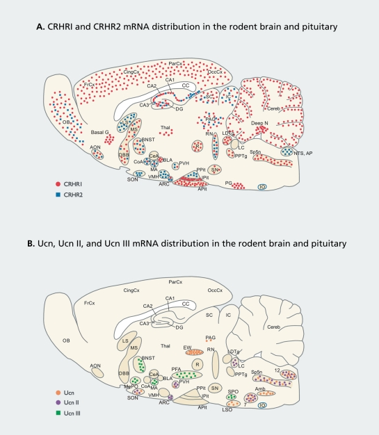
“The presented mRNA distribution is based on in situ hybridization studies.(12,14,16,20-23) The drawn sagittal sections are only 2D schematic representations and, therefore, cannot be neuroanatomically exact.”
Background
Research works printed but not linked: 11
- Holsboer F., 1984, N Engl J Med., 311, 1127
- Lederis K., 1982, Proc West Pharmacol Soc., 25, 223
- Owens MJ., 1991, Pharmacol Rev., 43, 425
- Okuyama S., 1999, J Pharmacol Exp Then., 289, 926
- Heinrichs SC., 2000, Soc Neurosci Abst., 26, 2149
- Fiorino DF., 2000, Soc Neurosci Abst., 26, 2265
- Valdez GR., 2001, Soc Neurosci Abst., 27.Abstract num
- Held K., 2001, J Psychiatr Res. in press
- Steckler T., 1999, Baillieres Clin Endocrinol Metab., 13, 107
- Davis M., 1992, The Amygdala. New York, NY: Wiley-Liss;, 255
- Gray TS., 1990, Corticotropin-releasing Factor: Basic and Clinical studies of a Neuropeptide. Boca Raton, Fia: CRC Press;, 53
Cited by
Sorted by citations
The neurobiology and control of anxious states
Millan, M. J. (2003) · Progress in Neurobiology · doi:10.1016/s0301-0082(03)00087-x · Cited by 978
Sex differences in anxiety and depression: Role of testosterone
McHenry, J. A., Carrier, N., Hull, E. M., & Kabbaj, M. (2013) · Frontiers in Neuroendocrinology · doi:10.1016/j.yfrne.2013.09.001 · Cited by 526
Pathophysiology of depression and mechanisms of treatment
Brigitta, B. (2002) · Dialogues in Clinical Neuroscience · doi:10.31887/dcns.2002.4.1/bbondy · Cited by 286
Pharmacological Treatment of Anxiety Disorders: The Role of the HPA Axis
Tafet, G. E., & Nemeroff, C. B. (2020) · Frontiers in Psychiatry · doi:10.3389/fpsyt.2020.00443 · Cited by 216
“These steroid hormones exert their effects through binding to mineralocorticoid receptors (MRs or type I) and glucocorticoid receptors (GRs or type II), constituting a hormone-receptor complex, which in turn may interact with specific DNA sequences located in the promoter region of target genes, termed glucocorticoid…”
BackgroundInfluentialSculpting the hippocampus from within: stress, spines, and CRH
Maras, P. M., & Baram, T. Z. (2012) · Trends in Neurosciences · doi:10.1016/j.tins.2012.01.005 · Cited by 183
“CRHR1 and CRHR2 differ in their distribution [37,43,44], as well as their role in mediating behavioral and endocrine responses to stress [45,46].”
BackgroundOxytocin and Social Relationships: From Attachment to Bond Disruption
Bosch, O. J., & Young, L. J. (2017) · Current Topics in Behavioral Neurosciences · doi:10.1007/7854_2017_10 · Cited by 162
“The brain CRF system, which consists of CRF and the urocortins 1–3 as well as of CRF receptor type 1 (CRF-R1), CRF-R2, and the CRF binding protein (Reul and Holsboer 2002), is the primary regulator of the HPA axis (Vale et al. 1981; Aguilera and Liu 2012).”
BackgroundExtra-adrenal glucocorticoid biosynthesis: implications for autoimmune and inflammatory disorders
Slominski, R. M., Tuckey, R. C., Manna, P. R., Jetten, A. M., Postlethwaite, A., Raman, C., & Slominski, A. T. (2020) · Genes & Immunity · doi:10.1038/s41435-020-0096-6 · Cited by 155
Stress, Definitions, Mechanisms, and Effects Outlined
Fink, G. (2016) · Elsevier eBooks · doi:10.1016/b978-0-12-800951-2.00001-7 · Cited by 166
Cavigelli, S. A., & Caruso, M. J. (2015) · Philosophical Transactions of the Royal Society B: Biological Sciences · doi:10.1098/rstb.2014.0103 · Cited by 112
Neuropeptide Regulation of Signaling and Behavior in the BNST
Kash, T. L., Pleil, K. E., Marcinkiewcz, C. A., Lowery‐Gionta, E. G., Crowley, N. A., Mazzone, C. M., Sugam, J. A., Hardaway, J. A., & McElligott, Z. A. (2014) · Molecules and Cells · doi:10.14348/molcells.2015.2261 · Cited by 105
“CRF binds to CRF-1 and CRF-2 receptors (CRF-1Rs and CRF-2Rs), which are G-protein coupled receptors (GPCRs) acting through a Gs-cAMP-PKA signaling mechanism (Arzt and Holsboer, 2006; Blank et al., 2003; Dautenberg and Hauger, 2002; Reul and Holsboer, 2002).”
BackgroundThe role of GPCRs in neurodegenerative diseases: avenues for therapeutic intervention
Huang, Y., Todd, N., & Thathiah, A. (2017) · Current Opinion in Pharmacology · doi:10.1016/j.coph.2017.02.001 · Cited by 104
Uribe-Mariño, A., Gassen, N. C., Wiesbeck, M. F., Balsevich, G., Santarelli, S., Solfrank, B., Dournes, C., Fries, G. R., Masana, M., Labermeier, C., et al. (2016) · Biological Psychiatry · doi:10.1016/j.biopsych.2016.03.2106 · Cited by 92
Picard, K., St‐Pierre, M., Vecchiarelli, H. A., Bordeleau, M., & Tremblay, M. (2021) · Neurochemistry International · doi:10.1016/j.neuint.2021.104987 · Cited by 79
Creutzberg, K. C., Sanson, A., Viola, T. W., Marchisella, F., Begni, V., Grassi‐Oliveira, R., & Riva, M. A. (2021) · Neuroscience & Biobehavioral Reviews · doi:10.1016/j.neubiorev.2021.04.032 · Cited by 65
Neural and endocrine mechanisms underlying stress-induced suppression of pulsatile LH secretion
McCosh, R. B., Breen, K. M., & Kauffman, A. S. (2019) · Molecular and Cellular Endocrinology · doi:10.1016/j.mce.2019.110579 · Cited by 65
“This disparity could reflect compensatory changes in whole-body knock-out animals during development, actions of the other CRHR1 agonist urocortin 1, or species differences.”
BackgroundInfluentialKlampfl, S. M., Neumann, I. D., & Bosch, O. J. (2013) · European Journal of Neuroscience · doi:10.1111/ejn.12274 · Cited by 63
Klampfl, S. M., Brunton, P. J., Bayerl, D. S., & Bosch, O. J. (2014) · Journal of Neuroscience · doi:10.1523/jneurosci.4220-13.2014 · Cited by 63
The Lateral Hypothalamus: An Uncharted Territory for Processing Peripheral Neurogenic Inflammation
Fakhoury, M., Salman, I., Najjar, W., Merhej, G., & Lawand, N. (2020) · Frontiers in Neuroscience · doi:10.3389/fnins.2020.00101 · Cited by 60
“Activation of the HPA axis occurs through the corticotropin releasing hormone (CRH), which binds to CRH1 and CRH2 receptors, whose activation regulate several behavioral and physiological processes including anxiety and sleep (Reul and Holsboer, 2002).”
BackgroundAntunes, C., Silva, J. D. D., Guerra‐Gomes, S., Alves, N. D., Ferreira, F., Loureiro‐Campos, E., Branco, M. R., Sousa, N., Reik, W., Pinto, L., et al. (2020) · Molecular Psychiatry · doi:10.1038/s41380-020-0695-7 · Cited by 60
“Regarding HPA axis, we confirmed by qRT-PCR the downregulation of corticotropin releasing hormone receptor type 2 (Crhr2), involved in stress-related disorders, such as anxiety and depression [36] (Fig.”
The Neurobiology of Love and Pair Bonding from Human and Animal Perspectives
Blumenthal, S. A., & Young, L. J. (2023) · Biology · doi:10.3390/biology12060844 · Cited by 59
Klampfl, S. M., Schramm, M. M., Gaßner, B. M., Hübner, K., Seasholtz, A. F., Brunton, P. J., Bayerl, D. S., & Bosch, O. J. (2018) · Neuropharmacology · doi:10.1016/j.neuropharm.2018.02.019 · Cited by 57
“The CRF system consists of four ligands, CRF (protein: CRF, gene: Crh) and the urocortins 1e3, which bind with different affinities to the two CRF receptors (protein: CRFR, gene: Crhr): CRFR1 and CRFR2 (Reul and Holsboer, 2002a).”
Mom doesn’t care: When increased brain CRF system activity leads to maternal neglect in rodents
Klampfl, S. M., & Bosch, O. J. (2019) · Frontiers in Neuroendocrinology · doi:10.1016/j.yfrne.2019.01.001 · Cited by 54
Jiang, Y., Peng, T., Gaur, U., Silva, M., Little, P. J., Chen, Z., Qiu, W., Zhang, Y., & Zheng, W. (2019) · Frontiers in Cellular Neuroscience · doi:10.3389/fncel.2019.00290 · Cited by 55
Jeong, S., Chokkalla, A. K., Davis, C. K., & Vemuganti, R. (2023) · Molecular Psychiatry · doi:10.1038/s41380-023-02099-8 · Cited by 50
“In addition, Tet3 deletion in mice led to neonatal lethality, and conditional Tet3 deletion increased anxiety-like behavior with downregulation of corticotropin-releasing hormone receptor type 2, involved in stress-related disorders (Table 1) [67 – 69].”
Phillips, T. J., Reed, C. L., & Pastor, R. (2015) · Genes Brain & Behavior · doi:10.1111/gbb.12189 · Cited by 46
Chew, Y. L., Grundy, L. J., Brown, A. E., Beets, I., & Schafer, W. R. (2018) · Philosophical Transactions of the Royal Society B Biological Sciences · doi:10.1098/rstb.2017.0368 · Cited by 41
Weinstock, M. (2014) · Advances in neurobiology · doi:10.1007/978-1-4939-1372-5_1 · Cited by 36
“This has been attributed to the increased action of corticotropin-releasing factor (CRF; Keck 2006; Reul and Holsboer 2002).”
Breen, M. S., Wingo, A. P., Koen, N., Donald, K. A., Nicol, M., Zar, H. J., Ressler, K. J., Buxbaum, J. D., & Stein, D. J. (2018) · Brain, Behavior, and Immunity · doi:10.1016/j.bbi.2018.05.016 · Cited by 35
“, 2005) and numerous neuropeptides (Reul and Holsboer, 2002) (e.”
Raftogianni, A., Roth, L. C., García‐González, D., Bus, T., Kühne, C., Monyer, H., Spergel, D. J., Deussing, J. M., & Grinevich, V. (2018) · Frontiers in Molecular Neuroscience · doi:10.3389/fnmol.2018.00305 · Cited by 36
Klampfl, S. M., Brunton, P. J., Bayerl, D. S., & Bosch, O. J. (2015) · Psychoneuroendocrinology · doi:10.1016/j.psyneuen.2015.11.015 · Cited by 32
Demaestri, C., Gallo, M. E., Mazenod, E., Hong, A. T., Arora, H., Short, A. K., Stern, H. S., Baram, T. Z., & Bath, K. G. (2022) · Neurobiology of Stress · doi:10.1016/j.ynstr.2022.100484 · Cited by 39
Randesi, M., Contoreggi, N. H., Zhou, Y., Rubin, B. R., Bellamy, J. R., Yu, F., Gray, J. D., McEwen, B. S., Milner, T. A., & Kreek, M. J. (2019) · Neuroscience · doi:10.1016/j.neuroscience.2019.04.047 · Cited by 31
“Crhr1 and Crhr2 mRNA show distinct but overlapping distributions in the brain (Reul and Holsboer, 2002).”
BackgroundSchneider, M. O., Engel, A., Fasching, P. A., Häberle, L., Binder, E. B., Voigt, F., Grimm, J., Faschingbauer, F., Eichler, A., Dammer, U., et al. (2014) · BioMed Research International · doi:10.1155/2014/469278 · Cited by 29
Rosinger, Z. J., Jacobskind, J. S., Park, S. G., Justice, N. J., & Zuloaga, D. G. (2017) · Neuroscience · doi:10.1016/j.neuroscience.2017.08.016 · Cited by 29
“CRFR2 deletion leads to increased anxiety-like behaviors; it may be that CRFR2 is partially necessary for inhibition of stress-related responses (Kishimoto et al., 2000; Reul and Holsboer, 2002).”
BackgroundDepression and the Glutamate/GABA-Glutamine Cycle
Mamelak, M. (2024) · Current Neuropharmacology · doi:10.2174/1570159x22666240815120244 · Cited by 29
Pharmacogenomic assessment of herbal drugs in affective disorders
Sahoo, S., & Brijesh, S. (2018) · Biomedicine & Pharmacotherapy · doi:10.1016/j.biopha.2018.10.135 · Cited by 30
Karaman, M., Toraman, E., Sulukan, E., Baran, A., Bolat, İ., Yıldırım, S., Kankaynar, M., Ghosigharehagaji, A., Budak, H., & Ceyhun, S. B. (2022) · Environmental Toxicology and Pharmacology · doi:10.1016/j.etap.2022.104044 · Cited by 26
Nowacka-Chmielewska, M. M., Liśkiewicz, D., Barski, J. J., Obuchowicz, E., & Małecki, A. (2017) · Stress · doi:10.1080/10253890.2017.1376185 · Cited by 27
Short-term sleep fragmentation enhances anxiety-related behavior: The role of hormonal alterations
Grubač, Ž., Šutulović, N., Ademovič, A., Velimirović, M., Rašić-Marković, A., Μacut, D., Petronijević, N., Stanojlović, O. P., & Hrnčić, D. (2019) · PLoS ONE · doi:10.1371/journal.pone.0218920 · Cited by 25
Mitochondrial stress-induced GFRAL signaling controls diurnal food intake and anxiety-like behavior
Gil, C. I., Coull, B. M., Jonas, W., Lippert, R. N., Klaus, S., & Klaus, S. (2022) · Life Science Alliance · doi:10.26508/lsa.202201495 · Cited by 26
“Given the involvement of CRH in the control of the stress response and anxiety-like behavior (Reul & Holsboer, 2002), we aimed to further characterize behavioral implications of GFRAL-dependent increased hypo-thalamic CRH in TG mice.”
Chan, I. I., & Wu, A. M. S. (2024) · Biological Psychiatry Global Open Science · doi:10.1016/j.bpsgos.2024.100294 · Cited by 30
“Depletion of CRF1 signaling in mice led to reduced symptoms related to anxiety and depression (67), and elevated cortisol was observed in patients with comorbid major depression and anxiety, but not in patients with major depression alone (68).”
BackgroundChen, C. V., Brummet, J. L., Jordan, C. L., & Breedlove, S. M. (2015) · Endocrinology · doi:10.1210/en.2015-1417 · Cited by 21
Heitland, I., Groenink, L., van Gool, J. M. G., Domschke, K., Reif, A., & Baas, J. M. P. (2015) · Genes Brain & Behavior · doi:10.1111/gbb.12276 · Cited by 23
“A multitude of potential underlying mechanisms for this interplay have been described (recently: Fox & Lowry 2013); see also (Homberg & Contet 2009; Reul & Holsboer 2002; Valentino et al. 2010) for reviews).”
Klampfl, S. M., Schramm, M. M., Stinnett, G. S., Bayerl, D. S., Seasholtz, A. F., & Bosch, O. J. (2016) · Hormones and Behavior · doi:10.1016/j.yhbeh.2016.06.009 · Cited by 18
Klampfl, S. M., & Bosch, O. J. (2018) · Archives of Women s Mental Health · doi:10.1007/s00737-018-0897-z · Cited by 19
“The CRF system plays a key role in a diversity of behaviors accompanying stress, anxiety, and depression and in their underlying physiology (Bale and Vale 2004; Koob andHeinrichs 1999; Reul and Holsboer 2002).”
BackgroundEffects of dorsal periaqueductal gray CRF1- and CRF2-receptor stimulation in animal models of panic
Sergio, T. D. O., Spiacci, A., & Zangrossi, H. (2014) · Psychoneuroendocrinology · doi:10.1016/j.psyneuen.2014.07.026 · Cited by 18
Cortisol as a Target for Treating Mental Disorders: A Promising Avenue for Therapy
Patel, V. K., Vaishnaw, A., Shirbhate, E., Kore, R., Singh, V. P., Veerasamy, R., & Rajak, H. (2023) · Mini-Reviews in Medicinal Chemistry · doi:10.2174/0113895575262104230928042150 · Cited by 18
Bender, J. G., Engeholm, M., Ederer, M. S., Breu, J., Møller, T. C., Michalakis, S., Raskó, T., Wanker, E. E., Biel, M., Martinez, K. L., et al. (2015) · PLoS ONE · doi:10.1371/journal.pone.0136768 · Cited by 18
“The corticotropin-releasing hormone receptor type 1 (CRHR1) is an important regulator of neuroendocrine, behavioral, and autonomic response to stress [1].”
BackgroundHarlé, G., Kaminski, S., Dubayle, D., Frippiat, J., & Ropars, A. (2018) · Scientific Reports · doi:10.1038/s41598-017-18401-y · Cited by 17
“CRH is known for having a greater affinity for CRHR1 than for CRHR2 (Kd of 3 nM and 10–40 nM, respectively) 37 .”
Hernández‐Díaz, Y., Mendoza, A. D. G., González‐Castro, T. B., Tovilla‐Zárate, C. A., Juárez‐Rojop, I. E., López‐Narváez, M. L., & Nicolini, H. (2021) · Genes · doi:10.3390/genes12101608 · Cited by 19
“CRHR1 plays a critical role in the acute phase of stress-induced HPA response and CRHR2 is involved in the recovery phase [12,13].”
BackgroundTenk, J., Rostás, I., Füredi, N., Mikó, A., Solymár, M., Soós, S., Gaszner, B., Feller, D., Székely, M., Pétervári, E., et al. (2017) · GeroScience · doi:10.1007/s11357-017-9962-1 · Cited by 17
Ratajczak, P., Martyński, J., Zięba, J. K., Świło, K., Kopciuch, D., Paczkowska, A., Zaprutko, T., & Kus, K. (2024) · Pharmaceutics · doi:10.3390/pharmaceutics16091144 · Cited by 19
Hsieh, M.-T., Liang, S. H., Yang, Y., Kuo, T., Lin, T., Wang, T., Chen, V. C., & Wu, M.-H. (2019) · Journal of Affective Disorders · doi:10.1016/j.jad.2019.04.037 · Cited by 14
Simon, B., Thury, A. Á., Török, L., Földesi, I., Csabafi, K., & Bagosi, Z. (2023) · Alcohol · doi:10.1016/j.alcohol.2023.05.004 · Cited by 14
Daniels, S. D., & Boison, D. (2023) · Neuropharmacology · doi:10.1016/j.neuropharm.2023.109756 · Cited by 15
Metabolic underpinnings of cancer-related fatigue
Zhang, X., & Perry, R. J. (2024) · American Journal of Physiology-Endocrinology and Metabolism · doi:10.1152/ajpendo.00378.2023 · Cited by 19
The Irritable Bowel Syndrome: How Stress Can Affect the Amygdala Activity and the Brain-Gut Axis
Bonaz, B. L., Sonia Pellissier, A., Sinniger, V., Clarençon, D., Peinnequin, A., & Canini, F. (2012) · InTech eBooks · doi:10.5772/52066 · Cited by 12
Diamantopoulou, A., Raftogianni, A., Stamatakis, A., Oitzl, M. S., & Stylianopoulou, F. (2012) · Psychoneuroendocrinology · doi:10.1016/j.psyneuen.2012.09.007 · Cited by 12
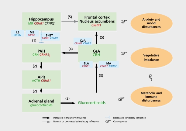
“The direct and indirect projections of the hippocampus and the CeA to the PVN suggest that stress response and related emotionality behaviors are regulated by such an interplay and the abundance of CRH-R1 in these regions may as well act, at least partially, as such a mediator (Reul and Holsboer, 2002).”
BackgroundGamma-Aminobutyric Acid Involvement in Depressive Illness
Anisman, H., Merali, Z., & Poulter, M. O. (2012) · Frontiers in neuroscience · doi:10.1201/b12215-5 · Cited by 11
A role of noradrenergic receptors in anxiolytic-like effect of high CRF in the rat frontal cortex
Śmiałowska, M., Zieba, B. A., & Domin, H. (2021) · Neuropeptides · doi:10.1016/j.npep.2021.102162 · Cited by 11
Dwivedi, Y. (2012) · CRC Press eBooks · doi:10.1201/b12215-10 · Cited by 11
Sex/Gender Differences in Depression and Anxiety Disorders
Park, H. Y. (2024) · Springer eBooks · doi:10.1007/978-981-97-0130-8_21 · Cited by 12
Pharmacogenetics of antidepressants and mood stabilizers
Serretti, A., Gibiino, S., & Olgiati, P. (2012) · Handbook of clinical neurology · doi:10.1016/b978-0-444-52002-9.00043-7 · Cited by 10
Wildfire-related smoke inhalation worsens cardiovascular risk in sleep disrupted rats
Martin, W. K., Schladweiler, M. C. J., Oshiro, W. M., Smoot, J. L., Fisher, A., Williams, W. C., Valdez, M. C., Miller, C. N., Jackson, T. W., Freeborn, D., et al. (2023) · Frontiers in Environmental Health · doi:10.3389/fenvh.2023.1166918 · Cited by 9
“A single sleep disruption and FA exposure increased hypothalamic expression of key hormonal receptors including Crhr2 and Nr3c2, which mediate stress and salt and water balance (63, 64), potentially indicating activation of the hypothalamicpituitary-adrenal stress axis (65–67), although circulating markers of these pr…”
BackgroundYang, Y., Chen, Y.-K., & Xie, M. (2024) · World Journal of Psychiatry · doi:10.5498/wjp.v14.i5.607 · Cited by 8
Luque‐García, A., Teruel‐Martí, V., Martínez‐Bellver, S., Adell, A., Cervera‐Ferri, A., & Martínez‐Ricós, J. (2018) · The Journal of Comparative Neurology · doi:10.1002/cne.24416 · Cited by 7
“…that mediate emotional behaviors, and are commonly related to alterations in the hypothalamic–pituitary–adrenal (HPA) axis, corticotropin-releasing factor signaling and consequent glucocorticoid secretion (Nemeroff, 1988; Arborelius, Owens, Plotsky, & Nemeroff, 1999; Reul & Holsboer, 2002).”
BackgroundRaskind, M. A., Williams, T., Holmes, H. A., Hart, K. L., Crews, L., Poupore, E. L., Thomas, R. G., Darnell, J., Daniels, C., Goke, K., et al. (2023) · Alcoholism Clinical and Experimental Research · doi:10.1111/acer.14989 · Cited by 7
Glucocorticoid-induced depression – the role of the dopaminergic system and microRNAs
Skórzewska, A. (2021) · Postępy Psychiatrii i Neurologii · doi:10.5114/ppn.2021.110791 · Cited by 7
“GR receptors play an important role in response to a stress factor when the concentration of GCs is high [2, 9].”
BackgroundBalogh, B., Vecsernyés, M., Veres-Székely, A., Berta, G., Stayer‐Harci, A., Tarjányi, O., & Sétáló, G. (2022) · Molecular and Cellular Endocrinology · doi:10.1016/j.mce.2022.111610 · Cited by 7
“UCNs are central regulators of food uptake and stress-coping (Reul and Holsboer, 2002), while being overweight and overstressed are both known risk factors in human tumorigenesis, including the development of breast cancer.”
BackgroundTOB is an effector of the hippocampus-mediated acute stress response
Youssef, M., Hamada, H. T., Lai, S., Kiyama, Y., El‐Tabbal, M., Kiyonari, H., Nakano, K., Kühn, B., & Yamamoto, T. S. (2022) · Translational Psychiatry · doi:10.1038/s41398-022-02078-7 · Cited by 6
“Crhr2 is activated by Urocotins I, II (stresscopin-related peptide), and III (stresscopin), which belong to the corticotrophin-releasing factor (CRF)-related peptides [83].”
BackgroundResponse of CRH system in brain and gill of marine medaka to seawater acidification
Chen, H., Feng, Y., Cui, J., & Wang, X. (2024) · Fish Physiology and Biochemistry · doi:10.1007/s10695-024-01332-7 · Cited by 6
“Crh acts through Crhr1 or Crhr2 with different distributions and physiological functions (Flik et al. 2006; Reul and Holsboer 2002).”
Tenk, J., Rostás, I., Füredi, N., Mikó, A., Soós, S., Solymár, M., Gaszner, B., Székely, M., Pétervári, E., & Balaskó, M. (2016) · Peptides · doi:10.1016/j.peptides.2016.09.005 · Cited by 6
A brain-driven neural circuit contributes to tissue regeneration in joint cartilage
Huo, K., Ma, X., Xue, Y., Zhao, L., Wang, F., Wang, G., You, Y., Jiang, W., Wang, R., Li, Y., et al. (2026) · Annals of the Rheumatic Diseases · doi:10.1016/j.ard.2025.12.004 · Cited by 4
Rajeswari, J. J., Gilbert, G. N. Y., Khalid, E., & Vijayan, M. M. (2025) · Molecular and Cellular Endocrinology · doi:10.1016/j.mce.2025.112494 · Cited by 4
Effect of photoperiod on the anxiety of zebrafish
Zhang, Y., Tao, H., Shen, Y.-J., Chen, Q.-L., & Liu, Z. (2024) · Environmental Biology of Fishes · doi:10.1007/s10641-024-01645-9 · Cited by 4
“Corticotro-pin-releasing hormone (CRH), a neuropeptide, plays a critical role in stress-related behaviors such as anxiety, arousal, motor function, learning, and memory (Reul and Holsboer 2002; Xin et al. 2020; Faught and Vijayan 2022).”
Radahmadi, M., Izadi, M. S., Rayatpour, A., & Ghasemi, M. (2021) · Basic and Clinical Neuroscience Journal · doi:10.32598/bcn.12.1.2346.1 · Cited by 4
“145 CRH neuropeptide family) mRNA in the paraventricular nucleus and amygdala that are involved in stress-coping responses related to CRH (Reul & Holsboer, 2002).”
BackgroundSchmidt, B. L., Santos, M. J., Cariaga-Martínez, A. E., Hill, L., Barloese, M. C. J., & Hall, Z. J. (2017) · Cited by 4
Hen-Shoval, D., Indig-Naimer, T., Moshe, L., Kogan, N. M., Zaidan, H., Gaisler‐Salomon, I., Okun, E., Mechoulam, R., Shoval, G., Zalsman, G., et al. (2024) · Journal of Psychiatric Research · doi:10.1016/j.jpsychires.2024.04.033 · Cited by 3
Ma, W., Sui, D., Sun, W., Yu, P., Li, Y., Guo, M., Wang, H., Zhang, X., Yu, X., Fu, W., et al. (2024) · Phytotherapy Research · doi:10.1002/ptr.8327 · Cited by 2
“The pathogenesis of anxiety disorders is linked to the hyper-activity of the HPA axis (Reul and Holsboer 2002).”
Kenney, L., Tekin, M., DeLeon, D., Marshall, S., & Smith, S. S. (2025) · Brain Research · doi:10.1016/j.brainres.2025.149874 · Cited by 3
Upadhyay, R. K., Barman, D., & Bhardwaj, K. (2026) · Nonlinear Dynamics · doi:10.1007/s11071-026-12223-2 · Cited by 2
Senevirathne, G., & Shubin, N. H. (2024) · Open Biology · doi:10.1098/rsob.240111
“I and VIM.2 [40,41]), extracellular matrix organizing and connective tissue-strengthening (e.g. COL9A1 , COL8A1 and CHAD [42,43]) and stress regulation ( CRCH.1 [44]).”
BackgroundThe role of the CRF-urocortin system in stress resilience
Henckens, M. J. A. G., Deussing, J. M., & Chen, A. (2019) · Elsevier eBooks · doi:10.1016/b978-0-12-813983-7.00016-1 · Cited by 1
Validation of Enhancer Regions in Primary Human Neural Progenitor Cells using Capture STARR-seqPreprint
Gaynor-Gillett, S. C., Cheng, L., Shi, M., Liu, J. J., Wang, G., Spector, M., Flaherty, M., Wall, M., Hwang, A., Gu, M., et al. (2024) · bioRxiv (Cold Spring Harbor Laboratory) · doi:10.1101/2024.03.14.585066 · Cited by 1
A Mechanistic Model for the HPA Axis Cortisol Paradox in PTSDPreprint
Danan, D., Grosskopf, Y., Hayut, Y., Toledano, Y., Doenyas‐Barak, K., Mayo, A., & Alon, U. S. (2025) · bioRxiv (Cold Spring Harbor Laboratory) · doi:10.1101/2025.10.13.681561 · Cited by 1
Klampfl, S. M. (2015) · University of Regensburg Publication Server (University of Regensburg) · doi:10.5283/epub.31369
Effects of Early, Elevated Prenatal Testosterone on the HPA Axis and Hormone Responsiveness in Mice
Wilson, H. A. (2019) · The Atrium (University of Guelph)
Tafet, G. E., & Feder, D. J. (2021) · Springer eBooks · doi:10.1007/978-3-030-61721-9_28
Senevirathne, G., & Shubin, N. H. (2021) · bioRxiv (Cold Spring Harbor Laboratory) · doi:10.1101/2021.10.04.462674
A REVIEW ON THERAPEUTIC UTILITY OF CRF1 RECEPTOR
(2024) · International Journal of Biology Pharmacy and Allied Sciences · doi:10.31032/ijbpas/2024/13.4.7970
ANALYSIS OF MODERN CONCEPTS OF NEURODEGENERATIVE PROPERTIES OF DEPRESSION
Yasenyavskaya, A. L., Tsibizova, A., & Самотруева, М. А. (2023) · Molekulyarnaya Meditsina (Molecular medicine) · doi:10.29296/24999490-2023-04-05
(2024) · Elsevier eBooks · doi:10.1016/b978-0-443-23565-8.09996-8
Anisman, H., Hayley, S. P., & Kusnecov, A. W. (2024) · Elsevier eBooks · doi:10.1016/b978-0-443-23565-8.00001-6
Stressor Processes and Effects on Neurobiological Functioning
Anisman, H., Hayley, S. P., & Kusnecov, A. (2024) · Elsevier eBooks · doi:10.1016/b978-0-443-23565-8.00018-1
Glucocorticoid Resistance Syndrome in 2 Patients With Diverse Genotype
Battiola, T., Viskochil, D. H., Wolken, K., & Couldwell, M. (2025) · JCEM Case Reports · doi:10.1210/jcemcr/luae243
The Neurobiology of Comorbidities
Kohshour, M. O., Navarro‐Flores, A., Budde, M., & Schulze, T. G. (2025) · Springer eBooks · doi:10.1007/978-3-031-81802-8_5
Kurihara, T., Sasanuma, M., Kato, M., Inoué, T., Okada, N., Shirayama, T., Tanikawa, Y., Kawahara, T., Sohya, K., Yasuda, H., et al. (2025) · Scientific Reports · doi:10.1038/s41598-025-03362-4
The Impacts of Binge Drinking and Hangover on the Social Brain: An Integrative Narrative Review
Bagosi, Z., Karasz, G., Thury, A. Á., Simon, B., Földesi, I., & Csabafi, K. (2025) · Biomedicines · doi:10.3390/biomedicines13112802
“…CRH is synthesized and released from the PVN of the hypothalamus, and, after reaching the median eminence, it stimulates the secretion of adrenocorticotrophic hormone (ACTH) in the anterior pituitary, which in turn stimulates the secretion of corticosteroids in the adrenal cortex [125].”
Metadata
- Authors
- Reul, J. M. H. M., & Holsboer, F.
- Affiliations
- Max Planck Institute of Psychiatry (DE)
- Corresponding author
- Reul, Johannes M. H. M.
- Venue
- Dialogues in Clinical Neuroscience
- Publisher
- Informa UK Limited
- Published
- 2002-03-31
- Volume
- 4
- Issue
- 1
- Pages
- 31-46
- ISSN
- 1294-8322 (ISSN-L)
- 1958-5969
- PMC
- PMC3181666
- OpenAlex
- W4294249858
- Cited by
- 103
- References
- 85
- Citations by year
- 2026: 3 · 2025: 8 · 2024: 16 · 2023: 8 · 2022: 5 · 2021: 8 · 2020: 5 · 2019: 9 · 2018: 8 · 2017: 6 · 2016: 4 · 2015: 7 · 2014: 5 · 2013: 3 · 2012: 6
- FWCI
- 0.28
- Type
- journal article
- Language
- en
- Open access
- diamond
- doi.org
- Licence
- Locations
- Dialogues in Clinical Neuroscience (publishedVersion, cc-by-nc-nd)
- Europe PMC (PubMed Central) (publishedVersion, cc-by-nc-nd)
- PubMed Central (publishedVersion, cc-by-nc-nd)
- PubMed (publishedVersion)
- Bristol Research (University of Bristol) (publishedVersion)
- PubMed
- Topics
- Life Sciences › Neuroscience › Behavioral Neuroscience › Stress Responses and Cortisol
- Life Sciences › Neuroscience › Cellular and Molecular Neuroscience › Neuropeptides and Animal Physiology
- Life Sciences › Neuroscience › Endocrine and Autonomic Systems › Neuroendocrine regulation and behavior
- Keywords
- corticotropin-releasing factor, CRH receptors, urocortin II, stress response, anxiety, depression, stress coping, anxiolysis, neuroendocrine stress response, neuropeptides
About these checks
Registry, retraction, Cites and statistics results are computed, not judged, with one exception: a printed reference that no index lists is read once by a model before it counts as a citation error. Claims are extracted by a model and kept only when their quote appears verbatim in the text; each cited source is then checked, with a model’s help, for whether it says what the claim relies on. Those verdicts can be wrong — read the claim’s quote and the cited source before relying on one.
Statistics and arithmetic are recomputed, not judged.
Confidence bands are shown only for fields calibrated on our annotated test set. None is calibrated yet, so no band is shown.
What counts as a claim ›How we measure confidence ›How we check this ›
Are you an author, or do you see a problem with this page? Request a review
