

Microphysiological systems as models for immunologically ‘cold’ tumors
Open accessThe DOI matches its Crossref record; 206 of the 210 references it cites were found in a registry or an index; 210 of the 287 sources its claims cite were checked for support.
Abstract
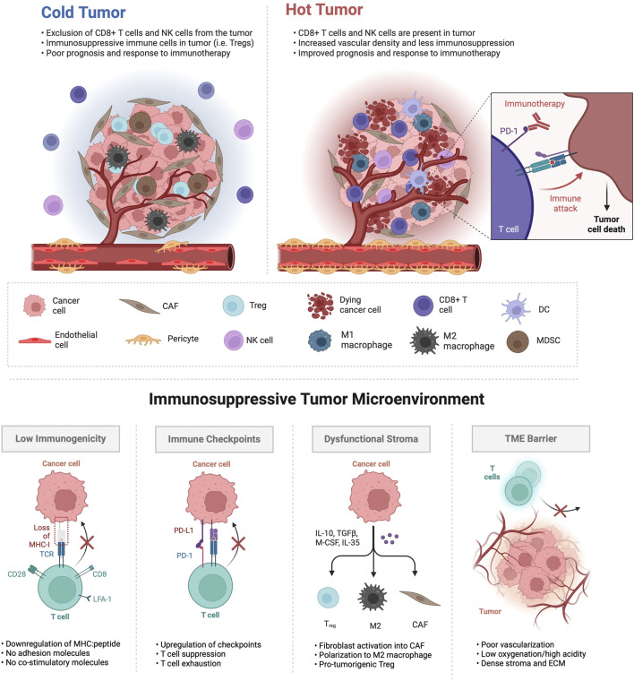
The tumor microenvironment (TME) is a diverse milieu of cells including cancerous and non-cancerous cells such as fibroblasts, pericytes, endothelial cells and immune cells. The intricate cellular interactions within the TME hold a central role in shaping the dynamics of cancer progression, influencing pivotal aspects such as tumor initiation, growth, invasion, response to therapeutic interventions, and the emergence of drug resistance. In immunologically ‘cold’ tumors, the TME is marked by a scarcity of infiltrating immune cells, limited antigen presentation in the absence of potent immune-stimulating signals, and an abundance of immunosuppressive factors. While strategies targeting the TME as a therapeutic avenue in ‘cold’ tumors have emerged, there is a pressing need for novel approaches that faithfully replicate the complex cellular and non-cellular interactions in order to develop targeted therapies that can effectively stimulate immune responses and improve therapeutic outcomes in patients. Microfluidic devices offer distinct advantages over traditional in vitro 3D co-culture models and in vivo animal models, as they better recapitulate key characteristics of the TME and allow for precise, controlled insights into the dynamic interplay between various immune, stromal and cancerous cell types at any timepoint. This review aims to underscore the pivotal role of microfluidic systems in advancing our understanding of the TME and presents current microfluidic model systems that aim to dissect tumor-stromal, tumor-immune and immune-stromal cellular interactions in various ‘cold’ tumors. Understanding the intricacies of the TME in ‘cold’ tumors is crucial for devising effective targeted therapies to reinvigorate immune responses and overcome the challenges of current immunotherapy approaches.
Overview
CoChat Summary
TL;DR
This review argues that microphysiological systems (MPS)/tumor chips can better model immunologically ‘cold’ tumors than traditional 2D or animal models by recapitulating tumor-stromal, tumor-immune, and immune-stromal interactions. It surveys MPS for ovarian, pancreatic, renal, and prostate cancers and concludes the field is nascent, with few models incorporating full vasculature and immune complexity.
Key findings
- Immunologically ‘cold’ tumors are characterized by immune exclusion or exhaustion, desmoplastic stroma, poor perfusion/hypoxia, and enrichment of immunosuppressive cells such as Tregs, M2 TAMs, MDSCs, and CAFs; fewer than 15% of patients respond to immune checkpoint inhibitor therapy.
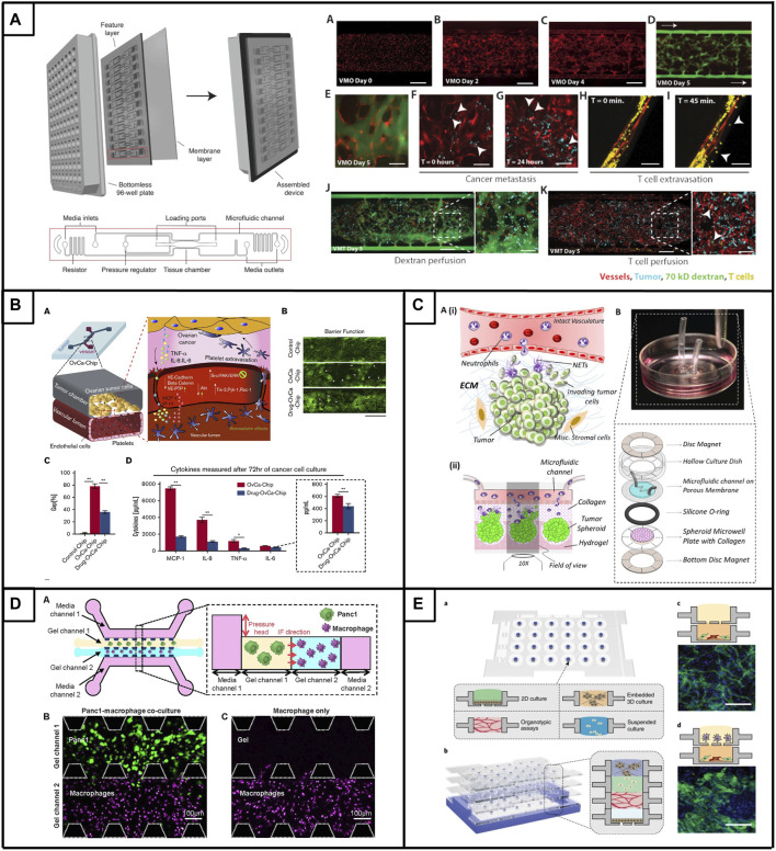
- MPS can reproduce key TME features: a vascularized micro-tumor model enables de novo perfusable microvascular networks and T-cell extravasation; ovarian PDX spheroids show enhanced formation, proliferation, and viability under microfluidic conditions compared with larger-scale 3D cultures or Matrigel; sequential fibroblast addition to ovarian spheroids yields the highest vascularization, and CAR T cells are recruited, extravasate, and produce a strong IFN-γ response.
- In PDAC, endothelial cells significantly reduce T-cell infiltration while pancreatic stellate cells increase it; activated T cells show a 50% increase in migration toward cancer cells. In RCC, high-density collagen limits CAR-T cell infiltration; in prostate, LNCaP versus C-2 cells polarize macrophages toward M1 versus M2 phenotypes and alter endothelial networks.
- The review cites disease-burden figures including about 20,000 new ovarian cancer cases and more than 12,000 deaths in 2024; PDAC median survival under 6 months and 5-year survival below 4%; metastatic RCC 5-year survival under 12%; and prostate cancer projected to claim more than 35,000 lives this year.
Methods
- Narrative review; no primary experiments are reported. It synthesizes published microfluidic/tumor-chip studies for immunologically ‘cold’ tumors, focusing on ovarian cancer, pancreatic ductal adenocarcinoma, renal cell carcinoma, and prostate cancer.
- Tables 1–4 summarize selected MPS by cancer model, key device features, cell types, and key findings.
- The main analysis is qualitative synthesis of device designs, cellular components, and reported outcomes; no systematic search strategy or meta-analysis is described.
Limitations
- Stated in the paper: few MPS exist for immunologically ‘cold’ tumors, especially those focusing on T-cell modulation; many models lack essential cellular and acellular TME components, physiologically relevant vascularization, and complex cellular crosstalk.
- Stated in the paper: adoption faces challenges including limited cell sourcing, accessibility to non-expert users, regulatory approvals for precision medicine use, and difficulties with intricate biofabrication protocols and throughput.
- Evident from the text: as a narrative review, it does not report a systematic search, inclusion criteria, or quantitative synthesis, so conclusions depend on the cited studies and are not independently validated.
Model-written summary. Check it against the paper before relying on it.
Integrity signals
Registry
Cites
Statistics
Figures
Verification
Claims the paper makes
159 Valid · 15 Not valid · 45 Not checked · 42 Own results and interpretations
At least one cited source doesn’t support this claim or points to a different paper, and none confirmed it — see the verdicts below.
In the paper
“In contrast, co-culturing PDAC cells with PSCs markedly increased T cell infiltration, emphasizing the influence of the TME on immune cell behavior.”
§ Microphysiological systems as models for immunologically ‘cold’ tumors > 3 Current microphysiological systems of ‘cold’ tumors > 3.2 Pancreatic cancer
What the sources say
- Disputedcites Mollica et al., 2021 matched to “A 3D pancreatic tumor model to study T cell infiltration” (2021) sourceExperimental — accuracy not yet measured
The Discussion (§3) explicitly says that adding PSCs to cancer cells did not increase activated T-cell infiltration, with or without endothelial cells. The Results (§2.3) report 86.73 infiltrating activated T cells with cancer cells plus PSCs versus 75.35 with cancer cells alone, but the paper does not characterize that difference as a marked increase. (LLM, conf=0.90)
Checked by a reasoning model — may be wrong.
plainstates a cause- Asserted: passes — The authors put it forward as true.
- Checkable: passes — It can be checked against evidence.
- Load-bearing: passes — The paper’s conclusions depend on it.
- Based: passes — The paper gives a reason why it holds.
- Rests on an assumption not shown here.
- Disputed
The cited sources support only part of this claim — read each source’s verdict below.
In the paper
“Additionally, immunologically ‘cold’ tumors are characterized by the presence of immunosuppressive cell populations such as tumor-associated macrophages (TAMs), myeloid-derived suppressor cells (MDSCs) and regulatory T cells (Tregs) (Hegde et al., 2016; Liu et al., 2021; Guha et al., 2022).”
§ Microphysiological systems as models for immunologically ‘cold’ tumors > 1 Introduction
What the sources say
- Not checked — no full text of the sourcecites Hegde et al., 2016 matched to “The Where, the When, and the How of Immune Monitoring for Cancer Immunotherapies in the Era of Checkpoint Inhibition” (2016) source
- Could not verifycites Liu et al., 2021 matched to “Tumor-on-a-chip: from bioinspired design to biomedical application” (2021) sourceExperimental — accuracy not yet measured
The Introduction (page unavailable) says tumors contain assorted immune and inflammatory cells, but does not characterize immunologically ‘cold’ tumors or identify TAMs, MDSCs, and Tregs as their characteristic populations. Searches for ‘cold tumors,’ ‘macrophages,’ ‘regulatory T cells,’ and ‘immunosuppressive’ found no passage addressing that claim. (LLM, conf=0.74)
- Partly supportedcites Guha et al., 2022 matched to “Assessing the future of solid tumor immunotherapy” (2022) sourceExperimental — accuracy not yet measured
Section 4.1 identifies MDSCs and Tregs as contributors to the immunosuppressive environment of solid tumors and mentions pro-tumorigenic macrophages. But Section 4.2 describes “cold” tumors by low neo-antigen expression and T-cell infiltration; it does not identify those three cell populations as characteristics of cold tumors specifically. Page numbers are unavailable in our copy. (LLM, conf=0.86)
plainstates an association- Asserted: passes — The authors put it forward as true.
- Checkable: passes — It can be checked against evidence.
- Load-bearing: unclear — It is unclear whether the paper’s conclusions depend on it.
- Based: passes — The paper gives a reason why it holds.
The cited sources support only part of this claim — read each source’s verdict below.
In the paper
“Notably, these physiologically relevant platforms have previously shown promise in drug discovery and testing by faithfully recapitulating pathophysiological human drug responses (Asghar et al., 2015; Benam et al., 2016; Hachey et al., 2021); hence offering the potential to advance the development of personalized therapy approaches.”
§ Microphysiological systems as models for immunologically ‘cold’ tumors > 1 Introduction
What the sources say
- Partly supportedcites Asghar et al., 2015 matched to “Engineering cancer microenvironments for in vitro 3-D tumor models” (2015) sourceExperimental — accuracy not yet measured
The Introduction and Abstract describe 3-D tumor models as promising alternatives for drug screening because they can better mimic tumor microenvironments. The Spheroids and hanging drops section reports drug responses in cultured human cancer cells, but the paper does not establish that these platforms faithfully reproduce human drug responses in patients. The citing sentence overstates that aspect of the evidence. (LLM, conf=0.84)
- Not checked — no full text of the sourcecites Benam et al., 2016 matched to “Matched-comparative modeling of normal and diseased human airway responses using a microengineered breathing lung chip” (2016) source
- Partly supportedcites Hachey et al., 2021 matched to “An in vitro vascularized micro-tumor model of human colorectal cancer recapitulates in vivo responses to standard-of-care therapy” (2021) sourceExperimental — accuracy not yet measured
The Results section reports that the human colorectal-cancer micro-tumor model closely matched FOLFOX responses in mouse xenografts, supporting its promise for drug testing. The Discussion also proposes its use in drug discovery and validation. The paper does not establish that the platform faithfully reproduces drug responses observed in human patients; its in vivo response comparison is with xenografts. (LLM, conf=0.86)
plainstates a cause- Asserted: passes — The authors put it forward as true.
- Checkable: passes — It can be checked against evidence.
- Load-bearing: passes — The paper’s conclusions depend on it.
- Based: passes — The paper gives a reason why it holds.
- Partly supported
The cited sources support only part of this claim — read each source’s verdict below.
In the paper
“Immunosuppressive cell types, such as TAMs, regulatory T cells, MDSCs, and cancer-associated fibroblasts (CAFs) play key roles in establishing an immunosuppressive environment in the TME and interfering with cytotoxic T cell activation (Wang et al., 2023; Xiao et al., 2023).”
§ Microphysiological systems as models for immunologically ‘cold’ tumors > 2 Defining the ‘cold’ tumor microenvironment
What the sources say
- Not matchedcites Wang et al., 2023
An author–year citation the paper’s reference list doesn’t settle. We don’t search these by name.
- Partly supportedcites Xiao et al., 2023 matched to “Desmoplastic stroma restricts T cell extravasation and mediates immune exclusion and immunosuppression in solid tumors” (2023) sourceExperimental — accuracy not yet measured
The Introduction identifies CAFs as components of an immunosuppressive tumor microenvironment, and the Results describe TAMs and regulatory T cells as potentially immunosuppressive. The Discussion links CAFs to hindered T-cell recruitment and reports fewer immunosuppressive myeloid cells after CAF-targeted treatment. Our copy does not identify MDSCs specifically or establish that all four listed cell types interfere with cytotoxic T-cell activation. (LLM, conf=0.87)
plainstates a cause- Asserted: passes — The authors put it forward as true.
- Checkable: passes — It can be checked against evidence.
- Load-bearing: passes — The paper’s conclusions depend on it.
- Based: passes — The paper gives a reason why it holds.
- Not matched
The cited sources support only part of this claim — read each source’s verdict below.
In the paper
“The vasculature in tumors exhibits structural disorganization and functional abnormalities, particularly in ‘cold’ tumors (Choi and Jung, 2023).”
§ Microphysiological systems as models for immunologically ‘cold’ tumors > 2 Defining the ‘cold’ tumor microenvironment
What the sources say
- Partly supportedcites Choi and Jung, 2023 matched to “Normalization of the tumor microenvironment by harnessing vascular and immune modulation to achieve enhanced cancer therapy” (2023) sourceExperimental — accuracy not yet measured
The Vascular normalization > Background section describes tumor vessels as irregular, leaky, and poorly perfused, supporting the structural and functional abnormalities. It does not say these abnormalities are particularly characteristic of ‘cold’ tumors; the only exact mention of cold tumors found is in a reference title. Page numbers are unavailable in our copy. (LLM, conf=0.86)
plainstates an association- Asserted: passes — The authors put it forward as true.
- Checkable: passes — It can be checked against evidence.
- Load-bearing: unclear — It is unclear whether the paper’s conclusions depend on it.
- Based: passes — The paper gives a reason why it holds.
- Partly supported
120 more claims · 66 with their cited sources checked
Create a free account to see all 261 claims and to verify the 2 references still open.
- It may state two separate claims as one. 121
- Probably not a substantial claim. 32
Self-check
Percentages, sums and reported differences re-computed from the paper’s own numbers.
Each contradiction between two passages listed here was confirmed by an independent check.
Rated minor: it would not touch a result — a value no result rests on, a p-value that stays on the same side of the significance level, or a conflict that is clerical, resolved elsewhere in the paper, or outside its own findings.
In the paper
§ Microphysiological systems as models for immunologically ‘cold’ tumors > 3 Current microphysiological systems of ‘cold’ tumors > 3.4 Prostate cancer
“HUVECs, THP-1 (monocytes), nHDFs, LNCap or C4-2 prostate cancer cells”
§ Microphysiological systems as models for immunologically ‘cold’ tumors > 3 Current microphysiological systems of ‘cold’ tumors > 3.4 Prostate cancer
“Accordingly, TH1 monocytes, epithelial cells and C4-2 (androgen-independent) or LNCaP tumor cells were prepared in separate layers. Subsequently, TH1 monocytes were stacked onto the prostate cancer cell layer, leading to the organotypical differentiation of TAMs (od-TAMs).”
Citations
Cites
Sung, H., Ferlay, J., Siegel, R. L., Laversanne, M., Soerjomataram, I., Jemal, A., & Bray, F. (2021) · CA: A Cancer Journal for Clinicians · doi:10.3322/caac.21660 · Cited by 108,699 · cited 1×
“Cancer ranks as the second leading cause of death worldwide and has claimed the lives of approximately 10 million people in 2020 (Sung et al., 2021; Siegel et al., 2023).”
BackgroundPD-1 and PD-L1 Immune Checkpoint Blockade to Treat Breast Cancer
Hartkopf, A. D., Taran, F.-A., Wallwiener, M., Walter, C. B., Krämer, B., Grischke, E.-M., & Brucker, S. Y. (2016) · Breast Care · doi:10.1159/000453569 · Cited by 30,845
Siegel, R. L., Miller, K. D., Wagle, N. S., & Jemal, A. (2023) · CA: A Cancer Journal for Clinicians · doi:10.3322/caac.21763 · Cited by 16,303
SEER Cancer Statistics Review, 1975-2003
Ries, L. A. G., Harkins, D., Krapcho, M. F., Mariotto, A. B., Miller, B., Feuer, E. J., Clegg, L. X., Eisner, M. P., Horner, M., Howlader, N., et al. (2006) · Digital Archive @ GSU · Cited by 11,783
Siegel, R. L., Giaquinto, A. N., & Jemal, A. (2024) · CA: A Cancer Journal for Clinicians · doi:10.3322/caac.21820 · Cited by 9,533
Predictive correlates of response to the anti-PD-L1 antibody MPDL3280A in cancer patients
Herbst, R. S., Soria, J.-C., Kowanetz, M., Fine, G. D., Hamid, O., Gordon, M. S., Sosman, J. A., McDermott, D. F., Powderly, J. D., Gettinger, S. N., et al. (2014) · Nature · doi:10.1038/nature14011 · Cited by 5,231
The evolving tumor microenvironment: From cancer initiation to metastatic outgrowth
de Visser, K. E., & Joyce, J. A. (2023) · Cancer Cell · doi:10.1016/j.ccell.2023.02.016 · Cited by 4,059
CAR-T cell therapy: current limitations and potential strategies
Sterner, R. C., & Sterner, R. M. (2021) · Blood Cancer Journal · doi:10.1038/s41408-021-00459-7 · Cited by 2,926 · cited 2×
“To address this, various immunotherapeutic strategies have emerged, including adoptive cell transfer, primarily utilizing genetically modi fi ed T cells expressing chimeric antigen receptors (CAR T-cells) to recognize and eliminate cancer cells directly (Sterner and Sterner, 2021).”
BackgroundCancer is a preventable disease that requires major lifestyle changes
Anand, P., Kunnumakara, A. B., Sundaram, C., Harikumar, K. B., Tharakan, S. T., Lai, O. S., Sung, B., & Aggarwal, B. B. (2008) · Pharmaceutical Research · doi:10.1007/s11095-008-9661-9 · Cited by 2,632
Edmondson, R., Broglie, J. J., Adcock, A. F., & Yang, L. (2014) · ASSAY and Drug Development Technologies · doi:10.1089/adt.2014.573 · Cited by 2,515
Research works printed but not linked: 1
- Bonaventura P. Shekarian T. Alcazer V. Valladeau-Guilemond J. Valsesia-Wittmann S. Amigorena S. (2019b). Cold tumors: a therapeutic challenge for immunotherapy. Front. Immunol. 10, 168. 10.3389/fimmu.2019.00168 30800125 PMC6376112 · DOI · Paper page
Cited by
Zhang, W., Jin, C., Liu, S., Wan, X., Li, Y., Liu, J., Duan, Z., Ma, J., & Gao, Y. (2026) · Frontiers in Immunology · doi:10.3389/fimmu.2026.1757513 · Cited by 22
Gaebler, D., Hachey, S. J., & Hughes, C. C. W. (2024) · Frontiers in Bioengineering and Biotechnology · doi:10.3389/fbioe.2024.1462293 · Cited by 22
Self-citationDefeating lethal cancer: Interrupting the ecologic and evolutionary basis of death from malignancy
Pienta, K. J., Goodin, P. L., & Amend, S. R. (2025) · CA A Cancer Journal for Clinicians · doi:10.3322/caac.70000 · Cited by 19
“171 129,182 This 186–190 New cancer - eradication paradigms to alter the tumor ecosystem: Classical cancer therapeutics (e.g., chemotherapy, growth factor inhibitors, radiation) most often target the machinery that a cell uses to proliferate, i.e., seeks to intervene directly to reduce fitness,…”
Methods for processing and analyzing images of vascularized micro-organ and tumor systems
Hachey, S. J., Hatch, C. J., Gaebler, D., Forsythe, A. G., Ewald, M. L., Chopra, A. L., Chen, Z., Thapa, K., Hodanu, M., Fang, J. S., et al. (2025) · Frontiers in Bioengineering and Biotechnology · doi:10.3389/fbioe.2025.1585003 · Cited by 5
Self-citationWang, F., Wei, X., Yang, M., Lu, C., Yang, X., Deng, J., Chen, Z., & Zhou, Q. H. (2025) · Thoracic Cancer · doi:10.1111/1759-7714.70025 · Cited by 3
“The TME comprises both tumor and nontumor cells including immune cells, fibroblasts, and endothelial cells, and the activity of these cells, particularly immune cells, is closely linked to tumor growth [34, 35].”
Große, M., Burchardt, M., & Caetano-Pinto, P. (2025) · Future Pharmacology · doi:10.3390/futurepharmacol5030036 · Cited by 2
“Customizable and modular : Devices can be tailored for specific cancer types, immune interactions, or treatment scenarios [82,83].”
InfluentialLiu, N., Zhang, Y., Liu, Y., Dong, X., Li, Y. D., Geng, J., & Li, Y. (2026) · Journal of the Korean Gastric Cancer Association · doi:10.5230/jgc.2026.26.e25 · Cited by 1
“PDOs offer a powerful platform for this approach by enabling biobank construction, biomarker screening, and multi-omics integration, thereby advancing personalized treatments for gastric cancer.”
From Cold to Hot: Nanozyme‐Based Strategies for Reprogramming the Tumour Immunoenvironment
Wang, Y., Xu, M., Wang, Y., Lin, X., Zhang, Q., Lin, X., Wu, X., Zhang, C., Huang, W., & Shen, J. (2026) · Cell Proliferation · doi:10.1111/cpr.70235 · Cited by 2
Zeng, J., Li, Q., Li, K., Yang, L., Xu, L., Wang, W., Yang, K., Wei, Q., Wang, J., Zhu, C., et al. (2025) · Frontiers in Immunology · doi:10.3389/fimmu.2025.1605227
“Complex cellular interactions within the TME play a central role in cancer progression, affecting tumor initiation, growth, invasion, therapeutic response, and drug resistance (42).”
Combination therapy in neoadjuvant and metastatic settings in hot tumors: strengths and weaknesses
Stea, E. X., Glantzounis, G. K., Lykoudis, E. G., & Roukos, D. H. (2025) · Expert Review of Anticancer Therapy · doi:10.1080/14737140.2025.2572980 · Cited by 1
“Beyond tumor type and stage at diagnosis [4], classification into hot (immune-inflamed), immune-excluded and cold (immune-desert) is crucial for decision-making for anticancer immunotherapy [6,7].”
Lee, C.-J., Kim, Y., Kim, M., Rim, Y. A., & Ju, J. H. (2026) · Bioengineering & Translational Medicine · doi:10.1002/btm2.70162
“21,22 In this review, we therefore frame autoimmune diseases as programmable microenvironmental states: Section 2 outlines the conserved and disease-specific pathogenic axes of SSc, SLE, and RA.”
Metadata
- Authors
- Gaebler, D., Hachey, S. J., & Hughes, C. C. W.
- Affiliations
- University of California, Irvine (US)
- Corresponding author
- Hughes, Christopher C. W.
- Venue
- Frontiers in Cell and Developmental Biology
- Publisher
- Frontiers Media SA
- Published
- 2024-04-22
- Volume
- 12
- Pages
- 1389012-1389012
- ISSN
- 2296-634X (ISSN-L)
- PMC
- PMC11070549
- OpenAlex
- W4395006996
- Semantic Scholar
- 63209d366461bb447684cf1d13e896f9558c0d91
- Cited by
- 14
- References
- 210
- Citations by year
- 2026: 7 · 2025: 6 · 2024: 1
- FWCI
- 0.45
- Type
- journal article
- Language
- en
- Open access
- gold
- doi.org
- Licence
- Locations
- Frontiers in Cell and Developmental Biology (publishedVersion, cc-by)
- PubMed Central (publishedVersion, cc-by)
- PubMed (publishedVersion)
- eScholarship (California Digital Library) (submittedVersion, cc-by)
- DOAJ (DOAJ: Directory of Open Access Journals) (submittedVersion)
- Funders
- National Institutes of Health
- Topics
- Physical Sciences › Engineering › Biomedical Engineering › 3D Printing in Biomedical Research
- Health Sciences › Medicine › Oncology › Cancer Cells and Metastasis
- Physical Sciences › Engineering › Biomedical Engineering › Microfluidic and Capillary Electrophoresis Applications
- Keywords
- microphysiological systems, cold tumors, tumor microenvironment, microfluidics, tumor-stroma interactions, tumor-immune interactions, cancer immunotherapy, immunosuppression, 3D co-culture model, drug resistance
About these checks
Registry, retraction, Cites and statistics results are computed, not judged, with one exception: a printed reference that no index lists is read once by a model before it counts as a citation error. Claims are extracted by a model and kept only when their quote appears verbatim in the text; each cited source is then checked, with a model’s help, for whether it says what the claim relies on. Those verdicts can be wrong — read the claim’s quote and the cited source before relying on one.
The claim verdicts this report counts are experimental: their accuracy is not yet measured.
A language model drew the claims from the paper on 2026-10-07, under version 3 of the claim definition (model deepseek-v4.1-flash, prompt extract-v3).
A second model labelled each claim on 2026-10-07 (model jev-1.13-20260917, definition revision gold-guideline-v0.2).
Cited sources are checked with a language model’s help (gpt-6-sol).
Statistics and arithmetic are recomputed, not judged.
Confidence bands are shown only for fields calibrated on our annotated test set. None is calibrated yet, so no band is shown.
What counts as a claim ›How we measure confidence ›How we check this ›
Are you an author, or do you see a problem with this page? Request a review
Data sources
Bibliographic data: Semantic Scholar, OpenAlex, Crossref.
Contains information from the Semantic Scholar Academic Graph Datasets, made available under the ODC Attribution License (ODC-BY 1.0).
Semantic Scholar’s data is described in: Kinney, R., et al. (2023). The Semantic Scholar Open Data Platform. arXiv:2301.10140.
