

Microphysiological systems as models for immunologically ‘cold’ tumors
Open accessThe DOI matches its Crossref record; 206 of the 210 references it cites were found in a registry or an index; 210 of the 287 sources its claims cite were checked for support.
Abstract
The tumor microenvironment (TME) is a diverse milieu of cells including cancerous and non-cancerous cells such as fibroblasts, pericytes, endothelial cells and immune cells. The intricate cellular interactions within the TME hold a central role in shaping the dynamics of cancer progression, influencing pivotal aspects such as tumor initiation, growth, invasion, response to therapeutic interventions, and the emergence of drug resistance. In immunologically ‘cold’ tumors, the TME is marked by a scarcity of infiltrating immune cells, limited antigen presentation in the absence of potent immune-stimulating signals, and an abundance of immunosuppressive factors. While strategies targeting the TME as a therapeutic avenue in ‘cold’ tumors have emerged, there is a pressing need for novel approaches that faithfully replicate the complex cellular and non-cellular interactions in order to develop targeted therapies that can effectively stimulate immune responses and improve therapeutic outcomes in patients. Microfluidic devices offer distinct advantages over traditional in vitro 3D co-culture models and in vivo animal models, as they better recapitulate key characteristics of the TME and allow for precise, controlled insights into the dynamic interplay between various immune, stromal and cancerous cell types at any timepoint. This review aims to underscore the pivotal role of microfluidic systems in advancing our understanding of the TME and presents current microfluidic model systems that aim to dissect tumor-stromal, tumor-immune and immune-stromal cellular interactions in various ‘cold’ tumors. Understanding the intricacies of the TME in ‘cold’ tumors is crucial for devising effective targeted therapies to reinvigorate immune responses and overcome the challenges of current immunotherapy approaches.
Overview
CoChat Summary
TL;DR
This review argues that microphysiological systems (MPS)/tumor chips can better model immunologically ‘cold’ tumors than traditional 2D or animal models by recapitulating tumor-stromal, tumor-immune, and immune-stromal interactions. It surveys MPS for ovarian, pancreatic, renal, and prostate cancers and concludes the field is nascent, with few models incorporating full vasculature and immune complexity.
Key findings
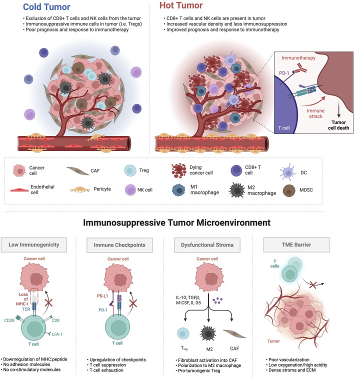
- Immunologically ‘cold’ tumors are characterized by immune exclusion or exhaustion, desmoplastic stroma, poor perfusion/hypoxia, and enrichment of immunosuppressive cells such as Tregs, M2 TAMs, MDSCs, and CAFs; fewer than 15% of patients respond to immune checkpoint inhibitor therapy.
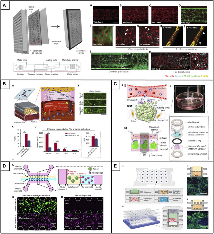
- MPS can reproduce key TME features: a vascularized micro-tumor model enables de novo perfusable microvascular networks and T-cell extravasation; ovarian PDX spheroids show enhanced formation, proliferation, and viability under microfluidic conditions compared with larger-scale 3D cultures or Matrigel; sequential fibroblast addition to ovarian spheroids yields the highest vascularization, and CAR T cells are recruited, extravasate, and produce a strong IFN-γ response.
- In PDAC, endothelial cells significantly reduce T-cell infiltration while pancreatic stellate cells increase it; activated T cells show a 50% increase in migration toward cancer cells. In RCC, high-density collagen limits CAR-T cell infiltration; in prostate, LNCaP versus C-2 cells polarize macrophages toward M1 versus M2 phenotypes and alter endothelial networks.
- The review cites disease-burden figures including about 20,000 new ovarian cancer cases and more than 12,000 deaths in 2024; PDAC median survival under 6 months and 5-year survival below 4%; metastatic RCC 5-year survival under 12%; and prostate cancer projected to claim more than 35,000 lives this year.
Methods
- Narrative review; no primary experiments are reported. It synthesizes published microfluidic/tumor-chip studies for immunologically ‘cold’ tumors, focusing on ovarian cancer, pancreatic ductal adenocarcinoma, renal cell carcinoma, and prostate cancer.
- Tables 1–4 summarize selected MPS by cancer model, key device features, cell types, and key findings.
- The main analysis is qualitative synthesis of device designs, cellular components, and reported outcomes; no systematic search strategy or meta-analysis is described.
Limitations
- Stated in the paper: few MPS exist for immunologically ‘cold’ tumors, especially those focusing on T-cell modulation; many models lack essential cellular and acellular TME components, physiologically relevant vascularization, and complex cellular crosstalk.
- Stated in the paper: adoption faces challenges including limited cell sourcing, accessibility to non-expert users, regulatory approvals for precision medicine use, and difficulties with intricate biofabrication protocols and throughput.
- Evident from the text: as a narrative review, it does not report a systematic search, inclusion criteria, or quantitative synthesis, so conclusions depend on the cited studies and are not independently validated.
Model-written summary. Check it against the paper before relying on it.
Integrity signals
Registry
Cites
Statistics
Figures
Verification
Claims the paper makes
159 Valid · 15 Not valid · 45 Not checked · 42 Own results and interpretations
At least one cited source doesn’t support this claim or points to a different paper, and none confirmed it — see the verdicts below.
In the paper
“In contrast, co-culturing PDAC cells with PSCs markedly increased T cell infiltration, emphasizing the influence of the TME on immune cell behavior.”
§ Microphysiological systems as models for immunologically ‘cold’ tumors > 3 Current microphysiological systems of ‘cold’ tumors > 3.2 Pancreatic cancer
What the sources say
- Disputedcites Mollica et al., 2021 matched to “A 3D pancreatic tumor model to study T cell infiltration” (2021) sourceExperimental — accuracy not yet measured
The Discussion (§3) explicitly says that adding PSCs to cancer cells did not increase activated T-cell infiltration, with or without endothelial cells. The Results (§2.3) report 86.73 infiltrating activated T cells with cancer cells plus PSCs versus 75.35 with cancer cells alone, but the paper does not characterize that difference as a marked increase. (LLM, conf=0.90)
Checked by a reasoning model — may be wrong.
plainstates a cause- Asserted: passes — The authors put it forward as true.
- Checkable: passes — It can be checked against evidence.
- Load-bearing: passes — The paper’s conclusions depend on it.
- Based: passes — The paper gives a reason why it holds.
- Rests on an assumption not shown here.
- Disputed
The cited sources support only part of this claim — read each source’s verdict below.
In the paper
“Additionally, immunologically ‘cold’ tumors are characterized by the presence of immunosuppressive cell populations such as tumor-associated macrophages (TAMs), myeloid-derived suppressor cells (MDSCs) and regulatory T cells (Tregs) (Hegde et al., 2016; Liu et al., 2021; Guha et al., 2022).”
§ Microphysiological systems as models for immunologically ‘cold’ tumors > 1 Introduction
What the sources say
- Not checked — no full text of the sourcecites Hegde et al., 2016 matched to “The Where, the When, and the How of Immune Monitoring for Cancer Immunotherapies in the Era of Checkpoint Inhibition” (2016) source
- Could not verifycites Liu et al., 2021 matched to “Tumor-on-a-chip: from bioinspired design to biomedical application” (2021) sourceExperimental — accuracy not yet measured
The Introduction (page unavailable) says tumors contain assorted immune and inflammatory cells, but does not characterize immunologically ‘cold’ tumors or identify TAMs, MDSCs, and Tregs as their characteristic populations. Searches for ‘cold tumors,’ ‘macrophages,’ ‘regulatory T cells,’ and ‘immunosuppressive’ found no passage addressing that claim. (LLM, conf=0.74)
- Partly supportedcites Guha et al., 2022 matched to “Assessing the future of solid tumor immunotherapy” (2022) sourceExperimental — accuracy not yet measured
Section 4.1 identifies MDSCs and Tregs as contributors to the immunosuppressive environment of solid tumors and mentions pro-tumorigenic macrophages. But Section 4.2 describes “cold” tumors by low neo-antigen expression and T-cell infiltration; it does not identify those three cell populations as characteristics of cold tumors specifically. Page numbers are unavailable in our copy. (LLM, conf=0.86)
plainstates an association- Asserted: passes — The authors put it forward as true.
- Checkable: passes — It can be checked against evidence.
- Load-bearing: unclear — It is unclear whether the paper’s conclusions depend on it.
- Based: passes — The paper gives a reason why it holds.
The cited sources support only part of this claim — read each source’s verdict below.
In the paper
“Notably, these physiologically relevant platforms have previously shown promise in drug discovery and testing by faithfully recapitulating pathophysiological human drug responses (Asghar et al., 2015; Benam et al., 2016; Hachey et al., 2021); hence offering the potential to advance the development of personalized therapy approaches.”
§ Microphysiological systems as models for immunologically ‘cold’ tumors > 1 Introduction
What the sources say
- Partly supportedcites Asghar et al., 2015 matched to “Engineering cancer microenvironments for in vitro 3-D tumor models” (2015) sourceExperimental — accuracy not yet measured
The Introduction and Abstract describe 3-D tumor models as promising alternatives for drug screening because they can better mimic tumor microenvironments. The Spheroids and hanging drops section reports drug responses in cultured human cancer cells, but the paper does not establish that these platforms faithfully reproduce human drug responses in patients. The citing sentence overstates that aspect of the evidence. (LLM, conf=0.84)
- Not checked — no full text of the sourcecites Benam et al., 2016 matched to “Matched-comparative modeling of normal and diseased human airway responses using a microengineered breathing lung chip” (2016) source
- Partly supportedcites Hachey et al., 2021 matched to “An in vitro vascularized micro-tumor model of human colorectal cancer recapitulates in vivo responses to standard-of-care therapy” (2021) sourceExperimental — accuracy not yet measured
The Results section reports that the human colorectal-cancer micro-tumor model closely matched FOLFOX responses in mouse xenografts, supporting its promise for drug testing. The Discussion also proposes its use in drug discovery and validation. The paper does not establish that the platform faithfully reproduces drug responses observed in human patients; its in vivo response comparison is with xenografts. (LLM, conf=0.86)
plainstates a cause- Asserted: passes — The authors put it forward as true.
- Checkable: passes — It can be checked against evidence.
- Load-bearing: passes — The paper’s conclusions depend on it.
- Based: passes — The paper gives a reason why it holds.
- Partly supported
The cited sources support only part of this claim — read each source’s verdict below.
In the paper
“Immunosuppressive cell types, such as TAMs, regulatory T cells, MDSCs, and cancer-associated fibroblasts (CAFs) play key roles in establishing an immunosuppressive environment in the TME and interfering with cytotoxic T cell activation (Wang et al., 2023; Xiao et al., 2023).”
§ Microphysiological systems as models for immunologically ‘cold’ tumors > 2 Defining the ‘cold’ tumor microenvironment
What the sources say
- Not matchedcites Wang et al., 2023
An author–year citation the paper’s reference list doesn’t settle. We don’t search these by name.
- Partly supportedcites Xiao et al., 2023 matched to “Desmoplastic stroma restricts T cell extravasation and mediates immune exclusion and immunosuppression in solid tumors” (2023) sourceExperimental — accuracy not yet measured
The Introduction identifies CAFs as components of an immunosuppressive tumor microenvironment, and the Results describe TAMs and regulatory T cells as potentially immunosuppressive. The Discussion links CAFs to hindered T-cell recruitment and reports fewer immunosuppressive myeloid cells after CAF-targeted treatment. Our copy does not identify MDSCs specifically or establish that all four listed cell types interfere with cytotoxic T-cell activation. (LLM, conf=0.87)
plainstates a cause- Asserted: passes — The authors put it forward as true.
- Checkable: passes — It can be checked against evidence.
- Load-bearing: passes — The paper’s conclusions depend on it.
- Based: passes — The paper gives a reason why it holds.
- Not matched
The cited sources support only part of this claim — read each source’s verdict below.
In the paper
“The vasculature in tumors exhibits structural disorganization and functional abnormalities, particularly in ‘cold’ tumors (Choi and Jung, 2023).”
§ Microphysiological systems as models for immunologically ‘cold’ tumors > 2 Defining the ‘cold’ tumor microenvironment
What the sources say
- Partly supportedcites Choi and Jung, 2023 matched to “Normalization of the tumor microenvironment by harnessing vascular and immune modulation to achieve enhanced cancer therapy” (2023) sourceExperimental — accuracy not yet measured
The Vascular normalization > Background section describes tumor vessels as irregular, leaky, and poorly perfused, supporting the structural and functional abnormalities. It does not say these abnormalities are particularly characteristic of ‘cold’ tumors; the only exact mention of cold tumors found is in a reference title. Page numbers are unavailable in our copy. (LLM, conf=0.86)
plainstates an association- Asserted: passes — The authors put it forward as true.
- Checkable: passes — It can be checked against evidence.
- Load-bearing: unclear — It is unclear whether the paper’s conclusions depend on it.
- Based: passes — The paper gives a reason why it holds.
- Partly supported
120 more claims · 66 with their cited sources checked
Create a free account to see all 261 claims and to verify the 2 references still open.
- It may state two separate claims as one. 121
- Probably not a substantial claim. 32
Self-check
Percentages, sums and reported differences re-computed from the paper’s own numbers.
Each contradiction between two passages listed here was confirmed by an independent check.
Rated minor: it would not touch a result — a value no result rests on, a p-value that stays on the same side of the significance level, or a conflict that is clerical, resolved elsewhere in the paper, or outside its own findings.
In the paper
§ Microphysiological systems as models for immunologically ‘cold’ tumors > 3 Current microphysiological systems of ‘cold’ tumors > 3.4 Prostate cancer
“HUVECs, THP-1 (monocytes), nHDFs, LNCap or C4-2 prostate cancer cells”
§ Microphysiological systems as models for immunologically ‘cold’ tumors > 3 Current microphysiological systems of ‘cold’ tumors > 3.4 Prostate cancer
“Accordingly, TH1 monocytes, epithelial cells and C4-2 (androgen-independent) or LNCaP tumor cells were prepared in separate layers. Subsequently, TH1 monocytes were stacked onto the prostate cancer cell layer, leading to the organotypical differentiation of TAMs (od-TAMs).”
Citations
Cites
Sung, H., Ferlay, J., Siegel, R. L., Laversanne, M., Soerjomataram, I., Jemal, A., & Bray, F. (2021) · CA: A Cancer Journal for Clinicians · doi:10.3322/caac.21660 · Cited by 108,699 · cited 1×
“Cancer ranks as the second leading cause of death worldwide and has claimed the lives of approximately 10 million people in 2020 (Sung et al., 2021; Siegel et al., 2023).”
BackgroundPD-1 and PD-L1 Immune Checkpoint Blockade to Treat Breast Cancer
Hartkopf, A. D., Taran, F.-A., Wallwiener, M., Walter, C. B., Krämer, B., Grischke, E.-M., & Brucker, S. Y. (2016) · Breast Care · doi:10.1159/000453569 · Cited by 30,845
Siegel, R. L., Miller, K. D., Wagle, N. S., & Jemal, A. (2023) · CA: A Cancer Journal for Clinicians · doi:10.3322/caac.21763 · Cited by 16,303
SEER Cancer Statistics Review, 1975-2003
Ries, L. A. G., Harkins, D., Krapcho, M. F., Mariotto, A. B., Miller, B., Feuer, E. J., Clegg, L. X., Eisner, M. P., Horner, M., Howlader, N., et al. (2006) · Digital Archive @ GSU · Cited by 11,783
Siegel, R. L., Giaquinto, A. N., & Jemal, A. (2024) · CA: A Cancer Journal for Clinicians · doi:10.3322/caac.21820 · Cited by 9,533
Predictive correlates of response to the anti-PD-L1 antibody MPDL3280A in cancer patients
Herbst, R. S., Soria, J.-C., Kowanetz, M., Fine, G. D., Hamid, O., Gordon, M. S., Sosman, J. A., McDermott, D. F., Powderly, J. D., Gettinger, S. N., et al. (2014) · Nature · doi:10.1038/nature14011 · Cited by 5,231
The evolving tumor microenvironment: From cancer initiation to metastatic outgrowth
de Visser, K. E., & Joyce, J. A. (2023) · Cancer Cell · doi:10.1016/j.ccell.2023.02.016 · Cited by 4,059
CAR-T cell therapy: current limitations and potential strategies
Sterner, R. C., & Sterner, R. M. (2021) · Blood Cancer Journal · doi:10.1038/s41408-021-00459-7 · Cited by 2,926 · cited 2×
“To address this, various immunotherapeutic strategies have emerged, including adoptive cell transfer, primarily utilizing genetically modi fi ed T cells expressing chimeric antigen receptors (CAR T-cells) to recognize and eliminate cancer cells directly (Sterner and Sterner, 2021).”
BackgroundCancer is a preventable disease that requires major lifestyle changes
Anand, P., Kunnumakara, A. B., Sundaram, C., Harikumar, K. B., Tharakan, S. T., Lai, O. S., Sung, B., & Aggarwal, B. B. (2008) · Pharmaceutical Research · doi:10.1007/s11095-008-9661-9 · Cited by 2,632
Edmondson, R., Broglie, J. J., Adcock, A. F., & Yang, L. (2014) · ASSAY and Drug Development Technologies · doi:10.1089/adt.2014.573 · Cited by 2,515
The immune contexture in cancer prognosis and treatment
Fridman, W. H., Zitvogel, L., Sautès–Fridman, C., & Kroemer, G. (2017) · Nature Reviews Clinical Oncology · doi:10.1038/nrclinonc.2017.101 · Cited by 2,263
Bagchi, S., Yuan, R., & Engleman, E. G. (2021) · Annual Review of Pathology: Mechanisms of Disease · doi:10.1146/annurev-pathol-042020-042741 · Cited by 2,189 · cited 2×
“(Bagchi et al., 2021).”
BackgroundTumor microenvironment as a therapeutic target in cancer
Xiao, Y., & Yu, D. (2021) · Pharmacology & Therapeutics · doi:10.1016/j.pharmthera.2020.107753 · Cited by 1,957 · cited 2×
“…of the TME and tumor immune status all contribute to determining tumor growth, metastasis, angiogenesis and therapy resistance (McMillin et al., 2013; Fontana et al., 2021; Rodrigues et al., 2021; Xiao and Yu, 2021), thereby critically in fl uencing the clinical outcome of cancer patients.”
BackgroundMetastatic patterns of prostate cancer: an autopsy study of 1,589 patients
Bubendorf, L., Schöpfer, A., Wagner, U., Sauter, G., Moch, H., Willi, N., Gasser, T. C., & Mihatsch, M. J. (2000) · Human Pathology · doi:10.1053/hp.2000.6698 · Cited by 1,895
Tumor microenvironment and therapeutic response
Wu, T., & Dai, Y. (2017) · Cancer Letters · doi:10.1016/j.canlet.2016.01.043 · Cited by 1,802
Tumor microenvironment complexity and therapeutic implications at a glance
Baghban, R., Roshangar, L., Jahanban-Esfahlan, R., Seidi, K., Ebrahimi-Kalan, A., Jaymand, M., Kolahian, S., Javaheri, T., & Zare, P. (2020) · Cell Communication and Signaling · doi:10.1186/s12964-020-0530-4 · Cited by 1,784
Deconstructing the third dimension – how 3D culture microenvironments alter cellular cues
Baker, B. M., & Chen, C. S. (2012) · Journal of Cell Science · doi:10.1242/jcs.079509 · Cited by 1,760 · cited 1×
“…treatment response of OC patients have relied heavily on in vitro techniques involving the growth of OC cell lines on tissue culture polystyrene, however, this leads to changes in cell morphology, phenotype, signaling and ultimately drug sensitivity (Baker and Chen, 2012; Brooks et al., 2019).”
BackgroundIs it time to start transitioning from 2D to 3D cell culture?
Jensen, C., & Teng, Y. (2020) · Frontiers in Molecular Biosciences · doi:10.3389/fmolb.2020.00033 · Cited by 1,640 · cited 2×
“…gene expression patterns and have the potential to also model complex cytokine and chemokine signaling networks, which may in fl uence immune cell exhaustion and therapeutic resistance (Powers et al., 2002; Holmes et al., 2011; Marushima et al., 2011; Edmondson et al., 2014; Jensen and Teng, 2020).”
BackgroundThe immune contexture and Immunoscore in cancer prognosis and therapeutic efficacy
Bruni, D., Angell, H. K., & Galon, J. (2020) · Nature reviews. Cancer · doi:10.1038/s41568-020-0285-7 · Cited by 1,586
Saxena, M., van der Burg, S. H., Melief, C. J. M., & Bhardwaj, N. (2021) · Nature reviews. Cancer · doi:10.1038/s41568-021-00346-0 · Cited by 1,581
Haslam, A., & Prasad, V. (2019) · JAMA Network Open · doi:10.1001/jamanetworkopen.2019.2535 · Cited by 1,413
Identification of unique neoantigen qualities in long-term survivors of pancreatic cancer
Initiative, A. P. C. G., Balachandran, V. P., Łuksza, M., Zhao, J. N., Makarov, V., Moral, J. A., Remark, R., Herbst, B., Askan, G., Bhanot, U., et al. (2017) · Nature · doi:10.1038/nature24462 · Cited by 1,191
Melief, C. J. M., van Hall, T., Arens, R., Ossendorp, F., & van der Burg, S. H. (2015) · Journal of Clinical Investigation · doi:10.1172/jci80009 · Cited by 1,164
Hyaluronan impairs vascular function and drug delivery in a mouse model of pancreatic cancer
Jacobetz, M. A., Chan, D. S., Neesse, A., Bapiro, T. E., Cook, N., Frese, K. K., Feig, C., Nakagawa, T., Caldwell, M. E., Zecchini, H. I., et al. (2013) · Gut · doi:10.1136/gutjnl-2012-302529 · Cited by 1,120
Cold Tumors: A Therapeutic Challenge for Immunotherapy
Bonaventura, P., Shekarian, T., Alcazer, V., Valladeau-Guilemond, J., Valsesia-Wittmann, S., Amigorena, S., Caux, C., & Depil, S. (2019) · Frontiers in Immunology · doi:10.3389/fimmu.2019.00168 · Cited by 1,108
Metabolic Dysregulation and Adipose Tissue Fibrosis: Role of Collagen VI
Khan, T., Muise, E. S., Iyengar, P., Wang, Z. V., Chandalia, M., Abate, N., Zhang, B. B., Bonaldo, P., Chua, S., & Scherer, P. E. (2009) · Molecular and Cellular Biology · doi:10.1128/mcb.01300-08 · Cited by 1,053
Lost in translation: animal models and clinical trials in cancer treatment.
(2014) · Europe PMC (PubMed Central) · Cited by 1,033
Hegde, P. S., Karanikas, V., & Evers, S. (2016) · Clinical Cancer Research · doi:10.1158/1078-0432.ccr-15-1507 · Cited by 1,000
Immune-checkpoint inhibitors: long-term implications of toxicity
Johnson, D. B., Nebhan, C. A., Moslehi, J. J., & Balko, J. M. (2022) · Nature Reviews Clinical Oncology · doi:10.1038/s41571-022-00600-w · Cited by 982 · cited 1×
“…may induce cytokine storms in patients, and T cell-engaging bispeci fi c antibodies and TCR/CAR-T engineered T cells, contingent on the target, could result in signi fi cant off-target tissue destruction, leading to associated morbidity and mortality (Sterner and Sterner, 2021; Johnson et al., 2022).”
BackgroundDavis, A. A., & Patel, V. G. (2019) · Journal for ImmunoTherapy of Cancer · doi:10.1186/s40425-019-0768-9 · Cited by 960
Three-dimensional culture systems in cancer research: Focus on tumor spheroid model
Nath, S., & Devi, G. R. (2016) · Pharmacology & Therapeutics · doi:10.1016/j.pharmthera.2016.03.013 · Cited by 919 · cited 1×
“…in vitro models to improve the drug development pipeline and explore more personalized treatment approaches in ovarian cancer has led researchers to combine cancer spheroids with 3D micro fl uidic approaches (Dadgar et al. (2020); Nath and Devi (2016); Cui et al. (2017); Gupta et al. (2022).”
BackgroundKirby, M., Hirst, C., & Crawford, E. D. (2011) · International Journal of Clinical Practice · doi:10.1111/j.1742-1241.2011.02799.x · Cited by 861
Park, S. E., Georgescu, A., & Huh, D. (2019) · Science · doi:10.1126/science.aaw7894 · Cited by 847 · cited 1×
“Furthermore, 3D micro fl uidic models serve as a platform for investigating the mechanistic interactions between tumor cells and immune cells, providing capabilities for high-resolution imaging and quanti fi cation of cell interactions (Park et al., 2019a; Chernyavska et al., 2023).”
BackgroundParaneoplastic thrombocytosis in ovarian cancer
Stone, R. L., Nick, A. M., McNeish, I. A., Balkwill, F., Han, H. D., Bottsford-Miller, J., Rupaimoole, R., Armaiz-Pena, G. N., Pecot, C. V., Coward, J., et al. (2012) · New England Journal of Medicine · doi:10.1056/nejmoa1110352 · Cited by 812
In Vitro Tumor Models: Advantages, Disadvantages, Variables, and Selecting the Right Platform
Katt, M. E., Placone, A. L., Wong, A. D., Xu, Z. S., & Searson, P. C. (2016) · Frontiers in Bioengineering and Biotechnology · doi:10.3389/fbioe.2016.00012 · Cited by 754 · cited 1×
“…to recapitulate cell-cell and cell-ECM interactions, failure to accurately model the perfusion of oxygen, nutrients, metabolites and drugs into the tumor mass, high costs, and complexity that is hard to deconvolve (Mak et al., 2014; Katt et al., 2016; Fontana et al., 2021; Rodrigues et al., 2021).”
BackgroundHypoxia and hypoxia-inducible factors: master regulators of metastasis
Lu, X., & Kang, Y. (2010) · Clinical Cancer Research · doi:10.1158/1078-0432.ccr-10-1360 · Cited by 723
Defining the role of the tumor vasculature in antitumor immunity and immunotherapy
Schaaf, M. B., Garg, A. D., & Agostinis, P. (2018) · Cell Death & Disease · doi:10.1038/s41419-017-0061-0 · Cited by 629
Giannoni, E., Bianchini, F., Masieri, L., Serni, S., Torre, E., Calorini, L., & Chiarugi, P. (2010) · Cancer Research · doi:10.1158/0008-5472.can-10-0785 · Cited by 596
Neutrophils in the tumor microenvironment
Powell, D. R., & Huttenlocher, A. (2016) · Trends in Immunology · doi:10.1016/j.it.2015.11.008 · Cited by 578 · cited 1×
“Neutrophils represent a signi fi cant portion of cells in the TME (Swierczak et al., 2015; Powell and Huttenlocher, 2016), and their abundance has been linked to adverse prognostic outcomes in cancer patients (Hiramatsu et al., 2018; Schiffmann et al., 2019; Yin et al., 2019; Kim et al., 2022).”
BackgroundTurning cold tumors hot: from molecular mechanisms to clinical applications
Zhang, J., Huang, D., Saw, P. E., & Song, E. (2022) · Trends in Immunology · doi:10.1016/j.it.2022.04.010 · Cited by 534 · cited 3×
“…into two general types: ‘ hot ’ tumors, distinguished by an immunosupportive TME and a positive response to immunotherapy, and ‘ cold ’ or non-immune reactive tumors exhibiting a poor response to immunotherapy and characterized by an immunosuppressive TME (Zhang et al., 2022; Benoit et al., 2023).”
BackgroundInfluentialOsteocytes: master orchestrators of bone
Schaffler, M. B., Cheung, W.-Y., Majeska, R., & Kennedy, O. (2014) · Calcified Tissue International · doi:10.1007/s00223-013-9790-y · Cited by 514
Advances in multicellular spheroids formation
Cui, X., Hartanto, Y., & Zhang, H. (2017) · Journal of The Royal Society Interface · doi:10.1098/rsif.2016.0877 · Cited by 508 · cited 1×
“…in vitro models to improve the drug development pipeline and explore more personalized treatment approaches in ovarian cancer has led researchers to combine cancer spheroids with 3D micro fl uidic approaches (Dadgar et al. (2020); Nath and Devi (2016); Cui et al. (2017); Gupta et al. (2022).”
BackgroundGillet, J.-P., Calcagno, A. M., Varma, S., Marino, M., Green, L. J., Vora, M. I., Patel, C., Orina, J. N., Eliseeva, T. A., Singal, V., et al. (2011) · Proceedings of the National Academy of Sciences · doi:10.1073/pnas.1111840108 · Cited by 477
The role of tumour-stromal interactions in modifying drug response: challenges and opportunities
McMillin, D. W., Negri, J. M., & Mitsiades, C. S. (2013) · Nature Reviews Drug Discovery · doi:10.1038/nrd3870 · Cited by 474
Cellular plasticity and the neuroendocrine phenotype in prostate cancer
Davies, A. H., Beltran, H., & Zoubeidi, A. (2018) · Nature Reviews Urology · doi:10.1038/nrurol.2018.22 · Cited by 431
Phenotypic analysis of prostate-infiltrating lymphocytes reveals TH17 and Treg skewing
Sfanos, K. S., Bruno, T. C., Maris, C. H., Xu, L., Thoburn, C. J., DeMarzo, A. M., Meeker, A. K., Isaacs, W. B., & Drake, C. G. (2008) · Clinical Cancer Research · doi:10.1158/1078-0432.ccr-07-5164 · Cited by 427
Piersma, S. J., Jordanova, E. S., van Poelgeest, M. I. E., Kwappenberg, K. M. C., van der Hulst, J. M., Drijfhout, J. W., Melief, C. J. M., Kenter, G. G., Fleuren, G. J., Offringa, R., et al. (2007) · Cancer Research · doi:10.1158/0008-5472.can-06-3388 · Cited by 414
3D In Vitro Model (R)evolution: Unveiling Tumor–Stroma Interactions
Rodrigues, J., Heinrich, M. A., Teixeira, L. M., & Prakash, J. (2021) · Trends in Cancer · doi:10.1016/j.trecan.2020.10.009 · Cited by 405 · cited 5×
“…of the TME and tumor immune status all contribute to determining tumor growth, metastasis, angiogenesis and therapy resistance (McMillin et al., 2013; Fontana et al., 2021; Rodrigues et al., 2021; Xiao and Yu, 2021), thereby critically in fl uencing the clinical outcome of cancer patients.”
BackgroundInfluentialIl-23 secreted by myeloid cells drives castration-resistant prostate cancer
Calcinotto, A., Spataro, C., Zagato, E., Di Mitri, D., Gil, V., Crespo, M., De Bernardis, G., Losa, M., Mirenda, M., Pasquini, E., et al. (2018) · Nature · doi:10.1038/s41586-018-0266-0 · Cited by 394
Kmiecik, J., Poli, A., Brons, N. H. C., Waha, A., Eide, G. E., Enger, P. Ø., Zimmer, J., & Chekenya, M. (2013) · Journal of Neuroimmunology · doi:10.1016/j.jneuroim.2013.08.013 · Cited by 389
3D microtumors in vitro supported by perfused vascular networks
Sobrino, A., Phan, D. T. T., Datta, R., Wang, X., Hachey, S. J., Romero-López, M., Gratton, E., Lee, A. P., George, S. C., & Hughes, C. C. W. (2016) · Scientific Reports · doi:10.1038/srep31589 · Cited by 387
Self-citationMultiorgan-on-a-Chip: A Systemic Approach To Model and Decipher Inter-Organ Communication
Picollet-D’hahan, N., Zuchowska, A., Lemeunier, I., & Le Gac, S. (2021) · Trends in Biotechnology · doi:10.1016/j.tibtech.2020.11.014 · Cited by 379
Vascularized cancer on a chip: the effect of perfusion on growth and drug delivery of tumor spheroid
Nashimoto, Y., Okada, R., Hanada, S., Arima, Y., Nishiyama, K., Miura, T., & Yokokawa, R. (2020) · Biomaterials · doi:10.1016/j.biomaterials.2019.119547 · Cited by 332
Microfluidic system for formation of PC-3 prostate cancer co-culture spheroids
Hsiao, A. Y., Torisawa, Y.-S., Tung, Y.-C., Sud, S., Taichman, R. S., Pienta, K. J., & Takayama, S. (2009) · Biomaterials · doi:10.1016/j.biomaterials.2009.02.047 · Cited by 323
Engineering cancer microenvironments for in vitro 3-D tumor models
Asghar, W., El Assal, R., Shafiee, H., Pitteri, S., Paulmurugan, R., & Demirci, U. (2015) · Materials Today · doi:10.1016/j.mattod.2015.05.002 · Cited by 316
Shirure, V. S., Bi, Y., Curtis, M. B., Lezia, A., Goedegebuure, M. M., Goedegebuure, S. P., Aft, R., Fields, R. C., & George, S. C. (2018) · Lab on a Chip · doi:10.1039/c8lc00596f · Cited by 316
Benam, K. H., Novak, R., Nawroth, J., Hirano-Kobayashi, M., Ferrante, T. C., Choe, Y., Prantil-Baun, R., Weaver, J. C., Bahinski, A., Parker, K. K., et al. (2016) · Cell Systems · doi:10.1016/j.cels.2016.10.003 · Cited by 309
Migrating into the tumor: a roadmap for T cells
van der Woude, L. L., Gorris, M. A. J., Halilovic, A., Figdor, C. G., & de Vries, I. J. M. (2017) · Trends in cancer · doi:10.1016/j.trecan.2017.09.006 · Cited by 307
miR200-regulated CXCL12β promotes fibroblast heterogeneity and immunosuppression in ovarian cancers
Givel, A.-M., Kieffer, Y., Scholer-Dahirel, A., Sirven, P., Cardon, M., Pelon, F., Magagna, I., Gentric, G., Costa, A., Bonneau, C., et al. (2018) · Nature Communications · doi:10.1038/s41467-018-03348-z · Cited by 295
Haslam, A., Gill, J., & Prasad, V. (2020) · JAMA Network Open · doi:10.1001/jamanetworkopen.2020.0423 · Cited by 283 · cited 1×
“…of this therapy is dependent on various factors such as the expression of immune checkpoint markers (i.e., PD-L1, CTLA-4 and LAG3) by the tumor cells, the TME and the in fi ltration of functional, tumor-killing cytotoxic T cells (Bonaventura et al., 2019a; Haslam et al., 2020; Wang et al., 2023).”
BackgroundXiao, Z., Todd, L., Huang, L., Noguera-Ortega, E., Lu, Z., Huang, L., Kopp, M., Li, Y., Pattada, N., Zhong, W., et al. (2023) · Nature Communications · doi:10.1038/s41467-023-40850-5 · Cited by 272 · cited 1×
“Immunosuppressive cell types, such as TAMs, regulatory T cells, MDSCs, and cancer-associated fi broblasts (CAFs) play key roles in establishing an immunosuppressive environment in the TME and interfering with cytotoxic T cell activation (Wang et al., 2023; Xiao et al., 2023).”
BackgroundTumor-on-a-chip: from bioinspired design to biomedical application
Liu, X., Fang, J., Huang, S., Wu, X., Xie, X., Wang, J., Liu, F., Zhang, M., Peng, Z., & Hu, N. (2021) · Microsystems & Nanoengineering · doi:10.1038/s41378-021-00277-8 · Cited by 246
Platelets increase the proliferation of ovarian cancer cells
Cho, M. S., Bottsford-Miller, J., Vasquez, H. G., Stone, R., Zand, B., Kroll, M. H., Sood, A. K., & Afshar-Kharghan, V. (2012) · Blood · doi:10.1182/blood-2012-06-438598 · Cited by 245
Hot and cold tumors: Immunological features and the therapeutic strategies
Wang, L., Geng, H., Liu, Y., Liu, L., Chen, Y., Wu, F., Liu, Z., Ling, S., Wang, Y., & Zhou, L. (2023) · MedComm · doi:10.1002/mco2.343 · Cited by 241
Ahmed, N., Thompson, E. W., & Quinn, M. A. (2007) · Journal of Cellular Physiology · doi:10.1002/jcp.21240 · Cited by 237
The efficacy of PD-1/PD-L1 blockade in cold cancers and future perspectives
Majidpoor, J., & Mortezaee, K. (2021) · Clinical Immunology · doi:10.1016/j.clim.2021.108707 · Cited by 236
Functional behavior of primary rat liver cells in a three-dimensional perfused microarray bioreactor
Powers, M. J., Janigian, D. M., Wack, K. E., Baker, C. S., Stolz, D. B., & Griffith, L. G. (2002) · Tissue Engineering · doi:10.1089/107632702760184745 · Cited by 234
Rizvi, I., Gurkan, U. A., Tasoglu, S., Alagic, N., Celli, J. P., Mensah, L. B., Mai, Z., Demirci, U., & Hasan, T. (2013) · Proceedings of the National Academy of Sciences · doi:10.1073/pnas.1216989110 · Cited by 234
Adipose extracellular matrix remodelling in obesity and insulin resistance
Lin, D., Chun, T.-H., & Kang, L. (2016) · Biochemical Pharmacology · doi:10.1016/j.bcp.2016.05.005 · Cited by 231
The morphological and molecular features of the epithelial-to-mesenchymal transition
Moreno-Bueno, G., Peinado, H., Molina, P., Olmeda, D., Cubillo, E., Santos, V., Palacios, J., Portillo, F., & Cano, A. (2009) · Nature Protocols · doi:10.1038/nprot.2009.152 · Cited by 221
Organ-on-a-Chip for Cancer and Immune Organs Modeling
Sun, W., Luo, Z., Lee, J., Kim, H., Lee, K., Tebon, P., Feng, Y., Dokmeci, M. R., Sengupta, S., & Khademhosseini, A. (2019) · Advanced Healthcare Materials · doi:10.1002/adhm.201801363 · Cited by 221
Neutrophils: important contributors to tumor progression and metastasis
Swierczak, A., Mouchemore, K. A., Hamilton, J. A., & Anderson, R. L. (2015) · Cancer and Metastasis Reviews · doi:10.1007/s10555-015-9594-9 · Cited by 220
Acquired chemotherapy resistance in ovarian cancer
Christie, E. L., & Bowtell, D. D. L. (2017) · Annals of Oncology · doi:10.1093/annonc/mdx446 · Cited by 220
Tumor microenvironment in ovarian cancer: function and therapeutic strategy
Yang, Y., Yang, Y., Yang, J., Zhao, X., & Wei, X. (2020) · Frontiers in Cell and Developmental Biology · doi:10.3389/fcell.2020.00758 · Cited by 218
Evolution of platinum resistance in high-grade serous ovarian cancer
Cooke, S. L., & Brenton, J. D. (2011) · The Lancet Oncology · doi:10.1016/s1470-2045(11)70123-1 · Cited by 213
BERRUTI, A., DOGLIOTTI, L., BITOSSI, R., FASOLIS, G., GORZEGNO, G., BELLINA, M., TORTA, M., PORPIGLIA, F., FONTANA, D., & ANGELI, A. (2000) · Journal of Urology · doi:10.1016/s0022-5347(05)67149-2 · Cited by 212
Pasero, C., Gravis, G., Guerin, M., Granjeaud, S., Thomassin-Piana, J., Rocchi, P., Paciencia-Gros, M., Poizat, F., Bentobji, M., Azario-Cheillan, F., et al. (2016) · Cancer Research · doi:10.1158/0008-5472.can-15-1965 · Cited by 209
Below, C. R., Kelly, J., Brown, A., Humphries, J. D., Hutton, C., Xu, J., Lee, B. Y., Cintas, C., Zhang, X., Hernandez-Gordillo, V., et al. (2022) · Nature Materials · doi:10.1038/s41563-021-01085-1 · Cited by 194
Ge, R., Wang, Z., & Cheng, L. (2022) · npj Precision Oncology · doi:10.1038/s41698-022-00272-w · Cited by 192 · cited 4×
“Tumorigenesis and therapy resistance development of prostate cancer is driven by changes in the TME (Ge et al., 2022).”
BackgroundInfluentialNeufeld, L., Yeini, E., Reisman, N., Shtilerman, Y., Ben-Shushan, D., Pozzi, S., Madi, A., Tiram, G., Eldar-Boock, A., Ferber, S., et al. (2021) · Science Advances · doi:10.1126/sciadv.abi9119 · Cited by 189
Choi, Y., & Jung, K. (2023) · Experimental & Molecular Medicine · doi:10.1038/s12276-023-01114-w · Cited by 188 · cited 2×
“The vasculature in tumors exhibits structural disorganization and functional abnormalities, particularly in ‘ cold ’ tumors (Choi and Jung, 2023).”
BackgroundDatta, A., Deng, S., Gopal, V., Yap, K. C.-H., Halim, C. E., Lye, M. L., Ong, M. S., Tan, T. Z., Sethi, G., Hooi, S. C., et al. (2021) · Cancers · doi:10.3390/cancers13081882 · Cited by 184 · cited 1×
“…step in the progression of ovarian cancer metastasis is the epithelial-to-mesenchymal transition (EMT), which is characterized by the loss of E-cadherin, cytoskeletal remodeling and changes in proliferation, motility and apoptosis (Ahmed et al., 2007; Moreno-Bueno et al., 2009; Datta et al., 2021).”
BackgroundBiomarkers of response to PD-1 pathway blockade
Li, H., van der Merwe, P. A., & Sivakumar, S. (2022) · British Journal of Cancer · doi:10.1038/s41416-022-01743-4 · Cited by 183
A biomimetic pancreatic cancer on-chip reveals endothelial ablation via ALK7 signaling
Nguyen, D.-H. T., Lee, E., Alimperti, S., Norgard, R. J., Wong, A., Lee, J. J.-K., Eyckmans, J., Stanger, B. Z., & Chen, C. S. (2019) · Science Advances · doi:10.1126/sciadv.aav6789 · Cited by 172
Aleman, J., & Skardal, A. (2019) · Biotechnology and Bioengineering · doi:10.1002/bit.26871 · Cited by 160
The effects of monocytes on tumor cell extravasation in a 3D vascularized microfluidic model
Boussommier-Calleja, A., Atiyas, Y., Haase, K., Headley, M., Lewis, C., & Kamm, R. D. (2019) · Biomaterials · doi:10.1016/j.biomaterials.2018.03.005 · Cited by 158
Straehla, J. P., Hajal, C., Safford, H. C., Offeddu, G. S., Boehnke, N., Dacoba, T. G., Wyckoff, J., Kamm, R. D., & Hammond, P. T. (2022) · Proceedings of the National Academy of Sciences · doi:10.1073/pnas.2118697119 · Cited by 152
Aung, A., Kumar, V., Theprungsirikul, J., Davey, S. K., & Varghese, S. (2020) · Cancer Research · doi:10.1158/0008-5472.can-19-0342 · Cited by 146
Lai, B. F. L., Lu, R. X. Z., Hu, Y., Davenport Huyer, L., Dou, W., Wang, E. Y., Radulovich, N., Tsao, M. S., Sun, Y., & Radisic, M. (2020) · Advanced Functional Materials · doi:10.1002/adfm.202000545 · Cited by 142
Bioengineered 3D models of human pancreatic cancer recapitulate in vivo tumour biology
Osuna de la Peña, D., Trabulo, S. M. D., Collin, E., Liu, Y., Sharma, S., Tatari, M., Behrens, D., Erkan, M., Lawlor, R. T., Scarpa, A., et al. (2021) · Nature Communications · doi:10.1038/s41467-021-25921-9 · Cited by 138
Patient-derived pancreatic cancer-on-a-chip recapitulates the tumor microenvironment
Haque, M. R., Wessel, C. R., Leary, D. D., Wang, C., Bhushan, A., & Bishehsari, F. (2022) · Microsystems & Nanoengineering · doi:10.1038/s41378-022-00370-6 · Cited by 137
Targeting the tumor microenvironment in renal cell cancer biology and therapy
Heidegger, I., Pircher, A., & Pichler, R. (2019) · Frontiers in Oncology · doi:10.3389/fonc.2019.00490 · Cited by 133 · cited 4×
“Clear cell renal cell carcinoma (ccRCC), representing about 75% of RCC cases, exhibits nests of malignant epithelial cells with clear cytoplasm surrounded by an extensive, arborizing vasculature (Heidegger et al., 2019).”
BackgroundInfluentialZhai, J., Gu, X., Liu, Y., Hu, Y., Jiang, Y., & Zhang, Z. (2023) · Frontiers in Pharmacology · doi:10.3389/fphar.2023.1152934 · Cited by 131
Mori, S., Kiuchi, S., Ouchi, A., Hase, T., & Murase, T. (2014) · International Journal of Biological Sciences · doi:10.7150/ijbs.8672 · Cited by 129 · cited 1×
“Importantly, these two ECM components are especially enriched in adipose tissue and the peritoneum (Shibata et al., 1997; Witz et al., 2001; Khan et al., 2009; Mori et al., 2014; Lin et al., 2016).”
BackgroundApplications of tumor chip technology
Hachey, S. J., & Hughes, C. C. W. (2018) · Lab on a Chip · doi:10.1039/c8lc00330k · Cited by 128 · cited 1×
“These chips incorporate cell line-derived or patient-derived cancer cells, spheroids or cancer organoids embedded in a 3D matrix, allowing the precise recapitulation of spatial aspects of in vivo tumor architecture (Hachey and Hughes, 2018).”
BackgroundSelf-citationHachey, S. J., Movsesyan, S., Nguyen, Q. H., Burton-Sojo, G., Tankazyan, A., Wu, J., Hoang, T., Zhao, D., Wang, S., Hatch, M. M., et al. (2021) · Lab on a Chip · doi:10.1039/d0lc01216e · Cited by 124
Kim, H., Chung, H., Kim, J., Choi, D., Shin, Y., Kang, Y. G., Kim, B., Seo, S., Chung, S., & Seok, S. H. (2019) · Advanced Science · doi:10.1002/advs.201900195 · Cited by 117
Yin, X., Wu, L. Y., Yang, H., & Yang, H. (2019) · Medicine · doi:10.1097/md.0000000000017475 · Cited by 113
Characterizing the role of monocytes in T cell cancer immunotherapy using a 3d microfluidic model
Lee, S. W. L., Adriani, G., Ceccarello, E., Pavesi, A., Tan, A. T., Bertoletti, A., Kamm, R. D., & Wong, S. C. (2018) · Frontiers in Immunology · doi:10.3389/fimmu.2018.00416 · Cited by 109
Shim, S., Belanger, M. C., Harris, A. R., Munson, J. M., & Pompano, R. R. (2019) · Lab on a Chip · doi:10.1039/c8lc00957k · Cited by 108
The intercell dynamics of T cells and dendritic cells in a lymph node-on-a-chip flow device
Moura Rosa, P., Gopalakrishnan, N., Ibrahim, H., Haug, M., & Halaas, Ø. (2016) · Lab on a Chip · doi:10.1039/c6lc00702c · Cited by 106
Poh, A. R., & Ernst, M. (2021) · Cancers · doi:10.3390/cancers13122860 · Cited by 106
A 3D human renal cell carcinoma-on-a-chip for the study of tumor angiogenesis
Miller, C. P., Tsuchida, C., Zheng, Y., Himmelfarb, J., & Akilesh, S. (2018) · Neoplasia · doi:10.1016/j.neo.2018.02.011 · Cited by 105 · cited 1×
“Ina 2018 study, primary human ccRCC cells from six donors were combined with normal-adjacent renal cortex cells fl anking a human EC-lined vessel in a vascularized, fl ow-directed, 3D culture system known as ‘ ccRCC-on-a-chip ’ (Miller et al., 2018).”
BackgroundAssessing the future of solid tumor immunotherapy
Guha, P., Heatherton, K. R., O’Connell, K. P., Alexander, I. S., & Katz, S. C. (2022) · Biomedicines · doi:10.3390/biomedicines10030655 · Cited by 105
The Landscape of Immune Cells Infiltrating in Prostate Cancer
Wu, Z., Chen, H., Luo, W., Zhang, H., Li, G., Zeng, F., & Deng, F. (2020) · Frontiers in Oncology · doi:10.3389/fonc.2020.517637 · Cited by 104
In vitro 3d cultures to model the tumor microenvironment
Fontana, F., Marzagalli, M., Sommariva, M., Gagliano, N., & Limonta, P. (2021) · Cancers · doi:10.3390/cancers13122970 · Cited by 103
Newly developed 3D in vitro models to study tumor–immune interaction
Mu, P., Zhou, S., Lv, T., Xia, F., Shen, L., Wan, J., Wang, Y., Zhang, H., Cai, S., Peng, J., et al. (2023) · Journal of Experimental & Clinical Cancer Research · doi:10.1186/s13046-023-02653-w · Cited by 103 · cited 2×
“…every tumor type, and incorporated a range of immune cell types, from various sources such as cell lines, organoids, peripheral blood mononuclear cells, and patient-derived tumor-in fi ltrating lymphocytes (Biselli et al., 2017; Lee et al., 2018; Boussommier-Calleja et al., 2019; Mu et al., 2023).”
BackgroundRole of Platelet-Derived Tgfβ1 in the Progression of Ovarian Cancer
Hu, Q., Hisamatsu, T., Haemmerle, M., Cho, M. S., Pradeep, S., Rupaimoole, R., Rodriguez-Aguayo, C., Lopez-Berestein, G., Wong, S. T. C., Sood, A. K., et al. (2017) · Clinical Cancer Research · doi:10.1158/1078-0432.ccr-16-3272 · Cited by 101
Cortesi, F., Delfanti, G., Grilli, A., Calcinotto, A., Gorini, F., Pucci, F., Lucianò, R., Grioni, M., Recchia, A., Benigni, F., et al. (2018) · Cell Reports · doi:10.1016/j.celrep.2018.02.058 · Cited by 100
Dadgar, N., Gonzalez-Suarez, A. M., Fattahi, P., Hou, X., Weroha, J. S., Gaspar-Maia, A., Stybayeva, G., & Revzin, A. (2020) · Microsystems & Nanoengineering · doi:10.1038/s41378-020-00201-6 · Cited by 99
Surendran, V., Rutledge, D., Colmon, R., & Chandrasekaran, A. (2021) · Biofabrication · doi:10.1088/1758-5090/abe1cf · Cited by 99
Composition of the extracellular matrix of the peritoneum
Witz, C. (2001) · Journal of the Society for Gynecologic Investigation · doi:10.1016/s1071-5576(01)00122-8 · Cited by 96 · cited 1×
“Importantly, these two ECM components are especially enriched in adipose tissue and the peritoneum (Shibata et al., 1997; Witz et al., 2001; Khan et al., 2009; Mori et al., 2014; Lin et al., 2016).”
BackgroundMhawech-Fauceglia, P., Yan, L., Sharifian, M., Ren, X., Liu, S., Kim, G., Gayther, S. A., Pejovic, T., & Lawrenson, K. (2015) · Cancer Microenvironment · doi:10.1007/s12307-014-0153-7 · Cited by 95
Reconfigurable open microfluidics for studying the spatiotemporal dynamics of paracrine signalling
Yu, J., Berthier, E., Craig, A., de Groot, T. E., Sparks, S., Ingram, P. N., Jarrard, D. F., Huang, W., Beebe, D. J., & Theberge, A. B. (2019) · Nature Biomedical Engineering · doi:10.1038/s41551-019-0421-4 · Cited by 95
Hu, S., Li, L., Yeh, S., Cui, Y., Li, X., Chang, H., Jin, J., & Chang, C. (2014) · Molecular Oncology · doi:10.1016/j.molonc.2014.07.013 · Cited by 94
Wang, Y., Wu, D., Wu, G., Wu, J., Lu, S., Lo, J., He, Y., Zhao, C., Zhao, X., Zhang, H., et al. (2020) · Theranostics · doi:10.7150/thno.38736 · Cited by 93
Engineering approaches for studying immune-tumor cell interactions and immunotherapy
Shelton, S. E., Nguyen, H. T., Barbie, D. A., & Kamm, R. D. (2021) · iScience · doi:10.1016/j.isci.2020.101985 · Cited by 93
Organs on chip approach: a tool to evaluate cancer -immune cells interactions
Biselli, E., Agliari, E., Barra, A., Bertani, F. R., Gerardino, A., De Ninno, A., Mencattini, A., Di Giuseppe, D., Mattei, F., Schiavoni, G., et al. (2017) · Scientific Reports · doi:10.1038/s41598-017-13070-3 · Cited by 91 · cited 1×
“…every tumor type, and incorporated a range of immune cell types, from various sources such as cell lines, organoids, peripheral blood mononuclear cells, and patient-derived tumor-in fi ltrating lymphocytes (Biselli et al., 2017; Lee et al., 2018; Boussommier-Calleja et al., 2019; Mu et al., 2023).”
BackgroundSaha, B., Mathur, T., Tronolone, J. J., Chokshi, M., Lokhande, G. K., Selahi, A., Gaharwar, A. K., Afshar-Kharghan, V., Sood, A. K., Bao, G., et al. (2021) · Science Advances · doi:10.1126/sciadv.abg5283 · Cited by 90
3d organ-on-a-chip: the convergence of microphysiological systems and organoids
Baptista, L. S., Porrini, C., Kronemberger, G. S., Kelly, D. J., & Perrault, C. M. (2022) · Frontiers in Cell and Developmental Biology · doi:10.3389/fcell.2022.1043117 · Cited by 90
Mannino, R. G., Santiago-Miranda, A. N., Pradhan, P., Qiu, Y., Mejias, J. C., Neelapu, S. S., Roy, K., & Lam, W. A. (2017) · Lab on a Chip · doi:10.1039/c6lc01204c · Cited by 89
Roberts, C. M., Cardenas, C., & Tedja, R. (2019) · Cancers · doi:10.3390/cancers11081083 · Cited by 89
Vickman, R. E., Broman, M. M., Lanman, N. A., Franco, O. E., Sudyanti, P. A. G., Ni, Y., Ji, Y., Helfand, B. T., Petkewicz, J., Paterakos, M. C., et al. (2020) · The Prostate · doi:10.1002/pros.23929 · Cited by 88
Yang, Y., Attwood, K., Bshara, W., Mohler, J. L., Guru, K., Xu, B., Kalinski, P., & Chatta, G. (2021) · The Prostate · doi:10.1002/pros.24068 · Cited by 88
New Strategy for Promoting Vascularization in Tumor Spheroids in a Microfluidic Assay
Wan, Z., Floryan, M. A., Coughlin, M. F., Zhang, S., Zhong, A. X., Shelton, S. E., Wang, X., Xu, C., Barbie, D. A., & Kamm, R. D. (2023) · Advanced Healthcare Materials · doi:10.1002/adhm.202201784 · Cited by 84
Yang, B., Li, X., Zhang, W., Fan, J., Zhou, Y., Li, W., Yin, J., Yang, X., Guo, E., Li, X., et al. (2022) · Cell Reports Medicine · doi:10.1016/j.xcrm.2022.100856 · Cited by 84
Kerns, S. J., Belgur, C., Petropolis, D., Kanellias, M., Barrile, R., Sam, J., Weinzierl, T., Fauti, T., Freimoser-Grundschober, A., Eckmann, J., et al. (2021) · eLife · doi:10.7554/elife.67106 · Cited by 82
Bevacizumab and ovarian cancer
García, A. A., & Singh, H. (2012) · Therapeutic Advances in Medical Oncology · doi:10.1177/1758834012467661 · Cited by 79 · cited 1×
“Ovarian cancer (OC) is the deadliest gynecological disease worldwide and ranks as the fi fth leading cause of cancer-related death in the US (Garcia and Singh, 2013).”
BackgroundWilliams, D. S., Mouradov, D., Jorissen, R. N., Newman, M. R., Amini, E., Nickless, D. K., Teague, J. A., Fang, C. G., Palmieri, M., Parsons, M. J., et al. (2019) · Gut · doi:10.1136/gutjnl-2017-315664 · Cited by 79
Tumour-infiltrating neutrophils counteract anti-VEGF therapy in metastatic colorectal cancer
Schiffmann, L. M., Fritsch, M., Gebauer, F., Günther, S. D., Stair, N. R., Seeger, J. M., Thangarajah, F., Dieplinger, G., Bludau, M., Alakus, H., et al. (2019) · British Journal of Cancer · doi:10.1038/s41416-018-0198-3 · Cited by 79
Immune-infiltrated kidney organoid-on-chip model for assessing T cell bispecific antibodies
Kroll, K. T., Mata, M. M., Homan, K. A., Micallef, V., Carpy, A., Hiratsuka, K., Morizane, R., Moisan, A., Gubler, M., Walz, A.-C., et al. (2023) · Proceedings of the National Academy of Sciences · doi:10.1073/pnas.2305322120 · Cited by 79
An, D., Kim, K., & Kim, J. (2014) · Biomolecules & Therapeutics · doi:10.4062/biomolther.2014.078 · Cited by 77
A microfluidic platform for drug screening in a 3D cancer microenvironment
Pandya, H. J., Dhingra, K., Prabhakar, D., Chandrasekar, V., Natarajan, S. K., Vasan, A. S., Kulkarni, A., & Shafiee, H. (2017) · Biosensors and Bioelectronics · doi:10.1016/j.bios.2017.03.054 · Cited by 76
Beer, M., Kuppalu, N., Stefanini, M., Becker, H., Schulz, I., Manoli, S., Schuette, J., Schmees, C., Casazza, A., Stelzle, M., et al. (2017) · Scientific Reports · doi:10.1038/s41598-017-01256-8 · Cited by 75
Engineering microphysiological immune system responses on chips
Miller, C. P., Shin, W., Ahn, E. H., Kim, H. J., & Kim, D.-H. (2020) · Trends in biotechnology · doi:10.1016/j.tibtech.2020.01.003 · Cited by 74
The Role of Stellate Cells in Pancreatic Ductal Adenocarcinoma: Targeting Perspectives
Wu, Y., Zhang, C., Jiang, K., Werner, J., Bazhin, A. V., & D’Haese, J. G. (2021) · Frontiers in Oncology · doi:10.3389/fonc.2020.621937 · Cited by 71
Cheng, J.-C., Auersperg, N., & Leung, P. C. K. (2012) · PLoS ONE · doi:10.1371/journal.pone.0034071 · Cited by 66 · cited 1×
“…(especially laminin), dysplasia, loss of cellular polarity of fallopian tube epithelial cells, and release of several soluble factors that regulate ROS-induced stress, anoikis, invasion and cell growth (Cheng et al., 2012; Alsina-Sanchis et al., 2016; Huang et al., 2016; Micek et al., 2020).”
BackgroundTumor-on-a-chip platforms to study cancer–immune system crosstalk in the era of immunotherapy
Parlato, S., Grisanti, G., Sinibaldi, G., Peruzzi, G., Casciola, C. M., & Gabriele, L. (2021) · Lab on a Chip · doi:10.1039/d0lc00799d · Cited by 66
Yee, C., Dickson, K.-A., Muntasir, M. N., Ma, Y., & Marsh, D. J. (2022) · Frontiers in Bioengineering and Biotechnology · doi:10.3389/fbioe.2022.836984 · Cited by 66 · cited 1×
“In addition, it has been shown that established cell lines do not capture disease heterogeneity across patients and hence are lacking in clinical relevance (Gillet et al., 2011; Yee et al., 2022).”
BackgroundCancer-on-chip models for metastasis: importance of the tumor microenvironment
Jouybar, M., de Winde, C. M., Wolf, K., Friedl, P., Mebius, R. E., & den Toonder, J. M. J. (2024) · Trends in Biotechnology · doi:10.1016/j.tibtech.2023.10.001 · Cited by 66 · cited 2×
“In the future, these organotypic platforms might serve as diverse metastatic organs for examining the immune system ’ s role in the premetastatic niche and its contribution to facilitating metastasis (Jouybar et al., 2023).”
BackgroundBedeschi, M., Marino, N., Cavassi, E., Piccinini, F., & Tesei, A. (2023) · Cells · doi:10.3390/cells12050802 · Cited by 65
Drifka, C. R., Eliceiri, K. W., Weber, S. M., & Kao, W. J. (2013) · Lab on a Chip · doi:10.1039/c3lc50487e · Cited by 64
Saha, B., Mathur, T., Handley, K. F., Hu, W., Afshar-Kharghan, V., Sood, A. K., & Jain, A. (2020) · Blood Advances · doi:10.1182/bloodadvances.2020001632 · Cited by 63
Kim, D., Hwang, K. S., Seo, E. U., Seo, S., Lee, B. C., Choi, N., Choi, J., & Kim, H. N. (2022) · Advanced Healthcare Materials · doi:10.1002/adhm.202102581 · Cited by 63
(1997) · PubMed · Cited by 62
Immunological classification of tumor types and advances in precision combination immunotherapy
Ren, X., Guo, S., Guan, X., Kang, Y., Liu, J., & Yang, X. (2022) · Frontiers in Immunology · doi:10.3389/fimmu.2022.790113 · Cited by 62
A microfluidic cancer-on-chip platform predicts drug response using organotypic tumor slice culture
Chakrabarty, S., Quiros-Solano, W. F., Kuijten, M. M. P., Haspels, B., Mallya, S., Lo, C. S. Y., Othman, A., Silvestri, C., van de Stolpe, A., Gaio, N., et al. (2022) · Cancer Research · doi:10.1158/0008-5472.can-21-0799 · Cited by 61
A microfluidic-based PDAC organoid system reveals the impact of hypoxia in response to treatment
Geyer, M., Schreyer, D., Gaul, L.-M., Pfeffer, S., Pilarsky, C., & Queiroz, K. (2023) · Cell Death Discovery · doi:10.1038/s41420-023-01334-z · Cited by 61
Lee, S. W. L., Seager, R. J., Litvak, F., Spill, F., Sieow, J. L., Leong, P. H., Kumar, D., Tan, A. S. M., Wong, S. C., Adriani, G., et al. (2020) · Integrative Biology · doi:10.1093/intbio/zyaa007 · Cited by 60
Bridging the gap between tumor-on-chip and clinics: a systematic review of 15 years of studies
Bouquerel, C., Dubrova, A., Hofer, I., Phan, D. T. T., Bernheim, M., Ladaigue, S., Cavaniol, C., Maddalo, D., Cabel, L., Mechta-Grigoriou, F., et al. (2023) · Lab on a Chip · doi:10.1039/d3lc00531c · Cited by 58
The TGFβ pathway stimulates ovarian cancer cell proliferation by increasing IGF1R levels
Alsina‐Sanchis, E., Figueras, A., Lahiguera, Á., Vidal, A., Casanovas, O., Graupera, M., Villanueva, A., & Viñals, F. (2016) · International Journal of Cancer · doi:10.1002/ijc.30233 · Cited by 57
Finkernagel, F., Reinartz, S., Schuldner, M., Malz, A., Jansen, J. M., Wagner, U., Worzfeld, T., Graumann, J., von Strandmann, E. P., & Müller, R. (2019) · Theranostics · doi:10.7150/thno.37549 · Cited by 55
Recent advances in the treatment of epithelial ovarian cancer
Harries, M., & Kaye, S. B. (2001) · Expert Opinion on Investigational Drugs · doi:10.1517/13543784.10.9.1715 · Cited by 54 · cited 2×
“In addition, up to 75% of patients who initially respond to therapy, relapse and effective second line treatment therapies are lacking (Harries and Kaye, 2001; Cooke and Brenton, 2011; Brooks et al., 2019).”
BackgroundMicrofluidic technologies for immunotherapy studies on solid tumours
Paterson, K., Zanivan, S. R., Glasspool, R. M., Coffelt, S. B., & Zagnoni, M. (2021) · Lab on a Chip · doi:10.1039/d0lc01305f · Cited by 54
Hiramatsu, S., Tanaka, H., Nishimura, J., Sakimura, C., Tamura, T., Toyokawa, T., Muguruma, K., Yashiro, M., Hirakawa, K., & Ohira, M. (2018) · BMC Immunology · doi:10.1186/s12865-018-0251-2 · Cited by 53
Koens, R., Tabata, Y., Serrano, J. C., Aratake, S., Yoshino, D., Kamm, R. D., & Funamoto, K. (2020) · APL Bioengineering · doi:10.1063/1.5127069 · Cited by 53
Ibrahim, L. I., Hajal, C., Offeddu, G. S., Gillrie, M. R., & Kamm, R. D. (2022) · Biomaterials · doi:10.1016/j.biomaterials.2022.121728 · Cited by 53
Holmes, T. D., El-Sherbiny, Y. M., Davison, A., Clough, S. L., Blair, G. E., & Cook, G. P. (2011) · The Journal of Immunology · doi:10.4049/jimmunol.1000951 · Cited by 52
An omentum-inspired 3d peg hydrogel for identifying ecm-drivers of drug resistant ovarian cancer
Brooks, E. A., Gencoglu, M. F., Corbett, D. C., Stevens, K. R., & Peyton, S. R. (2019) · APL Bioengineering · doi:10.1063/1.5091713 · Cited by 51
Lymph Nodes-On-Chip: Promising Immune Platforms for Pharmacological and Toxicological Applications
Shanti, A., Hallfors, N., Petroianu, G. A., Planelles, L., & Stefanini, C. (2021) · Frontiers in Pharmacology · doi:10.3389/fphar.2021.711307 · Cited by 50
Xie, H., Li, C., Dang, Q., Chang, L. S., & Li, L. (2016) · Oncotarget · doi:10.18632/oncotarget.6372 · Cited by 49
Single cell-derived spheroids capture the self-renewing subpopulations of metastatic ovarian cancer
Velletri, T., Villa, C. E., Cilli, D., Barzaghi, B., Lo Riso, P., Lupia, M., Luongo, R., López-Tobón, A., De Simone, M., Bonnal, R. J. P., et al. (2022) · Cell Death & Differentiation · doi:10.1038/s41418-021-00878-w · Cited by 48
Tan, K., & Naylor, M. J. (2022) · Frontiers in Oncology · doi:10.3389/fonc.2022.876451 · Cited by 48
Mechanical stimulation modulates osteocyte regulation of cancer cell phenotype
Verbruggen, S. W., Thompson, C. L., Duffy, M. P., Lunetto, S., Nolan, J., Pearce, O. M. T., Jacobs, C. R., & Knight, M. M. (2021) · Cancers · doi:10.3390/cancers13122906 · Cited by 44
miRNAs as therapeutic tools and biomarkers for prostate cancer
Arrighetti, N., & Beretta, G. L. (2021) · Pharmaceutics · doi:10.3390/pharmaceutics13030380 · Cited by 43
Organ-on-a-chip models for development of cancer immunotherapies
Chernyavska, M., Masoudnia, M., Valerius, T., & Verdurmen, W. P. R. (2023) · Cancer Immunology Immunotherapy · doi:10.1007/s00262-023-03572-7 · Cited by 43 · cited 1×
“Furthermore, 3D micro fl uidic models serve as a platform for investigating the mechanistic interactions between tumor cells and immune cells, providing capabilities for high-resolution imaging and quanti fi cation of cell interactions (Park et al., 2019a; Chernyavska et al., 2023).”
BackgroundShen, P., Jia, Y., Zhou, W., Zheng, W., Wu, Y., Qu, S., Du, S., Wang, S., Shi, H., Sun, J., et al. (2023) · Acta Pharmaceutica Sinica B · doi:10.1016/j.apsb.2023.04.010 · Cited by 42
Virumbrales-Muñoz, M., Chen, J., Ayuso, J., Lee, M., Abel, E. J., & Beebe, D. J. (2020) · Lab on a Chip · doi:10.1039/d0lc00252f · Cited by 39
Benoit, A., Vogin, G., Duhem, C., Berchem, G., & Janji, B. (2023) · Cells · doi:10.3390/cells12131787 · Cited by 39
Huang, H., Hsu, C., Chu, S., Chen, P., Ding, D., Chang, M., & Chu, T. (2016) · The Journal of Pathology · doi:10.1002/path.4807 · Cited by 38
A biomimetic tumor model of heterogeneous invasion in pancreatic ductal adenocarcinoma
Bradney, M. J., Venis, S. M., Yang, Y., Konieczny, S. F., & Han, B. (2020) · Small · doi:10.1002/smll.201905500 · Cited by 38 · cited 2×
“To address these challenges, a chip-based biomimetic ductal TME was developed to replicate the microanatomy of PDAC (Bradney et al., 2020).”
Method3D fluid-dynamic ovarian cancer model resembling systemic drug administration for efficacy assay
Marrella, A. (2020) · ALTEX · doi:10.14573/altex.2003131 · Cited by 36 · cited 1×
“Spheroids represent a useful tool for maintaining patient-speci fi c tumors in vitro and functional and genomic similarities between primary serous OC tumor tissue or ascites and matched spheroids have been shown (Dadgar et al., 2020; Marrella, 2020; Velletri et al., 2022).”
MethodOrgan-Chip Models: Opportunities for Precision Medicine in Pancreatic Cancer
Haque, M. R., Rempert, T. H., Al-Hilal, T. A., Wang, C., Bhushan, A., & Bishehsari, F. (2021) · Cancers · doi:10.3390/cancers13174487 · Cited by 36 · cited 1×
“PancreaticDuctal Adenocarcinoma (PDAC) is projected tobecome the second leading cause of cancer-related deaths by 2030, exhibiting a high mortality rate with a median survival time of less than 6 months and 5-year survival rates below 4% (Haque et al., 2021).”
BackgroundHachey, S. J., Sobrino, A., Lee, J. G., Jafari, M. D., Klempner, S. J., Puttock, E. J., Edwards, R. A., Lowengrub, J. S., Waterman, M. L., Zell, J. A., et al. (2022) · Translational research · doi:10.1016/j.trsl.2022.11.011 · Cited by 35
Self-citationRecent advances in vascularized tumor-on-a-chip
Huang, C. B. X., & Tu, T.-Y. (2023) · Frontiers in Oncology · doi:10.3389/fonc.2023.1150332 · Cited by 34 · cited 1×
“Metastasis to the peritoneum is accompanied by an abnormal accumulation of ascitic fl uid rich in cells and proteins, and this is associated with a poor median patient survival (Finkernagel et al., 2019; Huang and Tu, 2023).”
BackgroundA Pathway to Personalizing Therapy for Metastases Using Liver-on-a-Chip Platforms
Khazali, A. S., Clark, A. M., & Wells, A. (2017) · Stem Cell Reviews and Reports · doi:10.1007/s12015-017-9735-3 · Cited by 32
A 3D pancreatic tumor model to study T cell infiltration
Mollica, H., Teo, Y. J., Tan, A. S. M., Tan, D. Z. M., Decuzzi, P., Pavesi, A., & Adriani, G. (2021) · Biomaterials Science · doi:10.1039/d1bm00210d · Cited by 32
Schüller, H. E. K., Kim, J., Park, J., Oh, J. M., Choi, Y., Gözüaçık, D., & Cho, Y. (2017) · Scientific Reports · doi:10.1038/s41598-017-02172-7 · Cited by 31
Park, S. M., Eom, S., Hong, H., Yoon, J., Lee, S. J., Kim, B. C., Kim, H. W., & Kim, D. S. (2018) · Microelectronic Engineering · doi:10.1016/j.mee.2018.10.010 · Cited by 31
Human stroma and epithelium co-culture in a microfluidic model of a human prostate gland
Jiang, L., Ivich, F., Tahsin, S., Tran, M., Frank, S. B., Miranti, C. K., & Zohar, Y. (2019) · Biomicrofluidics · doi:10.1063/1.5126714 · Cited by 31
Tgf-beta in the natural history of prostate cancer
Ahel, J. (2019) · Acta Clinica Croatica · doi:10.20471/acc.2019.58.01.17 · Cited by 30
Rönnau, C. G. H., Fussek, S., Smit, F. P., Aalders, T. W., van Hooij, O., Pinto, P. M. C., Burchardt, M., Schalken, J. A., & Verhaegh, G. W. (2021) · World Journal of Urology · doi:10.1007/s00345-021-03723-4 · Cited by 28
Yanai, Y., Kosaka, T., Mikami, S., Hongo, H., Yasumizu, Y., Takeda, T., Matsumoto, K., Miyauchi, J., Kitano, S., & Oya, M. (2021) · Scientific Reports · doi:10.1038/s41598-021-01900-4 · Cited by 28
Models of renal cell carcinoma used to investigate molecular mechanisms and develop new therapeutics
Shapiro, D. D., Virumbrales-Muñoz, M., Beebe, D. J., & Abel, E. J. (2022) · Frontiers in Oncology · doi:10.3389/fonc.2022.871252 · Cited by 28
Recent advances in microfluidic-based cancer immunotherapy-on-a-chip strategies
Ngo, T. K. N., Kuo, C., & Tu, T. (2023) · Biomicrofluidics · doi:10.1063/5.0108792 · Cited by 27
Marushima, H., Shibata, S.-I., Asakura, T., Matsuura, T., Maehashi, H., Ishii, Y., Eda, H., Aoki, K., Iida, Y., Morikawa, T., et al. (2011) · International Journal of Oncology · doi:10.3892/ijo.2011.1142 · Cited by 26
Raghubar, A. M., Matigian, N. A., Crawford, J., Francis, L., Ellis, R., Healy, H. G., Kassianos, A. J., Ng, M. S. Y., Roberts, M. J., Wood, S., et al. (2023) · npj Precision Oncology · doi:10.1038/s41698-023-00441-5 · Cited by 26
Microfluidic-based human prostate-cancer-on-chip
Jiang, L., Khawaja, H., Tahsin, S., Clarkson, T. A., Miranti, C. K., & Zohar, Y. (2024) · Frontiers in Bioengineering and Biotechnology · doi:10.3389/fbioe.2024.1302223 · Cited by 26
Zhang, S., Wang, J., Bilen, M. A., Lin, S.-H., Stupp, S. I., & Satcher, R. L. (2009) · Clinical & Experimental Metastasis · doi:10.1007/s10585-009-9289-0 · Cited by 24
PDAC as an immune evasive disease: can 3D model systems aid to tackle this clinical problem?
Narayanan, S., Vicent, S., & Ponz-Sarvisé, M. (2021) · Frontiers in Cell and Developmental Biology · doi:10.3389/fcell.2021.787249 · Cited by 24
Guo, N., Yang, A., Farooq, F. B., Kalaria, S., Moss, E., DeVorkin, L., Lesperance, M., Bénard, F., Wilson, D. C., Tinker, A. V., et al. (2023) · Scientific Reports · doi:10.1038/s41598-023-30655-3 · Cited by 24
Hachey, S. J., Hatch, C. J., Gaebler, D., Mocherla, A., Nee, K., Kessenbrock, K., & Hughes, C. C. W. (2024) · Breast Cancer Research · doi:10.1186/s13058-023-01760-y · Cited by 24
Self-citationOliver, C. R., Westerhof, T. M., Castro, M. G., & Merajver, S. D. (2020) · Journal of Visualized Experiments · doi:10.3791/61654 · Cited by 23
Structure-based design of CDC42 effector interaction inhibitors for the treatment of cancer
Jahid, S., Ortega, J. A., Vuong, L. M., Acquistapace, I. M., Hachey, S. J., Flesher, J. L., La Serra, M. A., Brindani, N., La Sala, G., Manigrasso, J., et al. (2022) · Cell Reports · doi:10.1016/j.celrep.2022.110641 · Cited by 21 · cited 1×
“The model has been adapted to multiple cancer types, including immunologically ‘ cold ’ or ‘ mixed ’ tumors such as breast cancer, MSS subtype colorectal cancer and ovarian cancer, which display varying degrees of vascular disruption (Jahid et al., 2022; Hachey et al., 2024), Hachey et al. (2021)).”
MethodSelf-citationA layered cancer-on-a-chip system for anticancer drug screening and disease modeling
Flont, M., Dybko, A., & Jastrzębska, E. (2023) · The Analyst · doi:10.1039/d3an00959a · Cited by 21 · cited 2×
“…for the majority of human tissues and organs, and have also been established to study a diverse range of malignancies, including cancer (Baptista et al., 2022; Hachey et al. (2021; 2024); Jiang et al., 2024; Haque et al., 2022; Shen et al., 2023; Flont et al., 2023; Straehla et al., 2022).”
BackgroundMicrophysiological model of renal cell carcinoma to inform anti-angiogenic therapy
Virumbrales-Muñoz, M., Ayuso, J. M., Loken, J. R., Denecke, K. M., Rehman, S., Skala, M. C., Abel, E. J., & Beebe, D. J. (2022) · Biomaterials · doi:10.1016/j.biomaterials.2022.121454 · Cited by 20
Miller, C. P., Fung, M., Jaeger-Ruckstuhl, C. A., Xu, Y., Warren, E. H., Akilesh, S., & Tykodi, S. S. (2023) · Neoplasia · doi:10.1016/j.neo.2023.100948 · Cited by 19
You-on-a-chip’ for precision medicine
Low, L. A., & Tagle, D. A. (2018) · Expert Review of Precision Medicine and Drug Development · doi:10.1080/23808993.2018.1456333 · Cited by 18 · cited 1×
“However, the adoption of this technology faces challenges, including addressing concerns about limited cell sourcing, expanding accessibility to non-expert users, and obtaining regulatory approvals to fully leverage tissue chip data for precision medicine initiatives (Low and Tagle, 2018).”
BackgroundGupta, P., Miller, A. F., Olayanju, A., Madhuri, T. K., & Velliou, E. G. (2022) · Cancers · doi:10.3390/cancers14051274 · Cited by 18
PDAC-on-chip for in vitro modeling of stromal and pancreatic cancer cell crosstalk
Sgarminato, V., Marasso, S. L., Cocuzza, M., Scordo, G., Ballesio, A., Ciardelli, G., & Tonda-Turo, C. (2023) · Biomaterials Science · doi:10.1039/d2bm00881e · Cited by 17
Research works printed but not linked: 1
- Bonaventura P. Shekarian T. Alcazer V. Valladeau-Guilemond J. Valsesia-Wittmann S. Amigorena S. (2019b). Cold tumors: a therapeutic challenge for immunotherapy. Front. Immunol. 10, 168. 10.3389/fimmu.2019.00168 30800125 PMC6376112 · DOI · Paper page
Cited by
Zhang, W., Jin, C., Liu, S., Wan, X., Li, Y., Liu, J., Duan, Z., Ma, J., & Gao, Y. (2026) · Frontiers in Immunology · doi:10.3389/fimmu.2026.1757513 · Cited by 22
Gaebler, D., Hachey, S. J., & Hughes, C. C. W. (2024) · Frontiers in Bioengineering and Biotechnology · doi:10.3389/fbioe.2024.1462293 · Cited by 22
Self-citationDefeating lethal cancer: Interrupting the ecologic and evolutionary basis of death from malignancy
Pienta, K. J., Goodin, P. L., & Amend, S. R. (2025) · CA A Cancer Journal for Clinicians · doi:10.3322/caac.70000 · Cited by 19
“171 129,182 This 186–190 New cancer - eradication paradigms to alter the tumor ecosystem: Classical cancer therapeutics (e.g., chemotherapy, growth factor inhibitors, radiation) most often target the machinery that a cell uses to proliferate, i.e., seeks to intervene directly to reduce fitness,…”
Methods for processing and analyzing images of vascularized micro-organ and tumor systems
Hachey, S. J., Hatch, C. J., Gaebler, D., Forsythe, A. G., Ewald, M. L., Chopra, A. L., Chen, Z., Thapa, K., Hodanu, M., Fang, J. S., et al. (2025) · Frontiers in Bioengineering and Biotechnology · doi:10.3389/fbioe.2025.1585003 · Cited by 5
Self-citationWang, F., Wei, X., Yang, M., Lu, C., Yang, X., Deng, J., Chen, Z., & Zhou, Q. H. (2025) · Thoracic Cancer · doi:10.1111/1759-7714.70025 · Cited by 3
“The TME comprises both tumor and nontumor cells including immune cells, fibroblasts, and endothelial cells, and the activity of these cells, particularly immune cells, is closely linked to tumor growth [34, 35].”
Metadata
- Authors
- Gaebler, D., Hachey, S. J., & Hughes, C. C. W.
- Affiliations
- University of California, Irvine (US)
- Corresponding author
- Hughes, Christopher C. W.
- Venue
- Frontiers in Cell and Developmental Biology
- Publisher
- Frontiers Media SA
- Published
- 2024-04-22
- Volume
- 12
- Pages
- 1389012-1389012
- ISSN
- 2296-634X (ISSN-L)
- PMC
- PMC11070549
- OpenAlex
- W4395006996
- Semantic Scholar
- 63209d366461bb447684cf1d13e896f9558c0d91
- Cited by
- 14
- References
- 210
- Citations by year
- 2026: 7 · 2025: 6 · 2024: 1
- FWCI
- 0.45
- Type
- journal article
- Language
- en
- Open access
- gold
- doi.org
- Licence
- Locations
- Frontiers in Cell and Developmental Biology (publishedVersion, cc-by)
- PubMed Central (publishedVersion, cc-by)
- PubMed (publishedVersion)
- eScholarship (California Digital Library) (submittedVersion, cc-by)
- DOAJ (DOAJ: Directory of Open Access Journals) (submittedVersion)
- Funders
- National Institutes of Health
- Topics
- Physical Sciences › Engineering › Biomedical Engineering › 3D Printing in Biomedical Research
- Health Sciences › Medicine › Oncology › Cancer Cells and Metastasis
- Physical Sciences › Engineering › Biomedical Engineering › Microfluidic and Capillary Electrophoresis Applications
- Keywords
- microphysiological systems, cold tumors, tumor microenvironment, microfluidics, tumor-stroma interactions, tumor-immune interactions, cancer immunotherapy, immunosuppression, 3D co-culture model, drug resistance
About these checks
Registry, retraction, Cites and statistics results are computed, not judged, with one exception: a printed reference that no index lists is read once by a model before it counts as a citation error. Claims are extracted by a model and kept only when their quote appears verbatim in the text; each cited source is then checked, with a model’s help, for whether it says what the claim relies on. Those verdicts can be wrong — read the claim’s quote and the cited source before relying on one.
The claim verdicts this report counts are experimental: their accuracy is not yet measured.
A language model drew the claims from the paper on 2026-10-07, under version 3 of the claim definition (model deepseek-v4.1-flash, prompt extract-v3).
A second model labelled each claim on 2026-10-07 (model jev-1.13-20260917, definition revision gold-guideline-v0.2).
Cited sources are checked with a language model’s help (gpt-6-sol).
Statistics and arithmetic are recomputed, not judged.
Confidence bands are shown only for fields calibrated on our annotated test set. None is calibrated yet, so no band is shown.
What counts as a claim ›How we measure confidence ›How we check this ›
Are you an author, or do you see a problem with this page? Request a review
Data sources
Bibliographic data: Semantic Scholar, OpenAlex, Crossref.
Contains information from the Semantic Scholar Academic Graph Datasets, made available under the ODC Attribution License (ODC-BY 1.0).
Semantic Scholar’s data is described in: Kinney, R., et al. (2023). The Semantic Scholar Open Data Platform. arXiv:2301.10140.
