

The DOI matches its Crossref record; 80 of the 80 references it cites were found in a registry or an index.
Differential Phagocytic Properties of CD45low Microglia and CD45high Brain Mononuclear Phagocytes—Activation and Age-Related Effects
Open accessAbstract
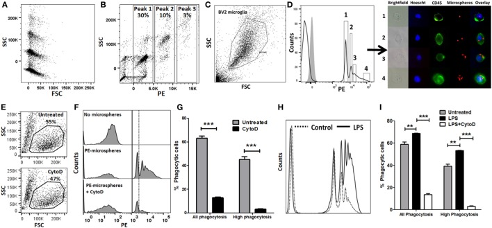
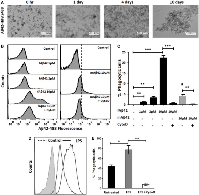
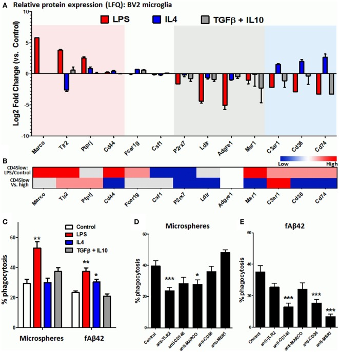
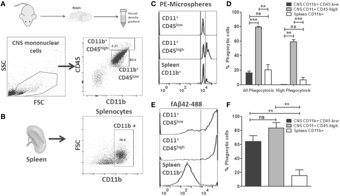
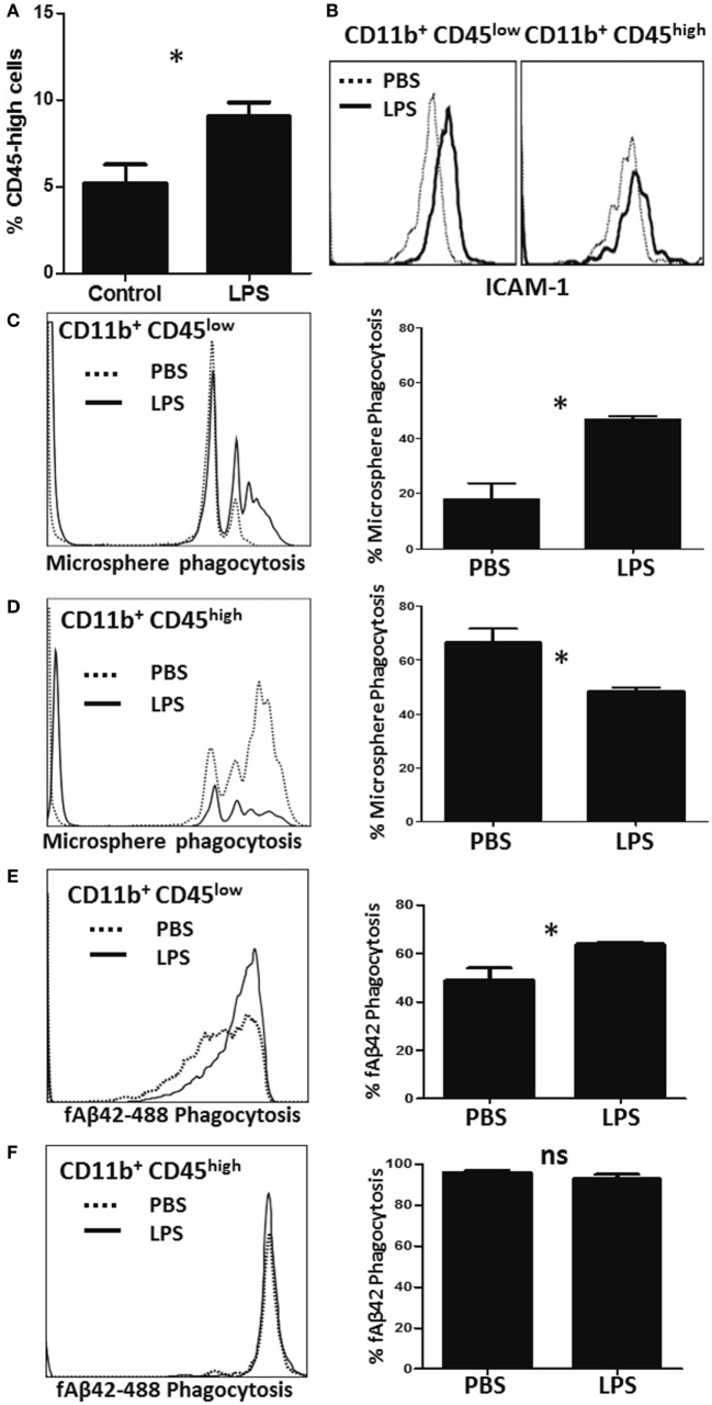
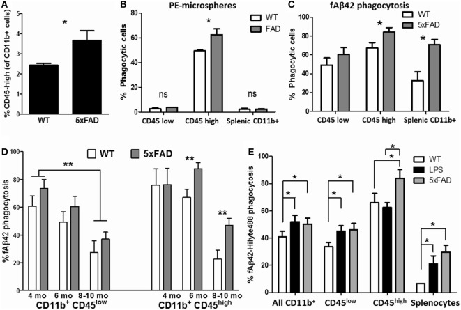
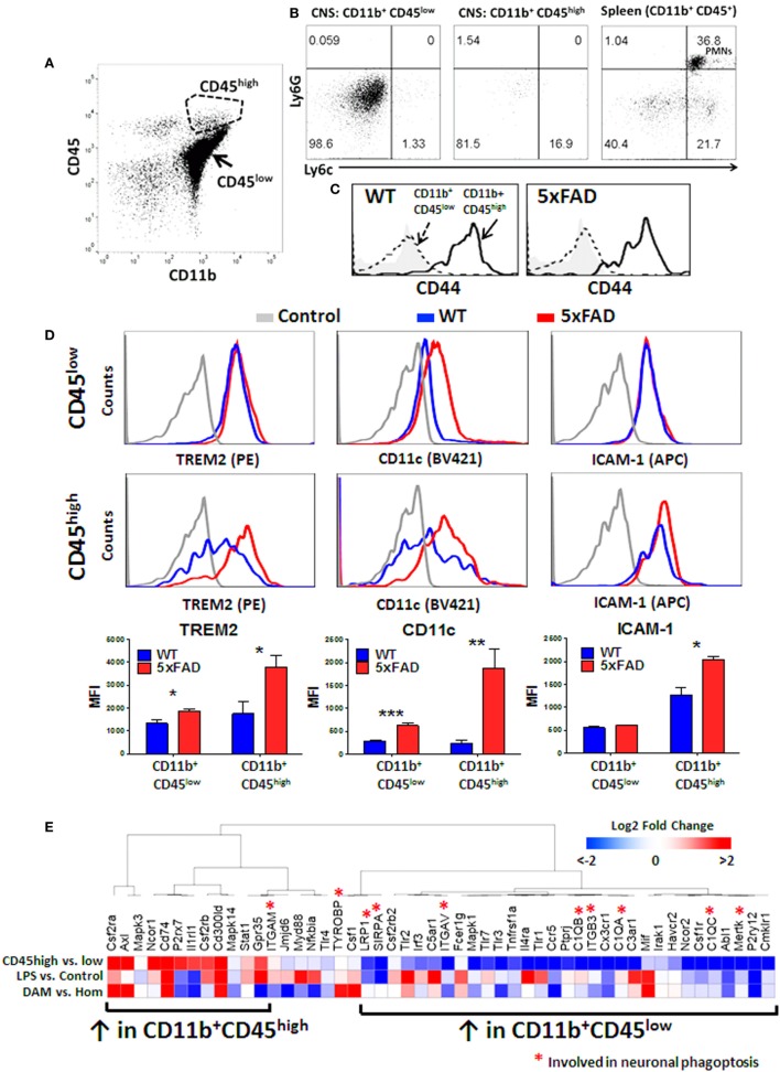
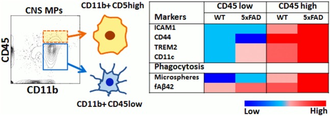
In the central nervous system (CNS), microglia are innate immune mononuclear phagocytes (CNS MPs) that can phagocytose infectious particles, apoptotic cells, neurons and pathological protein aggregates such as Aβ in Alzheimer’s disease (AD). While CD11b+CD45low microglia account for the majority of CNS MPs, a small population of CD11b+CD45high CNS MPs is also recognized in AD that surround Aβ plaques. These transcriptionally-distinct and pathologically-relevant CD45high cells have unclear origin and undefined phagocytic characteristics. We have comprehensively validated rapid flow cytometric assays of bulk-phase and amyloid β fibril (fAβ) phagocytosis and applied these to study acutely isolated CNS MPs. Using these methods, we provide novel insights into differential abilities of CD11b+ CD45low and CD45high CNS MPs to phagocytose macroparticles and fAβ under normal, acute and chronic neuroinflammatory states. CD45high CNS MPs also highly upregulate TREM2, CD11c and several disease-associated-microglia (DAM) signature genes, and have a higher phagocytic capacity for Aβ as compared to CD45low microglia in the 5xFAD mouse model of AD that becomes more apparent with ageing. Our data suggest an overall pro-phagocytic and protective role for CD11b+CD45high CNS MPs in neurodegeneration, which if promoted, could be beneficial.
Overview
The claims and statistics of this paper haven’t been checked yet.
Run the check — free, no account needed. It reads the open-access text and checks the sources it cites.
Integrity signals
Registry
Cites
Statistics
Figures
Verification
Claims the paper makes
The claims and statistics of this paper haven’t been checked yet.
Run the check — free, no account needed. It reads the open-access text and checks the sources it cites.
Claims other papers make about this one
Each is checked against this paper’s own text: does it say what the citing paper says it does?
In the citing paper
“We have found that Cytochalasin D inhibits the second peak of fluorescence in this assay, suggesting that the first peak represent non-specific binding of Aβ42 to cells while the second peak represents true uptake in an actin-dependent manner [31].”
From Identification and therapeutic modulation of a pro-inflammatory subset of disease-associated-microglia in Alzheimer’s disease (2018) · cites this paper as [31]
Self-check
Percentages, sums and reported differences re-computed from the paper’s own numbers.
The self-check needs the paper’s full text.
Citations
Cites
Amyloid oligomers exacerbate tau pathology in a mouse model of tauopathy
Selenica, M.-L. B., Brownlow, M., Jimenez, J. P., Lee, D. C., Pena, G., Dickey, C. A., Gordon, M. N., & Morgan, D. (2013) · Neurodegenerative Diseases · doi:10.1159/000337230 · Cited by 10,359
A Unique Microglia Type Associated with Restricting Development of Alzheimer’s Disease
Keren-Shaul, H., Spinrad, A., Weiner, A., Matcovitch-Natan, O., Dvir-Szternfeld, R., Ulland, T. K., David, E., Baruch, K., Lara-Astaiso, D., Toth, B., et al. (2017) · Cell · doi:10.1016/j.cell.2017.05.018 · Cited by 5,493
Cox, J., Hein, M. Y., Luber, C. A., Paron, I., Nagaraj, N., & Mann, M. (2014) · Molecular & Cellular Proteomics · doi:10.1074/mcp.m113.031591 · Cited by 5,180
Oakley, H., Cole, S. L., Logan, S., Maus, E., Shao, P., Craft, J., Guillozet-Bongaarts, A., Ohno, M., Disterhoft, J., Van Eldik, L., et al. (2006) · The Journal of Neuroscience · doi:10.1523/jneurosci.1202-06.2006 · Cited by 3,530 · cited 2×
“Therefore, we studied phagocytic profiles of acutely isolated CNS MPs in the 5xFAD mouse model of AD pathology in which rapid accumulation of Aβ plaques begins at 3 months of age followed by neurodegeneration and neurobehavioral deficits by 8–10 months of age (59).”
BackgroundGene Ontology Consortium: going forward
The Gene Ontology Consortium (2015) · Nucleic Acids Research · doi:10.1093/nar/gku1179 · Cited by 3,492
Hyman, B. T., Phelps, C. H., Beach, T. G., Bigio, E. H., Cairns, N. J., Carrillo, M. C., Dickson, D. W., Duyckaerts, C., Frosch, M. P., Masliah, E., et al. (2012) · Alzheimer's & Dementia · doi:10.1016/j.jalz.2011.10.007 · Cited by 3,092
Variant of TREM2 Associated with the Risk of Alzheimer's Disease
Jonsson, T., Stefansson, H., Steinberg, S., Jonsdottir, I., Jonsson, P. V., Snaedal, J., Bjornsson, S., Huttenlocher, J., Levey, A. I., Lah, J. J., et al. (2013) · New England Journal of Medicine · doi:10.1056/nejmoa1211103 · Cited by 2,558
Self-citationDifferential Roles of M1 and M2 Microglia in Neurodegenerative Diseases
Tang, Y., & Le, W. (2016) · Molecular Neurobiology · doi:10.1007/s12035-014-9070-5 · Cited by 2,190
Systemic LPS causes chronic neuroinflammation and progressive neurodegeneration
Qin, L., Wu, X., Block, M. L., Liu, Y., Breese, G. R., Hong, J., Knapp, D. J., & Crews, F. T. (2007) · Glia · doi:10.1002/glia.20467 · Cited by 2,144
Microglial M1/M2 polarization and metabolic states
Orihuela, R., McPherson, C. A., & Harry, G. J. (2016) · British Journal of Pharmacology · doi:10.1111/bph.13139 · Cited by 2,070
Cited by
Sorted by citations
Items 101–128 of 128
Gcm: a novel anti-inflammatory transcriptional cascade conserved from flies to humansPreprint
Pavlidaki, A., Panic, R., Monticelli, S., Riet, C., Yuasa, Y., Cattenoz, P. B., Nait‐Oumesmar, B., & Giangrande, A. (2022) · bioRxiv (Cold Spring Harbor Laboratory) · doi:10.1101/2022.05.29.493864 · Cited by 2
Morphofunctional changes in the neuronal environment in suicide
Кислов, М. А., Trusova, D. S., Николаевич, К. К., Zhiganova, M. S., & Maksimov, A. V. (2023) · Russian Journal of Forensic Medicine · doi:10.17816/fm723 · Cited by 1
Galectin-3 shapes microglial phenotype through endogenous and exogenous mechanisms
Camprubí-Ferrer, L., Yang, Y., Fernández‐Calle, R., Boza‐Serrano, A., García‐Revilla, J., Frontiñán-Rubio, J., & Deierborg, T. (2025) · Frontiers in Cellular Neuroscience · doi:10.3389/fncel.2025.1729776 · Cited by 1
Marques, E. T. A., Demers, M., D’Aiuto, L., Castanha, P. M. S., Yeung, J., Wood, J. A., Chowdari, K. V., Zheng, W., Yolken, R. H., & Nimgaonkar, V. L. (2022) · Current topics in behavioral neurosciences · doi:10.1007/7854_2022_383 · Cited by 1
Quantification of Microglial Engulfment of Synaptic Material Using Flow Cytometry
Ugursu, B., & Wolf, S. A. (2024) · Journal of Visualized Experiments · doi:10.3791/66639 · Cited by 1
Consistent specificity and efficiency of tamoxifen-mediated cre induction across agesPreprint
Kellogg, C. M., Pham, K. D., Ko, S., Cox, J. E. J., Machalinski, A. H., Stout, M. B., Sharpe, A. L., Beckstead, M. J., Chucair‐Elliott, A. J., Ocañas, S. R., et al. (2023) · bioRxiv (Cold Spring Harbor Laboratory) · doi:10.1101/2023.09.19.558482 · Cited by 1
Open Practices DisclosurePreprint
Chatard, A. (2017) · OSF Preprints (OSF Preprints)
Sudwarts, A., Ramesha, S., Gao, T., Ponnusamy, M., Wang, S., Hansen, M. T., Kozlova, A., Bitarafan, S., Kumar, P., Beaulieu‐Abdelahad, D., et al. (2023) · Figshare · doi:10.6084/m9.figshare.22609114
Self-citationUgursu, B., & Wolf, S. A. (2024) · doi:10.3791/66639-v
Sánchez-García, M. A., Lara-Ureña, N., March‐Díaz, R., Luis, C. O. S., Quiñones-Cañete, S., Barba-Reyes, J. M., Cabello-Rivera, D., Muñoz-Cabello, A. M., Mora-Romero, B., Romero‐Molina, C., et al. (2024) · bioRxiv (Cold Spring Harbor Laboratory) · doi:10.1101/2024.10.01.616066
Sudwarts, A., Ramesha, S., Gao, T., Ponnusamy, M., Wang, S., Hansen, M. T., Kozlova, A., Bitarafan, S., Kumar, P., Beaulieu‐Abdelahad, D., et al. (2022) · Figshare · doi:10.6084/m9.figshare.19728376
Self-citationSudwarts, A., Ramesha, S., Gao, T., Ponnusamy, M., Wang, S., Hansen, M. T., Kozlova, A., Bitarafan, S., Kumar, P., Beaulieu‐Abdelahad, D., et al. (2022) · Figshare · doi:10.6084/m9.figshare.19728382
Self-citationSudwarts, A., Ramesha, S., Gao, T., Ponnusamy, M., Wang, S., Hansen, M. T., Kozlova, A., Bitarafan, S., Kumar, P., Beaulieu‐Abdelahad, D., et al. (2022) · Figshare · doi:10.6084/m9.figshare.19728391
Self-citationSudwarts, A., Ramesha, S., Gao, T., Ponnusamy, M., Wang, S., Hansen, M. T., Kozlova, A., Bitarafan, S., Kumar, P., Beaulieu‐Abdelahad, D., et al. (2022) · Figshare · doi:10.6084/m9.figshare.19728373
Self-citationJu, H., Park, K. W., Kim, I.-D., Cave, J. W., & Cho, S. (2022) · Figshare · doi:10.6084/m9.figshare.20335340
Sudwarts, A., Ramesha, S., Gao, T., Ponnusamy, M., Wang, S., Hansen, M. T., Kozlova, A., Bitarafan, S., Kumar, P., Beaulieu‐Abdelahad, D., et al. (2022) · Figshare · doi:10.6084/m9.figshare.19728388
Self-citationSudwarts, A., Ramesha, S., Gao, T., Ponnusamy, M., Wang, S., Hansen, M. T., Kozlova, A., Bitarafan, S., Kumar, P., Beaulieu‐Abdelahad, D., et al. (2022) · Figshare · doi:10.6084/m9.figshare.19728385
Self-citationHonarpisheh, P., Lee, J., Banerjee, A., Blasco-Conesa, M. P., Honarpisheh, P., d’Aigle, J., Mamun, A. A., Ritzel, R. M., Chauhan, A., Ganesh, B. P., et al. (2020) · Figshare · doi:10.6084/m9.figshare.13316858
Sudwarts, A., Ramesha, S., Gao, T., Ponnusamy, M., Wang, S., Hansen, M. T., Kozlova, A., Bitarafan, S., Kumar, P., Beaulieu‐Abdelahad, D., et al. (2022) · Figshare · doi:10.6084/m9.figshare.19728379
Self-citationSudwarts, A., Ramesha, S., Gao, T., Ponnusamy, M., Wang, S., Hansen, M. T., Kozlova, A., Bitarafan, S., Kumar, P., Beaulieu‐Abdelahad, D., et al. (2022) · Figshare · doi:10.6084/m9.figshare.19728370
Self-citationJu, H., Park, K. W., Kim, I.-D., Cave, J. W., & Cho, S. (2022) · Figshare · doi:10.6084/m9.figshare.20335337
Sudwarts, A., Ramesha, S., Gao, T., Ponnusamy, M., Wang, S., Hansen, M. T., Kozlova, A., Bitarafan, S., Kumar, P., Beaulieu‐Abdelahad, D., et al. (2022) · Figshare · doi:10.6084/m9.figshare.19728367
Self-citationHonarpisheh, P., Lee, J., Banerjee, A., Blasco-Conesa, M. P., Honarpisheh, P., d’Aigle, J., Mamun, A. A., Ritzel, R. M., Chauhan, A., Ganesh, B. P., et al. (2020) · Figshare · doi:10.6084/m9.figshare.13316861
Kodali, M. C., Wang, Z., Lin, G., & Liao, F. (2026) · Research Square · doi:10.21203/rs.3.rs-8844780/v2
Li, R., Fan, J.-Q., & Lin, B. (2026) · Science Advances · doi:10.1126/sciadv.aea6467
Kim, D., Kim, Y. S., & Jeong, B. (2026) · Alzheimer s & Dementia Translational Research & Clinical Interventions · doi:10.1002/trc2.70288
Jaganathan, R., Vijayakumar, S., Chen, Y., Ye, J., Bakthavatchalam, P., Wankhar, D., Yang, C., Iyaswamy, A., & Li, M. (2026) · Neurobiology of Disease · doi:10.1016/j.nbd.2026.107555
Gaire, S., Gardner, R. S., Kyle, M. M., Qiu, X., Miller, W., Chin, L. S., & Zhao, L. (2026) · Molecular Neurobiology · doi:10.1007/s12035-026-06096-0
Metadata
- Authors
- Rangaraju, S., Raza, S. A., Li, N. X., Betarbet, R., Dammer, E. B., Duong, D., Lah, J. J., Seyfried, N. T., & Levey, A. I.
- Affiliations
- Emory University (US)
- Corresponding author
- Rangaraju, Srikant
- ORCID
- Venue
- Frontiers in Immunology
- Publisher
- Frontiers Media SA
- Published
- 2018-03-02
- Volume
- 9
- Pages
- 405-405
- ISSN
- 1664-3224 (ISSN-L)
- PMC
- PMC5840283
- OpenAlex
- W2792377614
- Cited by
- 126
- References
- 80
- Citations by year
- 2026: 9 · 2025: 8 · 2024: 12 · 2023: 18 · 2022: 35 · 2021: 13 · 2020: 19 · 2019: 8 · 2018: 3
- FWCI
- 4.82
- Type
- journal article
- Language
- en
- Open access
- gold
- doi.org
- Locations
- Frontiers in Immunology (publishedVersion, cc-by)
- Europe PMC (PubMed Central) (publishedVersion, cc-by)
- PubMed Central (publishedVersion, cc-by)
- PubMed (publishedVersion)
- DOAJ (DOAJ: Directory of Open Access Journals) (submittedVersion, cc-by-sa)
- Funders
- National Institutes of Health (P50 AG025688, NS099474-1, P30 NS055077, 1U01AG046161)
- National Science Foundation (CHE-1507932, NSF/DMR-BSF 1610377)
- American Brain Foundation
- Alzheimer’s Association
- Topics
- Life Sciences › Neuroscience › Neurology › Neuroinflammation and Neurodegeneration Mechanisms
- Life Sciences › Immunology and Microbiology › Immunology › Phagocytosis and Immune Regulation
- Life Sciences › Neuroscience › Cellular and Molecular Neuroscience › Alzheimer's disease research and treatments
- Keywords
- microglia, phagocytosis, amyloid-beta fibrils, Alzheimer's disease, disease-associated microglia, TREM2, CD11c, neuroinflammation, aging, 5xFAD mice, neurodegeneration
- MeSH terms
- Aging, Alzheimer Disease, Animals, Brain, Cell Line, Disease Models, Animal, Humans, Membrane Glycoproteins, Mice, Inbred C57BL, Mice, Mutant Strains, Phagocytes, Phagocytosis, Receptors, Immunologic, Mononuclear Phagocyte System, Amyloid beta-Peptides, Leukocyte Common Antigens, Microglia, Mice
About these checks
Registry, retraction, Cites and statistics results are computed, not judged, with one exception: a printed reference that no index lists is read once by a model before it counts as a citation error. Claims are extracted by a model and kept only when their quote appears verbatim in the text; each cited source is then checked, with a model’s help, for whether it says what the claim relies on. Those verdicts can be wrong — read the claim’s quote and the cited source before relying on one.
Statistics and arithmetic are recomputed, not judged.
Confidence bands are shown only for fields calibrated on our annotated test set. None is calibrated yet, so no band is shown.
What counts as a claim ›How we measure confidence ›How we check this ›
Are you an author, or do you see a problem with this page? Request a review
