

The DOI matches its Crossref record; 80 of the 80 references it cites were found in a registry or an index.
Differential Phagocytic Properties of CD45low Microglia and CD45high Brain Mononuclear Phagocytes—Activation and Age-Related Effects
Open accessAbstract
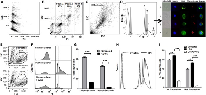
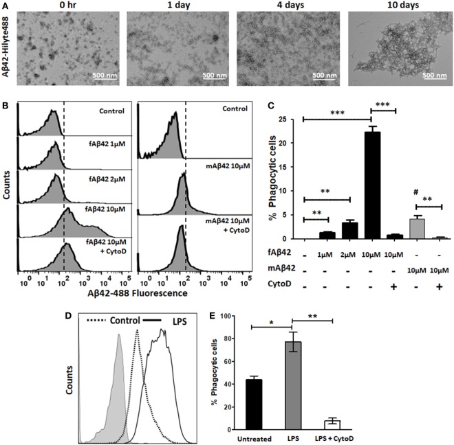
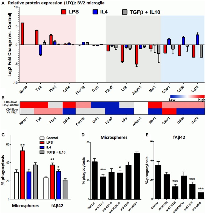
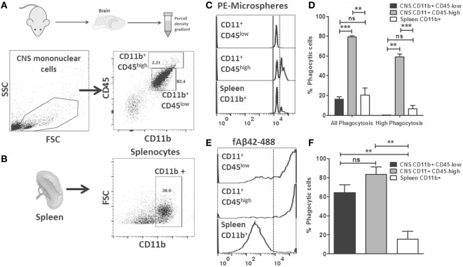
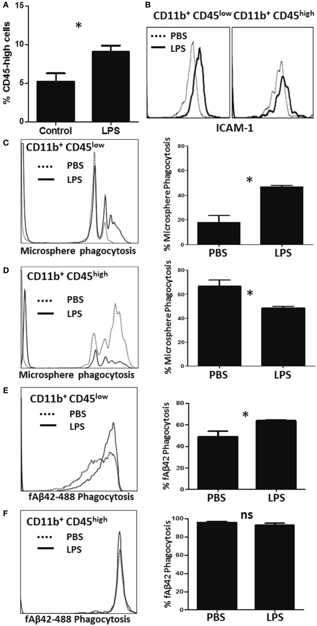
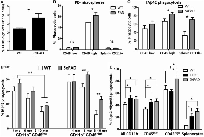
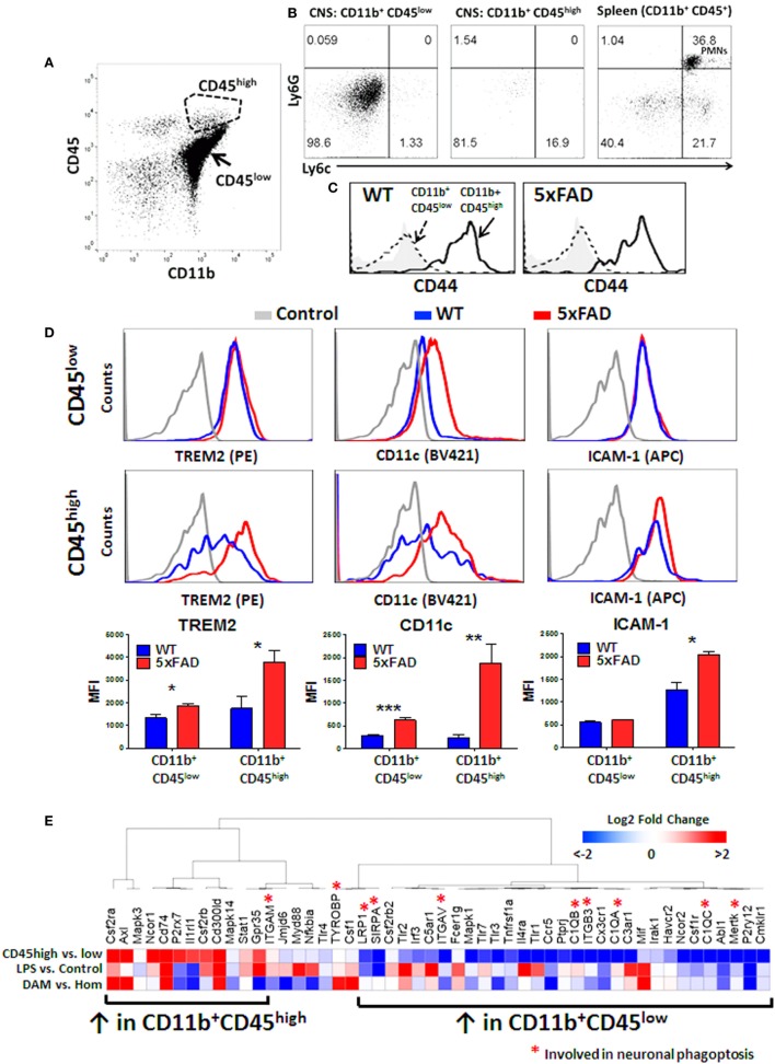
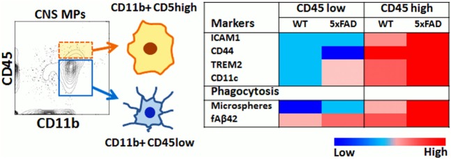
In the central nervous system (CNS), microglia are innate immune mononuclear phagocytes (CNS MPs) that can phagocytose infectious particles, apoptotic cells, neurons and pathological protein aggregates such as Aβ in Alzheimer’s disease (AD). While CD11b+CD45low microglia account for the majority of CNS MPs, a small population of CD11b+CD45high CNS MPs is also recognized in AD that surround Aβ plaques. These transcriptionally-distinct and pathologically-relevant CD45high cells have unclear origin and undefined phagocytic characteristics. We have comprehensively validated rapid flow cytometric assays of bulk-phase and amyloid β fibril (fAβ) phagocytosis and applied these to study acutely isolated CNS MPs. Using these methods, we provide novel insights into differential abilities of CD11b+ CD45low and CD45high CNS MPs to phagocytose macroparticles and fAβ under normal, acute and chronic neuroinflammatory states. CD45high CNS MPs also highly upregulate TREM2, CD11c and several disease-associated-microglia (DAM) signature genes, and have a higher phagocytic capacity for Aβ as compared to CD45low microglia in the 5xFAD mouse model of AD that becomes more apparent with ageing. Our data suggest an overall pro-phagocytic and protective role for CD11b+CD45high CNS MPs in neurodegeneration, which if promoted, could be beneficial.
Overview
The claims and statistics of this paper haven’t been checked yet.
Run the check — free, no account needed. It reads the open-access text and checks the sources it cites.
Integrity signals
Registry
Cites
Statistics
Figures
Verification
Claims the paper makes
The claims and statistics of this paper haven’t been checked yet.
Run the check — free, no account needed. It reads the open-access text and checks the sources it cites.
Claims other papers make about this one
Each is checked against this paper’s own text: does it say what the citing paper says it does?
In the citing paper
“We have found that Cytochalasin D inhibits the second peak of fluorescence in this assay, suggesting that the first peak represent non-specific binding of Aβ42 to cells while the second peak represents true uptake in an actin-dependent manner [31].”
From Identification and therapeutic modulation of a pro-inflammatory subset of disease-associated-microglia in Alzheimer’s disease (2018) · cites this paper as [31]
Self-check
Percentages, sums and reported differences re-computed from the paper’s own numbers.
The self-check needs the paper’s full text.
Citations
Cites
Amyloid oligomers exacerbate tau pathology in a mouse model of tauopathy
Selenica, M.-L. B., Brownlow, M., Jimenez, J. P., Lee, D. C., Pena, G., Dickey, C. A., Gordon, M. N., & Morgan, D. (2013) · Neurodegenerative Diseases · doi:10.1159/000337230 · Cited by 10,359
A Unique Microglia Type Associated with Restricting Development of Alzheimer’s Disease
Keren-Shaul, H., Spinrad, A., Weiner, A., Matcovitch-Natan, O., Dvir-Szternfeld, R., Ulland, T. K., David, E., Baruch, K., Lara-Astaiso, D., Toth, B., et al. (2017) · Cell · doi:10.1016/j.cell.2017.05.018 · Cited by 5,493
Cox, J., Hein, M. Y., Luber, C. A., Paron, I., Nagaraj, N., & Mann, M. (2014) · Molecular & Cellular Proteomics · doi:10.1074/mcp.m113.031591 · Cited by 5,180
Oakley, H., Cole, S. L., Logan, S., Maus, E., Shao, P., Craft, J., Guillozet-Bongaarts, A., Ohno, M., Disterhoft, J., Van Eldik, L., et al. (2006) · The Journal of Neuroscience · doi:10.1523/jneurosci.1202-06.2006 · Cited by 3,530 · cited 2×
“Therefore, we studied phagocytic profiles of acutely isolated CNS MPs in the 5xFAD mouse model of AD pathology in which rapid accumulation of Aβ plaques begins at 3 months of age followed by neurodegeneration and neurobehavioral deficits by 8–10 months of age (59).”
BackgroundGene Ontology Consortium: going forward
The Gene Ontology Consortium (2015) · Nucleic Acids Research · doi:10.1093/nar/gku1179 · Cited by 3,492
Hyman, B. T., Phelps, C. H., Beach, T. G., Bigio, E. H., Cairns, N. J., Carrillo, M. C., Dickson, D. W., Duyckaerts, C., Frosch, M. P., Masliah, E., et al. (2012) · Alzheimer's & Dementia · doi:10.1016/j.jalz.2011.10.007 · Cited by 3,092
Variant of TREM2 Associated with the Risk of Alzheimer's Disease
Jonsson, T., Stefansson, H., Steinberg, S., Jonsdottir, I., Jonsson, P. V., Snaedal, J., Bjornsson, S., Huttenlocher, J., Levey, A. I., Lah, J. J., et al. (2013) · New England Journal of Medicine · doi:10.1056/nejmoa1211103 · Cited by 2,558
Self-citationDifferential Roles of M1 and M2 Microglia in Neurodegenerative Diseases
Tang, Y., & Le, W. (2016) · Molecular Neurobiology · doi:10.1007/s12035-014-9070-5 · Cited by 2,190
Systemic LPS causes chronic neuroinflammation and progressive neurodegeneration
Qin, L., Wu, X., Block, M. L., Liu, Y., Breese, G. R., Hong, J., Knapp, D. J., & Crews, F. T. (2007) · Glia · doi:10.1002/glia.20467 · Cited by 2,144
Microglial M1/M2 polarization and metabolic states
Orihuela, R., McPherson, C. A., & Harry, G. J. (2016) · British Journal of Pharmacology · doi:10.1111/bph.13139 · Cited by 2,070
New tools for studying microglia in the mouse and human CNS
Bennett, M. L., Bennett, F. C., Liddelow, S. A., Ajami, B., Zamanian, J. L., Fernhoff, N. B., Mulinyawe, S. B., Bohlen, C. J., Adil, A., Tucker, A., et al. (2016) · Proceedings of the National Academy of Sciences · doi:10.1073/pnas.1525528113 · Cited by 1,804
Neuroinflammation and M2 Microglia: The Good, the Bad, and the Inflamed
Cherry, J. D., Olschowka, J. A., & O’Banion, M. K. (2014) · Journal of Neuroinflammation · doi:10.1186/1742-2094-11-98 · Cited by 1,748
TREM2 Lipid Sensing Sustains the Microglial Response in an Alzheimer’s Disease Model
Wang, Y., Cella, M., Mallinson, K., Ulrich, J. D., Young, K. L., Robinette, M. L., Gilfillan, S., Krishnan, G. M., Sudhakar, S., Zinselmeyer, B. H., et al. (2015) · Cell · doi:10.1016/j.cell.2015.01.049 · Cited by 1,736
A polarizing question: do M1 and M2 microglia exist?
Ransohoff, R. M. (2016) · Nature Neuroscience · doi:10.1038/nn.4338 · Cited by 1,573
The microglial sensome revealed by direct RNA sequencing
Hickman, S. E., Kingery, N. D., Ohsumi, T. K., Borowsky, M. L., Wang, L.-C., Means, T. K., & El Khoury, J. (2013) · Nature Neuroscience · doi:10.1038/nn.3554 · Cited by 1,511
An environment-dependent transcriptional network specifies human microglia identity
Gosselin, D., Skola, D., Coufal, N. G., Holtman, I. R., Schlachetzki, J. C. M., Sajti, E., Jaeger, B. N., O’Connor, C., Fitzpatrick, C., Pasillas, M. P., et al. (2017) · Science · doi:10.1126/science.aal3222 · Cited by 1,220
ARUK Consortium, Sims, R., GERAD/PERADES, C., van der Lee, S. J., Naj, A. C., Bellenguez, C., Badarinarayan, N., Jakobsdottir, J., Kunkle, B. W., Boland, A., et al. (2017) · Nature Genetics · doi:10.1038/ng.3916 · Cited by 1,012
Microglial Phagocytosis of Live Neurons
Brown, G. C., & Neher, J. J. (2014) · Nature Reviews Neuroscience · doi:10.1038/nrn3710 · Cited by 853
TREM2-mediated early microglial response limits diffusion and toxicity of amyloid plaques
Wang, Y., Ulland, T. K., Ulrich, J. D., Song, W., Tzaferis, J. A., Hole, J. T., Yuan, P., Mahan, T. E., Shi, Y., Gilfillan, S., et al. (2016) · Journal of Experimental Medicine · doi:10.1084/jem.20151948 · Cited by 840
Splenic nerve is required for cholinergic antiinflammatory pathway control of TNF in endotoxemia
Rosas‐Ballina, M., Ochani, M., Parrish, W. R., Ochani, K., Harris, Y., Huston, J. M., Chavan, S. S., & Tracey, K. J. (2008) · Proceedings of the National Academy of Sciences · doi:10.1073/pnas.0803237105 · Cited by 762
Phagocytosis and the actin cytoskeleton
May, R. C., & Machesky, L. M. (2001) · Journal of Cell Science · doi:10.1242/jcs.114.6.1061 · Cited by 715
Phagocytosis of Microglia in the Central Nervous System Diseases
Fu, R., Shen, Q., Xu, P., Luo, J. J., & Tang, Y. (2014) · Molecular Neurobiology · doi:10.1007/s12035-013-8620-6 · Cited by 687
Jay, T. R., Miller, C. M., Cheng, P. J., Graham, L. C., Bemiller, S., Broihier, M. L., Xu, G., Margevicius, D., Karlo, J. C., Sousa, G. L., et al. (2015) · Journal of Experimental Medicine · doi:10.1084/jem.20142322 · Cited by 682 · cited 2×
“who also found that 5xFAD CD45high CNS MPs selectively upregulated TREM2 expression in an age-dependent manner (69).”
BackgroundChhor, V., Charpentier, T. L., Lebon, S., Oré, M. V., Lara‐Celador, I., Josserand, J., Degos, V., Jacotot, E. D., Hagberg, H., Sävman, K., et al. (2013) · Brain Behavior and Immunity · doi:10.1016/j.bbi.2013.02.005 · Cited by 676
Friedman, B. A., Srinivasan, K., Ayalon, G., Meilandt, W. J., Lin, H., Huntley, M. A., Cao, Y., Lee, S.-H., Haddick, P. C. G., Ngu, H., et al. (2018) · Cell Reports · doi:10.1016/j.celrep.2017.12.066 · Cited by 662 · cited 1×
“inflammatory activated Microglial states Microglia and CNS MPs adopt a diverse and complex array of activation states with distinct transcriptomic profiles that are now being revealed by RNA sequencing studies (12, 37, 51).”
BackgroundChiu, I. M., Morimoto, E. T. A., Goodarzi, H., Liao, J. T., O’Keeffe, S., Phatnani, H. P., Muratet, M., Carroll, M. C., Levy, S., Tavazoie, S., et al. (2013) · Cell Reports · doi:10.1016/j.celrep.2013.06.018 · Cited by 651
Quantitative proteomics reveals subset-specific viral recognition in dendritic cells
Luber, C. A., Cox, J., Lauterbach, H., Fancke, B., Selbach, M., Tschopp, J., Akira, S., Wiegand, M., Hochrein, H., O'Keeffe, M., et al. (2010) · Immunity · doi:10.1016/j.immuni.2010.01.013 · Cited by 576
Janus-faced microglia: beneficial and detrimental consequences of microglial phagocytosis
Sierra, A., Abiega, O., Shahraz, A., & Neumann, H. (2013) · Frontiers in Cellular Neuroscience · doi:10.3389/fncel.2013.00006 · Cited by 574
A Cell Surface Receptor Complex for Fibrillar β-Amyloid Mediates Microglial Activation
Bamberger, M. E., Harris, M. E., McDonald, D. R., Husemann, J., & Landreth, G. E. (2003) · The Journal of Neuroscience · doi:10.1523/jneurosci.23-07-02665.2003 · Cited by 501
Koenigsknecht-Talboo, J., & Landreth, G. E. (2005) · The Journal of Neuroscience · doi:10.1523/jneurosci.1808-05.2005 · Cited by 477
Seyfried, N. T., Dammer, E. B., Swarup, V., Nandakumar, D., Duong, D. M., Yin, L., Deng, Q., Nguyen, T., Hales, C. M., Wingo, T., et al. (2017) · Cell Systems · doi:10.1016/j.cels.2016.11.006 · Cited by 477
Self-citationA role for TREM2 ligands in the phagocytosis of apoptotic neuronal cells by microglia
Hsieh, C. L., Koike, M., Spusta, S. C., Niemi, E. C., Yenari, M., Nakamura, M. C., & Seaman, W. E. (2009) · Journal of Neurochemistry · doi:10.1111/j.1471-4159.2009.06042.x · Cited by 475
Microglial Phagocytosis of Fibrillar β-Amyloid through a β1Integrin-Dependent Mechanism
Koenigsknecht-Talboo, J., & Landreth, G. E. (2004) · Journal of Neuroscience · doi:10.1523/jneurosci.2557-04.2004 · Cited by 457
High-dimensional, single-cell characterization of the brain's immune compartment
Korin, B., Ben-Shaanan, T. L., Schiller, M., Dubovik, T., Azulay‐Debby, H., Boshnak, N., Koren, T., & Rolls, A. (2017) · Nature Neuroscience · doi:10.1038/nn.4610 · Cited by 438
Pavlov, V. A., Parrish, W. R., Rosas‐Ballina, M., Ochani, M., Gallowitsch‐Puerta, M., Ochani, K., Chavan, S. S., Al‐Abed, Y., & Tracey, K. J. (2008) · Brain Behavior and Immunity · doi:10.1016/j.bbi.2008.06.011 · Cited by 437
Chen, Z., Jalabi, W., Shpargel, K. B., Farabaugh, K. T., Dutta, R., Yin, X., Kidd, G. J., Bergmann, C. C., Stohlman, S. A., & Trapp, B. D. (2012) · The Journal of Neuroscience · doi:10.1523/jneurosci.0730-12.2012 · Cited by 432
Sleep Loss Promotes Astrocytic Phagocytosis and Microglial Activation in Mouse Cerebral Cortex
Bellesi, M., de Vivo, L., Chini, M., Gilli, F., Tononi, G., & Cirelli, C. (2017) · Journal of Neuroscience · doi:10.1523/jneurosci.3981-16.2017 · Cited by 431
Reactive astrocytes function as phagocytes after brain ischemia via ABCA1-mediated pathway
Morizawa, Y. M., Hirayama, Y., Ohno, N., Shibata, S., Shigetomi, E., Sui, Y., Nabekura, J., Sato, K., Okajima, F., Takebayashi, H., et al. (2017) · Nature Communications · doi:10.1038/s41467-017-00037-1 · Cited by 424
Casella, J. F., Flanagan, M. D., & Lin, S. (1981) · Nature · doi:10.1038/293302a0 · Cited by 423
Stansley, B., Post, J., & Hensley, K. (2012) · Journal of Neuroinflammation · doi:10.1186/1742-2094-9-115 · Cited by 419
TREM2 variants: new keys to decipher Alzheimer disease pathogenesis
Colonna, M., & Wang, Y. (2016) · Nature Reviews Neuroscience · doi:10.1038/nrn.2016.7 · Cited by 418
Marsh, S. E., Abud, E. M., Lakatos, A., Karimzadeh, A., Yeung, S. T., Davtyan, H., Fote, G. M., Lau, L., Weinger, J. G., Lane, T. E., et al. (2016) · Proceedings of the National Academy of Sciences · doi:10.1073/pnas.1525466113 · Cited by 411
Fadok, V. A., Warner, M. L., Bratton, D. L., & Henson, P. M. (1998) · PubMed · Cited by 406
Monocyte subpopulations and their differentiation patterns during infection
Strauss‐Ayali, D., Conrad, S. M., & Mosser, D. M. (2007) · Journal of Leukocyte Biology · doi:10.1189/jlb.0307191 · Cited by 359
Fadok, V. A., Warner, M. L., Bratton, D. L., & Henson, P. M. (1998) · The Journal of Immunology · doi:10.4049/jimmunol.161.11.6250 · Cited by 353
Microglial Physiology and Pathophysiology: Insights from Genome-wide Transcriptional Profiling
Crotti, A., & Ransohoff, R. M. (2016) · Immunity · doi:10.1016/j.immuni.2016.02.013 · Cited by 323
Jiang, T., Tan, L., Zhu, X.-C., Zhang, Q.-Q., Cao, L., Tan, M.-S., Gu, L.-Z., Wang, H.-F., Ding, Z.-Z., Zhang, Y.-D., et al. (2014) · Neuropsychopharmacology · doi:10.1038/npp.2014.164 · Cited by 320
Is the class A macrophage scavenger receptor (SR-A) multifunctional? — The mouse’s tale
Platt, N., & Gordon, S. (2001) · Journal of Clinical Investigation · doi:10.1172/jci13903 · Cited by 313
Is the class A macrophage scavenger receptor (SR-A) multifunctional? — The mouse’s tale
Platt, N., & Gordon, S. (2001) · Journal of Clinical Investigation · doi:10.1172/jci200113903 · Cited by 265
Differences in the Mode of Phagocytosis with Fc and C3 Receptors in Macrophages
Kaplan, G. (1977) · Scandinavian Journal of Immunology · doi:10.1111/j.1365-3083.1977.tb02153.x · Cited by 244
Intrahippocampal LPS injections reduce Aβ load in APP+PS1 transgenic mice
DiCarlo, G. (2001) · Neurobiology of Aging · doi:10.1016/s0197-4580(01)00292-5 · Cited by 223
Brain macrophages: questions of origin and interrelationship
Jordan, F. L., & Thomas, W. E. (1988) · Brain Research Reviews · doi:10.1016/0165-0173(88)90019-7 · Cited by 222
Arredouani, M. S., Palecanda, A., Koziel, H., Huang, Y.-C., Imrich, A., Sulahian, T. H., Ning, Y. Y., Yang, Z., Pikkarainen, T., Sankala, M., et al. (2005) · The Journal of Immunology · doi:10.4049/jimmunol.175.9.6058 · Cited by 222
MFG-E8 Mediates Primary Phagocytosis of Viable Neurons during Neuroinflammation
Fricker, M., Neher, J. J., Zhao, J.-W., Théry, C., Tolkovsky, A. M., & Brown, G. C. (2012) · The Journal of Neuroscience · doi:10.1523/jneurosci.4837-11.2012 · Cited by 218
Coppolino, M. G., Krause, M., Hagendorff, P., Monner, D. A., Trimble, W. S., Grinstein, S., Wehland, J., & Sechi, A. S. (2001) · Journal of Cell Science · doi:10.1242/jcs.114.23.4307 · Cited by 200
Das, A., Kim, S. H., Arifuzzaman, S., Yoon, T., Chai, J. C., Lee, Y. S., Park, K. S., Jung, K. H., & Chai, Y. G. (2016) · Journal of Neuroinflammation · doi:10.1186/s12974-016-0644-1 · Cited by 183
Greenberg, S., el Khoury, J., di Virgilio, F., Kaplan, E. M., & Silverstein, S. C. (1991) · The Journal of cell biology · doi:10.1083/jcb.113.4.757 · Cited by 171
Genetics ignite focus on microglial inflammation in Alzheimer’s disease
Malik, M., Parikh, I., Vasquez, J. B., Smith, C., Tai, L., Bu, G., LaDu, M. J., Fardo, D. W., Rebeck, G. W., & Estus, S. (2015) · Molecular Neurodegeneration · doi:10.1186/s13024-015-0048-1 · Cited by 161
Primary Phagocytosis of Neurons by Inflamed Microglia: Potential Roles in Neurodegeneration
Neher, J. J., Neniškytė, U., & Brown, G. C. (2012) · Frontiers in Pharmacology · doi:10.3389/fphar.2012.00027 · Cited by 157
Nuclear Receptors License Phagocytosis by Trem2+Myeloid Cells in Mouse Models of Alzheimer's Disease
Savage, J. C., Jay, T., Goduni, E., Quigley, C., Mariani, M. M., Malm, T., Ransohoff, R. M., Lamb, B. T., & Landreth, G. E. (2015) · The Journal of Neuroscience · doi:10.1523/jneurosci.4586-14.2015 · Cited by 154 · cited 1×
“The presence of CD45+ TREM2+ CNS MPs around Aβ plaques in AD mouse models suggest a pro-phagocytic and Aβ-clearing role for CD45high cells (68), but direct functional characterization of phagocytic properties of CD45high CNS MPs, in contrast to CD45low CNS MPs, has not been performed.”
BackgroundFricker, M., Oliva-Martín, M. J., & Brown, G. C. (2012) · Journal of Neuroinflammation · doi:10.1186/1742-2094-9-196 · Cited by 137
Woo, M., Yang, J., Beltran, C., & Cho, S. (2016) · Journal of Biological Chemistry · doi:10.1074/jbc.m116.750018 · Cited by 136
Ryan, T. M., Caine, J. M., Mertens, H. D. T., Kirby, N. M., Nigro, J., Breheney, K., Waddington, L. J., Streltsov, V. A., Curtain, C. C., Masters, C. L., et al. (2013) · PeerJ · doi:10.7717/peerj.73 · Cited by 133
Tse, S. M. L., Furuya, W., Gold, E., Schreiber, A. D., Sandvig, K., Inman, R. D., & Grinstein, S. (2003) · Journal of Biological Chemistry · doi:10.1074/jbc.m207966200 · Cited by 124
Isolation of highly enriched primary human microglia for functional studies
Rustenhoven, J., Park, T. I.-H., Schweder, P., Scotter, J., Correia, J., Smith, A. M., Gibbons, H. M., Oldfield, R. L., Bergin, P. S., Mee, E. W., et al. (2016) · Scientific Reports · doi:10.1038/srep19371 · Cited by 99
Multiparameter Flow Cytometry: Advances in High Resolution Analysis
O'Donnell, E. A., Ernst, D. N., & Hingorani, R. (2013) · Immune Network · doi:10.4110/in.2013.13.2.43 · Cited by 95
Microglia display modest phagocytic capacity for extracellular tau oligomers
Majerova, P., Zilkova, M., Kazmerova, Z., Kovac, A., Paholikova, K., Kovacech, B., Zilka, N., & Novak, M. (2014) · Journal of Neuroinflammation · doi:10.1186/s12974-014-0161-z · Cited by 90
Takahashi, T., Takahashi, K., Mernaugh, R. L., Tsuboi, N., Liu, H., & Daniel, T. O. (2006) · Blood · doi:10.1182/blood-2005-10-4296 · Cited by 82
Single-cell RNA sequencing identifies distinct mouse medial ganglionic eminence cell types
Chen, Y.-J. J., Friedman, B. A., Ha, C., Durinck, S., Liu, J., Rubenstein, J. L. R., Seshagiri, S., & Modrusan, Z. D. (2017) · Scientific Reports · doi:10.1038/srep45656 · Cited by 80
Migration of blood cells to β-amyloid plaques in Alzheimer's disease
Hohsfield, L. A., & Humpel, C. (2015) · Experimental Gerontology · doi:10.1016/j.exger.2015.03.002 · Cited by 79
Biswas, S., Zimman, A., Gao, D., Byzova, T. V., & Podrez, E. A. (2017) · Circulation Research · doi:10.1161/circresaha.117.311069 · Cited by 77
Noristani, H. N., Gerber, Y. N., Sabourin, J.-C., Corre, M. L., Lonjon, N., Mestre‐Francés, N., Hirbec, H., & Perrin, F. E. (2017) · Frontiers in Molecular Neuroscience · doi:10.3389/fnmol.2017.00090 · Cited by 75
Rangaraju, S., Raza, S. A., Pennati, A., Deng, Q., Dammer, E. B., Duong, D., Pennington, M. W., Tansey, M. G., Lah, J. J., Betarbet, R., et al. (2017) · Journal of Neuroinflammation · doi:10.1186/s12974-017-0906-6 · Cited by 75
Self-citationBell‐Temin, H., Culver‐Cochran, A. E., Chaput, D., Carlson, C., Kuehl, M. N., Burkhardt, B. R., Bickford, P. C., Liu, B., & Stevens, S. M. (2015) · Molecular & Cellular Proteomics · doi:10.1074/mcp.m115.053926 · Cited by 62
P2Y6 Receptor-Mediated Microglial Phagocytosis in Radiation-Induced Brain Injury
Xu, Y., Hu, W., Liu, Y., Xu, P., Li, Z., Wu, R., Shi, X., & Tang, Y. (2015) · Molecular Neurobiology · doi:10.1007/s12035-015-9282-3 · Cited by 61
Analysis of the Impact of CD200 on Phagocytosis
Lyons, A., Minogue, A. M., Jones, R. S., Fitzpatrick, O. M., Noonan, J., Campbell, V. A., & Lynch, M. A. (2016) · Molecular Neurobiology · doi:10.1007/s12035-016-0223-6 · Cited by 45
Imaging Microglia in Live Brain Slices and Slice CulturesPreprint
Kurpius, D., & Dailey, M. E. (2007) · Cold Spring Harbor Protocols · doi:10.1101/pdb.prot4852 · Cited by 30
Imaging Microglia in Brain Slices and Slice CulturesPreprint
Dailey, M. E., Eyo, U. B., Fuller, L. C., Hass, J., & Kurpius, D. (2013) · Cold Spring Harbor Protocols · doi:10.1101/pdb.prot079483 · Cited by 29
αvβ5 Integrin/FAK/PGC-1α Pathway Confers Protective Effects on Retinal Pigment Epithelium
Roggia, M. F., & Ueta, T. (2015) · PLoS ONE · doi:10.1371/journal.pone.0134870 · Cited by 26
Mutzke, E., Chomyshyn, E., Nguyen, K. C., Blahoianu, M., & Tayabali, A. F. (2015) · Analytical Biochemistry · doi:10.1016/j.ab.2015.04.034 · Cited by 13
Cited by
Sorted by citations
Rangaraju, S., Dammer, E. B., Raza, S. A., Rathakrishnan, P., Xiao, H., Gao, T., Duong, D. M., Pennington, M. W., Lah, J. J., Seyfried, N. T., et al. (2018) · Molecular Neurodegeneration · doi:10.1186/s13024-018-0254-8 · Cited by 430
Self-citationMicroglia: Agents of the CNS Pro-Inflammatory Response
Rodríguez-Gómez, J. A., Kavanagh, E., Engskog-Vlachos, P., Engskog, M. K. R., Herrera, A. J., Espinosa-Oliva, A. M., Joseph, B., Hajji, N., Venero, J. L., & Burguillos, M. A. (2020) · Cells · doi:10.3390/cells9071717 · Cited by 365
The choroid plexus links innate immunity to CSF dysregulation in hydrocephalus
Robert, S. M., Reeves, B. C., Kiziltug, E., Duy, P. Q., Karimy, J. K., Mansuri, M. S., Marlier, A., Allington, G., Greenberg, A. B. W., DeSpenza, T., et al. (2023) · Cell · doi:10.1016/j.cell.2023.01.017 · Cited by 162
Fairley, L. H., Lai, K. O., Wong, J. H., Chong, W. J., Vincent, A. S., D’Agostino, G. A., Wu, X., Naik, R. R., Jayaraman, A., Langley, S. R., et al. (2023) · Proceedings of the National Academy of Sciences · doi:10.1073/pnas.2209177120 · Cited by 132
SARS-CoV-2 Infection of Microglia Elicits Proinflammatory Activation and Apoptotic Cell Death
Jeong, G. U., Lyu, J., Kim, K., Chung, Y. C., Yoon, G. Y., Lee, S., Hwang, I., Shin, W.-H., Ko, J., Lee, J., et al. (2022) · Microbiology Spectrum · doi:10.1128/spectrum.01091-22 · Cited by 126
Metadata
- Authors
- Rangaraju, S., Raza, S. A., Li, N. X., Betarbet, R., Dammer, E. B., Duong, D., Lah, J. J., Seyfried, N. T., & Levey, A. I.
- Affiliations
- Emory University (US)
- Corresponding author
- Rangaraju, Srikant
- ORCID
- Venue
- Frontiers in Immunology
- Publisher
- Frontiers Media SA
- Published
- 2018-03-02
- Volume
- 9
- Pages
- 405-405
- ISSN
- 1664-3224 (ISSN-L)
- PMC
- PMC5840283
- OpenAlex
- W2792377614
- Cited by
- 126
- References
- 80
- Citations by year
- 2026: 9 · 2025: 8 · 2024: 12 · 2023: 18 · 2022: 35 · 2021: 13 · 2020: 19 · 2019: 8 · 2018: 3
- FWCI
- 4.82
- Type
- journal article
- Language
- en
- Open access
- gold
- doi.org
- Locations
- Frontiers in Immunology (publishedVersion, cc-by)
- Europe PMC (PubMed Central) (publishedVersion, cc-by)
- PubMed Central (publishedVersion, cc-by)
- PubMed (publishedVersion)
- DOAJ (DOAJ: Directory of Open Access Journals) (submittedVersion, cc-by-sa)
- Funders
- National Institutes of Health (P50 AG025688, NS099474-1, P30 NS055077, 1U01AG046161)
- National Science Foundation (CHE-1507932, NSF/DMR-BSF 1610377)
- American Brain Foundation
- Alzheimer’s Association
- Topics
- Life Sciences › Neuroscience › Neurology › Neuroinflammation and Neurodegeneration Mechanisms
- Life Sciences › Immunology and Microbiology › Immunology › Phagocytosis and Immune Regulation
- Life Sciences › Neuroscience › Cellular and Molecular Neuroscience › Alzheimer's disease research and treatments
- Keywords
- microglia, phagocytosis, amyloid-beta fibrils, Alzheimer's disease, disease-associated microglia, TREM2, CD11c, neuroinflammation, aging, 5xFAD mice, neurodegeneration
- MeSH terms
- Aging, Alzheimer Disease, Animals, Brain, Cell Line, Disease Models, Animal, Humans, Membrane Glycoproteins, Mice, Inbred C57BL, Mice, Mutant Strains, Phagocytes, Phagocytosis, Receptors, Immunologic, Mononuclear Phagocyte System, Amyloid beta-Peptides, Leukocyte Common Antigens, Microglia, Mice
About these checks
Registry, retraction, Cites and statistics results are computed, not judged, with one exception: a printed reference that no index lists is read once by a model before it counts as a citation error. Claims are extracted by a model and kept only when their quote appears verbatim in the text; each cited source is then checked, with a model’s help, for whether it says what the claim relies on. Those verdicts can be wrong — read the claim’s quote and the cited source before relying on one.
Statistics and arithmetic are recomputed, not judged.
Confidence bands are shown only for fields calibrated on our annotated test set. None is calibrated yet, so no band is shown.
What counts as a claim ›How we measure confidence ›How we check this ›
Are you an author, or do you see a problem with this page? Request a review
