

The DOI matches its Crossref record; 80 of the 80 references it cites were found in a registry or an index.
Differential Phagocytic Properties of CD45low Microglia and CD45high Brain Mononuclear Phagocytes—Activation and Age-Related Effects
Open accessAbstract
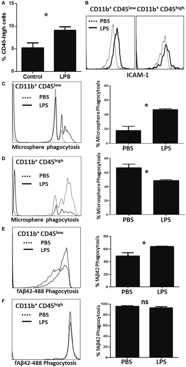
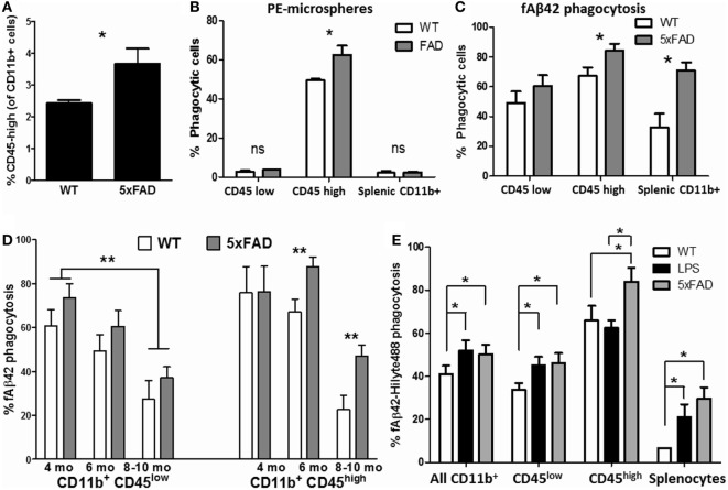
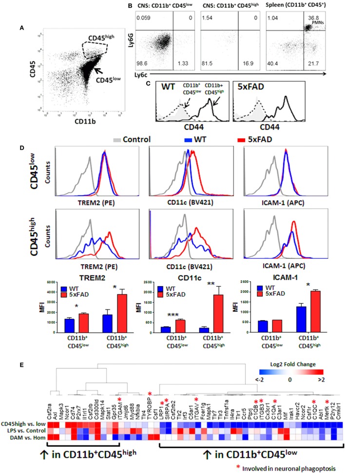
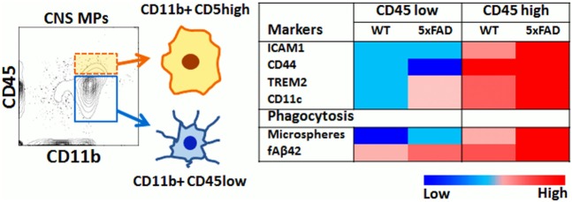
In the central nervous system (CNS), microglia are innate immune mononuclear phagocytes (CNS MPs) that can phagocytose infectious particles, apoptotic cells, neurons and pathological protein aggregates such as Aβ in Alzheimer’s disease (AD). While CD11b+CD45low microglia account for the majority of CNS MPs, a small population of CD11b+CD45high CNS MPs is also recognized in AD that surround Aβ plaques. These transcriptionally-distinct and pathologically-relevant CD45high cells have unclear origin and undefined phagocytic characteristics. We have comprehensively validated rapid flow cytometric assays of bulk-phase and amyloid β fibril (fAβ) phagocytosis and applied these to study acutely isolated CNS MPs. Using these methods, we provide novel insights into differential abilities of CD11b+ CD45low and CD45high CNS MPs to phagocytose macroparticles and fAβ under normal, acute and chronic neuroinflammatory states. CD45high CNS MPs also highly upregulate TREM2, CD11c and several disease-associated-microglia (DAM) signature genes, and have a higher phagocytic capacity for Aβ as compared to CD45low microglia in the 5xFAD mouse model of AD that becomes more apparent with ageing. Our data suggest an overall pro-phagocytic and protective role for CD11b+CD45high CNS MPs in neurodegeneration, which if promoted, could be beneficial.
Overview
The claims and statistics of this paper haven’t been checked yet.
Run the check — free, no account needed. It reads the open-access text and checks the sources it cites.
Integrity signals
Registry
Cites
Statistics
Figures
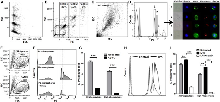
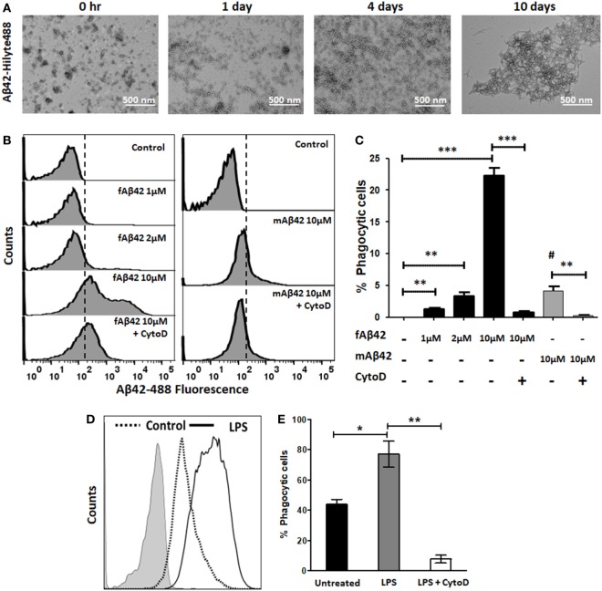
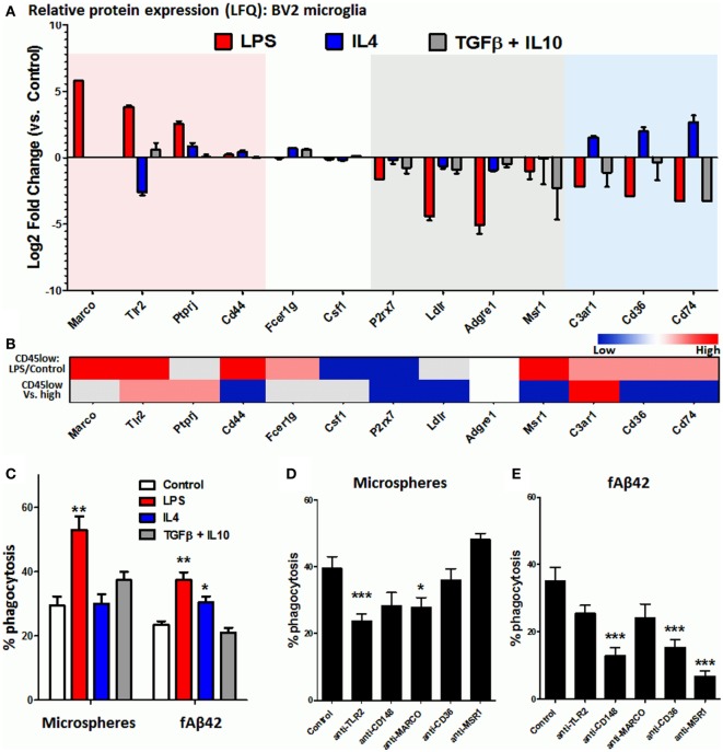
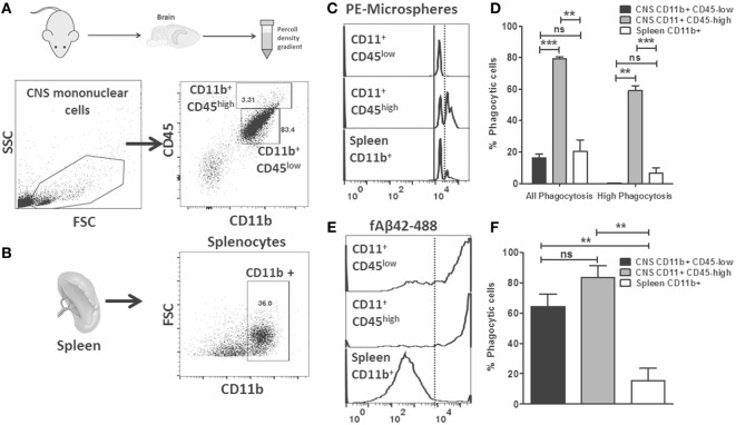
Verification
Claims the paper makes
The claims and statistics of this paper haven’t been checked yet.
Run the check — free, no account needed. It reads the open-access text and checks the sources it cites.
Claims other papers make about this one
Each is checked against this paper’s own text: does it say what the citing paper says it does?
In the citing paper
“We have found that Cytochalasin D inhibits the second peak of fluorescence in this assay, suggesting that the first peak represent non-specific binding of Aβ42 to cells while the second peak represents true uptake in an actin-dependent manner [31].”
From Identification and therapeutic modulation of a pro-inflammatory subset of disease-associated-microglia in Alzheimer’s disease (2018) · cites this paper as [31]
Self-check
Percentages, sums and reported differences re-computed from the paper’s own numbers.
The self-check needs the paper’s full text.
Citations
Cites
Amyloid oligomers exacerbate tau pathology in a mouse model of tauopathy
Selenica, M.-L. B., Brownlow, M., Jimenez, J. P., Lee, D. C., Pena, G., Dickey, C. A., Gordon, M. N., & Morgan, D. (2013) · Neurodegenerative Diseases · doi:10.1159/000337230 · Cited by 10,359
A Unique Microglia Type Associated with Restricting Development of Alzheimer’s Disease
Keren-Shaul, H., Spinrad, A., Weiner, A., Matcovitch-Natan, O., Dvir-Szternfeld, R., Ulland, T. K., David, E., Baruch, K., Lara-Astaiso, D., Toth, B., et al. (2017) · Cell · doi:10.1016/j.cell.2017.05.018 · Cited by 5,493
Cox, J., Hein, M. Y., Luber, C. A., Paron, I., Nagaraj, N., & Mann, M. (2014) · Molecular & Cellular Proteomics · doi:10.1074/mcp.m113.031591 · Cited by 5,180
Oakley, H., Cole, S. L., Logan, S., Maus, E., Shao, P., Craft, J., Guillozet-Bongaarts, A., Ohno, M., Disterhoft, J., Van Eldik, L., et al. (2006) · The Journal of Neuroscience · doi:10.1523/jneurosci.1202-06.2006 · Cited by 3,530 · cited 2×
“Therefore, we studied phagocytic profiles of acutely isolated CNS MPs in the 5xFAD mouse model of AD pathology in which rapid accumulation of Aβ plaques begins at 3 months of age followed by neurodegeneration and neurobehavioral deficits by 8–10 months of age (59).”
BackgroundGene Ontology Consortium: going forward
The Gene Ontology Consortium (2015) · Nucleic Acids Research · doi:10.1093/nar/gku1179 · Cited by 3,492
Hyman, B. T., Phelps, C. H., Beach, T. G., Bigio, E. H., Cairns, N. J., Carrillo, M. C., Dickson, D. W., Duyckaerts, C., Frosch, M. P., Masliah, E., et al. (2012) · Alzheimer's & Dementia · doi:10.1016/j.jalz.2011.10.007 · Cited by 3,092
Variant of TREM2 Associated with the Risk of Alzheimer's Disease
Jonsson, T., Stefansson, H., Steinberg, S., Jonsdottir, I., Jonsson, P. V., Snaedal, J., Bjornsson, S., Huttenlocher, J., Levey, A. I., Lah, J. J., et al. (2013) · New England Journal of Medicine · doi:10.1056/nejmoa1211103 · Cited by 2,558
Self-citationDifferential Roles of M1 and M2 Microglia in Neurodegenerative Diseases
Tang, Y., & Le, W. (2016) · Molecular Neurobiology · doi:10.1007/s12035-014-9070-5 · Cited by 2,190
Systemic LPS causes chronic neuroinflammation and progressive neurodegeneration
Qin, L., Wu, X., Block, M. L., Liu, Y., Breese, G. R., Hong, J., Knapp, D. J., & Crews, F. T. (2007) · Glia · doi:10.1002/glia.20467 · Cited by 2,144
Microglial M1/M2 polarization and metabolic states
Orihuela, R., McPherson, C. A., & Harry, G. J. (2016) · British Journal of Pharmacology · doi:10.1111/bph.13139 · Cited by 2,070
Cited by
Sorted by citations
Rangaraju, S., Dammer, E. B., Raza, S. A., Rathakrishnan, P., Xiao, H., Gao, T., Duong, D. M., Pennington, M. W., Lah, J. J., Seyfried, N. T., et al. (2018) · Molecular Neurodegeneration · doi:10.1186/s13024-018-0254-8 · Cited by 430
Self-citationMicroglia: Agents of the CNS Pro-Inflammatory Response
Rodríguez-Gómez, J. A., Kavanagh, E., Engskog-Vlachos, P., Engskog, M. K. R., Herrera, A. J., Espinosa-Oliva, A. M., Joseph, B., Hajji, N., Venero, J. L., & Burguillos, M. A. (2020) · Cells · doi:10.3390/cells9071717 · Cited by 365
The choroid plexus links innate immunity to CSF dysregulation in hydrocephalus
Robert, S. M., Reeves, B. C., Kiziltug, E., Duy, P. Q., Karimy, J. K., Mansuri, M. S., Marlier, A., Allington, G., Greenberg, A. B. W., DeSpenza, T., et al. (2023) · Cell · doi:10.1016/j.cell.2023.01.017 · Cited by 162
Fairley, L. H., Lai, K. O., Wong, J. H., Chong, W. J., Vincent, A. S., D’Agostino, G. A., Wu, X., Naik, R. R., Jayaraman, A., Langley, S. R., et al. (2023) · Proceedings of the National Academy of Sciences · doi:10.1073/pnas.2209177120 · Cited by 132
SARS-CoV-2 Infection of Microglia Elicits Proinflammatory Activation and Apoptotic Cell Death
Jeong, G. U., Lyu, J., Kim, K., Chung, Y. C., Yoon, G. Y., Lee, S., Hwang, I., Shin, W.-H., Ko, J., Lee, J., et al. (2022) · Microbiology Spectrum · doi:10.1128/spectrum.01091-22 · Cited by 126
Luk, B., Engevik, M. A., Ganesh, B. P., Lackey, E. P., Lin, T., Balderas, M. A., Major, A. M., Runge, J. K., Luna, R. A., Sillitoe, R. V., et al. (2020) · Scientific Reports · doi:10.1038/s41598-020-64173-3 · Cited by 128
Extracellular signal‐regulated kinase regulates microglial immune responses in Alzheimer’s disease
Chen, M. J., Ramesha, S., Weinstock, L. D., Gao, T., Ping, L., Xiao, H., Dammer, E. B., Duong, D. D., Levey, A. I., Lah, J. J., et al. (2021) · Journal of Neuroscience Research · doi:10.1002/jnr.24829 · Cited by 120
Self-citationBIN1 is a key regulator of proinflammatory and neurodegeneration-related activation in microglia
Sudwarts, A., Ramesha, S., Gao, T., Ponnusamy, M., Wang, S., Hansen, M. T., Kozlova, A., Bitarafan, S., Kumar, P., Beaulieu‐Abdelahad, D., et al. (2022) · Molecular Neurodegeneration · doi:10.1186/s13024-022-00535-x · Cited by 110
Self-citationHonarpisheh, P., Lee, J., Banerjee, A., Blasco-Conesa, M. P., Honarpisheh, P., d’Aigle, J., Mamun, A. A., Ritzel, R. M., Chauhan, A., Ganesh, B. P., et al. (2020) · Journal of Neuroinflammation · doi:10.1186/s12974-020-02019-5 · Cited by 104
Thomas, A. L., Lehn, M. A., Janssen, E. M., Hildeman, D. A., & Chougnet, C. A. (2022) · Scientific Reports · doi:10.1038/s41598-022-21920-y · Cited by 94
Chen, S.-W., Hung, Y.-S., Fuh, J., Chen, N.-J., Chu, Y., Chen, S.-C., Fann, M., & Wong, Y. (2021) · Stem Cell Reports · doi:10.1016/j.stemcr.2021.03.010 · Cited by 83
Decoding the temporal and regional specification of microglia in the developing human brain
Li, Y., Li, Z., Yang, M., Wang, F., Zhang, Y., Li, R., Li, Q., Gong, Y., Wang, B., Fan, B., et al. (2022) · Cell stem cell · doi:10.1016/j.stem.2022.02.004 · Cited by 82
Welcome, M. O. (2019) · NeuroMolecular Medicine · doi:10.1007/s12017-019-08547-5 · Cited by 77
Koizumi, T., Kerkhofs, D., Mizuno, T., Steinbusch, H. W. M., & Foulquier, S. (2019) · Frontiers in Neuroscience · doi:10.3389/fnins.2019.01291 · Cited by 74
Delbridge, A. R. D., Huh, D., Brickelmaier, M., Burns, J. C., Roberts, C., Challa, R., Raymond, N., Cullen, P., Carlile, T. M., Ennis, K. A., et al. (2020) · Frontiers in Cellular Neuroscience · doi:10.3389/fncel.2020.592005 · Cited by 72
Rayaprolu, S., Gao, T., Xiao, H., Ramesha, S., Weinstock, L. D., Shah, J., Duong, D. M., Dammer, E. B., Webster, J. A., Lah, J. J., et al. (2019) · doi:10.1101/802694 · Cited by 71
Self-citationNeurovascular unit, neuroinflammation and neurodegeneration markers in brain disorders
Kempuraj, D., Dourvetakis, K. D., Cohen, J., Valladares, D. S., Joshi, R. S., Kothuru, S. P., Anderson, T., Chinnappan, B., Cheema, A. K., Klimas, N. G., et al. (2024) · Frontiers in Cellular Neuroscience · doi:10.3389/fncel.2024.1491952 · Cited by 78
Mercurio, D., Fumagalli, S., Schäfer, M., Pedragosa, J., Ngassam, L. D. C., Wilhelmi, V., Winterberg, S., Planas, A. M., Weihe, E., & Simoni, M.-G. D. (2022) · Frontiers in Cellular Neuroscience · doi:10.3389/fncel.2022.820127 · Cited by 72
Gao, T., Jernigan, J. E., Raza, S. A., Dammer, E. B., Xiao, H., Seyfried, N. T., Levey, A. I., & Rangaraju, S. (2019) · Glia · doi:10.1002/glia.23678 · Cited by 68
Self-citationInterferon β-Mediated Protective Functions of Microglia in Central Nervous System Autoimmunity
Scheu, S., Ali, S., Mann‐Nüttel, R., Richter, L., Arolt, V., Dannlowski, U., Kuhlmann, T., Klotz, L., & Alferink, J. (2019) · International Journal of Molecular Sciences · doi:10.3390/ijms20010190 · Cited by 49
Ramesha, S., Rayaprolu, S., Bowen, C. A., Giver, C. R., Bitarafan, S., Nguyen, H. M., Gao, T., Chen, M. J., Nwabueze, N. V., Dammer, E. B., et al. (2021) · Proceedings of the National Academy of Sciences · doi:10.1073/pnas.2013545118 · Cited by 47
Self-citationSaber, M., Giordano, K. R., Hur, Y., Ortiz, J. B., Morrison, H., Godbout, J. P., Murphy, S. M., Lifshitz, J., & Rowe, R. K. (2020) · European Journal of Neuroscience · doi:10.1111/ejn.14611 · Cited by 46
Ju, H., Park, K. W., Kim, I., Cave, J. W., & Cho, S. (2022) · Journal of Neuroinflammation · doi:10.1186/s12974-022-02552-5 · Cited by 49
Divergent Functions of Tissue-Resident and Blood-Derived Macrophages in the Hemorrhagic Brain
Chang, C.-F., Goods, B. A., Askenase, M. H., Beatty, H. E., Osherov, A., DeLong, J. H., Hammond, M. D., Massey, J., Landreneau, M. J., Love, J. C., et al. (2021) · Stroke · doi:10.1161/strokeaha.120.032196 · Cited by 44
CD11bhigh B Cells Increase after Stroke and Regulate Microglia
Korf, J. M., Honarpisheh, P., Mohan, E. C., Banerjee, A., Blasco-Conesa, M. P., Honarpisheh, P., Guzman, G. U., Khan, R., Ganesh, B. P., Hazen, A. L., et al. (2022) · The Journal of Immunology · doi:10.4049/jimmunol.2100884 · Cited by 44
d -mannose suppresses oxidative response and blocks phagocytosis in experimental neuroinflammation
Wang, J., Motlagh, N. J., Wang, C., Wojtkiewicz, G. R., Schmidt, S., Chau, C., Narsimhan, R. P., Kuellenberg, E. G., Zhu, C., Linnoila, J., et al. (2021) · Proceedings of the National Academy of Sciences · doi:10.1073/pnas.2107663118 · Cited by 41
Kutsyr, O., Sánchez‐Sáez, X., Martínez‐Gil, N., de Juan, E., Lax, P., Maneu, V., & Cuenca, N. (2020) · Investigative Ophthalmology & Visual Science · doi:10.1167/iovs.61.10.1 · Cited by 42
Functional and Metabolic Characterization of Microglia Culture in a Defined Medium
Montilla, A., Zabala, A., Matute, C., & Domercq, M. (2020) · Frontiers in Cellular Neuroscience · doi:10.3389/fncel.2020.00022 · Cited by 40
Sharps, M. C., Baker, B., Guevara, T., Bischof, H., Jones, R. L., Greenwood, S. L., & Heazell, A. E. P. (2020) · American Journal of Reproductive Immunology · doi:10.1111/aji.13267 · Cited by 34
Wu, S.-Y., Sharma, S., Wu, K., Tyagi, A., Zhao, D., Deshpande, R. P., & Watabe, K. (2021) · Breast Cancer Research · doi:10.1186/s13058-021-01412-z · Cited by 34
RetractedHwang, D., Ishikawa, L. L. W., Seyedsadr, M., Mari, E. R., Kasimoglu, E., Sahin, Z., Boehm, A., Jang, S., Rasouli, J., Vaccaro, C., et al. (2022) · Blood Advances · doi:10.1182/bloodadvances.2022008234 · Cited by 28
Khan, M. A. S., & Chang, S. L. (2023) · Cells · doi:10.3390/cells12202475 · Cited by 30
Schroeter, C. B., Herrmann, A. M., Böck, S., Vogelsang, A., Eichler, S., Albrecht, P., Meuth, S. G., & Ruck, T. (2021) · Cells · doi:10.3390/cells10030651 · Cited by 27
Immunoproteasomes control activation of innate immune signaling and microglial function
Çetin, G., Studencka‐Turski, M., Venz, S., Schormann, E., Junker, H., Hammer, E., Völker, U., Ebstein, F., & Krüger, E. (2022) · Frontiers in Immunology · doi:10.3389/fimmu.2022.982786 · Cited by 26
Sunna, S. N., Bowen, C. A., Zeng, H., Rayaprolu, S., Kumar, P., Bagchi, P., Dammer, E. B., Guo, Q., Duong, D. M., Bitarafan, S., et al. (2023) · Molecular & Cellular Proteomics · doi:10.1016/j.mcpro.2023.100546 · Cited by 27
Self-citationAssaying Microglia Functions In Vitro
Maguire, E., Connor‐Robson, N., Shaw, B., O’Donoghue, R., Stöberl, N., & Hall‐Roberts, H. (2022) · Cells · doi:10.3390/cells11213414 · Cited by 31
Li, H., Zhang, P., Liu, C., Wang, Y., Deng, Y., Dong, W., & Yu, Y. W. (2022) · Cells · doi:10.3390/cells12010008 · Cited by 25
Gao, T., Raza, S. A., Ramesha, S., Nwabueze, N. V., Tomkins, A. J., Cheng, L., Xiao, H., Yepes, M., & Rangaraju, S. (2019) · Journal of Neuroinflammation · doi:10.1186/s12974-019-1510-8 · Cited by 23
Self-citationPathology of Amyloid-β (Aβ) Peptide Peripheral Clearance in Alzheimer’s Disease
Tsoy, A., Umbayev, B. A., Kassenova, A., Kaupbayeva, B., & Askarova, S. (2024) · International Journal of Molecular Sciences · doi:10.3390/ijms252010964 · Cited by 24
Cho, S. M., Vardi, A., Platt, N., & Futerman, A. H. (2018) · Journal of Neurochemistry · doi:10.1111/jnc.14483 · Cited by 21
Strategies and Tools for Studying Microglial-Mediated Synapse Elimination and Refinement
Morini, R., Bizzotto, M., Perrucci, F., Filipello, F., & Matteoli, M. (2021) · Frontiers in Immunology · doi:10.3389/fimmu.2021.640937 · Cited by 22
Welcome, M. O. (2018) · Dementia and Geriatric Cognitive Disorders Extra · doi:10.1159/000492491 · Cited by 22
Norte‐Muñoz, M., Gallego‐Ortega, A., Lucas‐Ruiz, F., González‐Riquelme, M. J., Changa-Espinoza, Y. I., Galindo‐Romero, C., Ponsaerts, P., Vidal‐Sanz, M., García‐Bernal, D., & Agudo‐Barriuso, M. (2022) · Stem Cell Research & Therapy · doi:10.1186/s13287-022-03129-y · Cited by 20
Bobotis, B. C., Halvorson, T., Carrier, M., & Tremblay, M. (2024) · Frontiers in Cellular Neuroscience · doi:10.3389/fncel.2024.1317125 · Cited by 22
Myeloid cell subpopulations compensate each other for Ccr2‐deficiency in glioblastoma
Bungert, A. D., Urbantat, R. M., Jelgersma, C., Bekele, B. M., Mueller, S. G., Mueller, A., Felsenstein, M., Dusatko, S., Blank, A., Ghori, A., et al. (2022) · Neuropathology and Applied Neurobiology · doi:10.1111/nan.12863 · Cited by 19
Immune response of BV-2 microglial cells is impacted by peroxisomal beta-oxidation
Tawbeh, A., Raas, Q., Tahri-Joutey, M., Keime, C., Kaiser, R., Trompier, D., Nasser, B., Bellanger, E., Dessard, M., Hamon, Y., et al. (2023) · Frontiers in Molecular Neuroscience · doi:10.3389/fnmol.2023.1299314 · Cited by 18
Modulation of Innate Immunity by Amyloidogenic Peptides
Westwell-Roper, C. Y., & Verchere, C. B. (2019) · Trends in Immunology · doi:10.1016/j.it.2019.06.005 · Cited by 17
SARS-CoV-2 Infection of Microglia Elicits Pro-inflammatory Activation and Apoptotic Cell DeathPreprint
Jeong, G. U., Lyu, J., Kim, K., Chung, Y. C., Yoon, G. Y., Lee, S., Hwang, I., Shin, W.-H., Ko, J., Lee, J., et al. (2022) · bioRxiv (Cold Spring Harbor Laboratory) · doi:10.1101/2022.01.04.475015 · Cited by 17
Decoding the role of zebrafish neuroglia in CNS disease modeling
Zabegalov, K. N., Wang, D., Yang, L., Wang, J., Hu, G., Serikuly, N., Alpyshov, E. T., Khatsko, S. L., Zhdanov, A., Demin, K. A., et al. (2020) · Brain Research Bulletin · doi:10.1016/j.brainresbull.2020.09.020 · Cited by 17
Li, H., Ye, T., Liu, X., Guo, R., Yang, X., Li, Y., Qi, D., Wei, Y., Zhu, Y., Wen, L., et al. (2023) · Journal of Pharmaceutical Analysis · doi:10.1016/j.jpha.2023.05.008 · Cited by 15
Novel Microglia Cell Line Expressing the Human EP2 Receptor
Rojas, A., Banik, A., Chen, D., Flood, K., Ganesh, T., & Dingledine, R. J. (2019) · ACS Chemical Neuroscience · doi:10.1021/acschemneuro.9b00311 · Cited by 15
Novel protocol for the isolation of highly purified neonatal murine microglia and astrocytes
Zelenka, L., Pägelow, D., Krüger, C., Seele, J., Ebner, F., Rausch, S., Rohde, M., Lehnardt, S., van Vorst, K., & Fulde, M. (2021) · Journal of Neuroscience Methods · doi:10.1016/j.jneumeth.2021.109420 · Cited by 15
Jäkel, L., Biemans, E. A. L. M., Klijn, C. J. M., Kuiperij, H. B., & Verbeek, M. M. (2020) · Molecular Neurobiology · doi:10.1007/s12035-020-01873-x · Cited by 15
Francistiová, L., Vörös, K., Lovász, Z., Dinnyés, A., & Kobolák, J. (2022) · Frontiers in Molecular Neuroscience · doi:10.3389/fnmol.2021.793769 · Cited by 14
Recent Progress in Multiple Sclerosis Treatment Using Immune Cells as Targets
Ma, X., Ma, R., Zhang, M., Qian, B., Wang, B. B., & Yang, W. (2023) · Pharmaceutics · doi:10.3390/pharmaceutics15030728 · Cited by 17
Advances in proteomic phenotyping of microglia in neurodegeneration
Sunna, S. N., Bowen, C. A., Ramelow, C. C., Santiago, J. V., Kumar, P., & Rangaraju, S. (2023) · PROTEOMICS · doi:10.1002/pmic.202200183 · Cited by 13
Self-citationChirico, F. D., Poeta, E., Babini, G., Piccolino, I., Monti, B., & Massenzio, F. (2022) · Frontiers in Cellular Neuroscience · doi:10.3389/fncel.2022.1038721 · Cited by 11
Chen, J., Chen, H., Wei, Q., Lu, Y., Wang, T., Pang, X., Xing, G., Chen, Z., Cao, X., & Yao, J. (2025) · Cell Death Discovery · doi:10.1038/s41420-025-02454-4 · Cited by 11
García‐Juárez, M., García-Rodríguez, A., Cruz-Carrillo, G., Flores-Maldonado, O. E., Becerril-García, M. A., Garza‐Ocañas, L., Torre-Villalvazo, I., & Camacho‐Morales, A. (2024) · Molecular Neurobiology · doi:10.1007/s12035-024-04340-z · Cited by 11
Sánchez-García, M. A., Lara-Ureña, N., March‐Díaz, R., Luis, C. O. S., Quiñones-Cañete, S., Mora-Romero, B., Cabello-Rivera, D., Cabello-Rivera, D., Romero‐Molina, C., Romero‐Molina, C., et al. (2025) · Science Advances · doi:10.1126/sciadv.adu2244 · Cited by 10
Zhang, X., Yuan, J., Zhang, S., Li, W., Xu, Y., Li, H., Zhang, L., Chen, X., Ding, W., Zhu, J., et al. (2022) · Journal of Cerebral Blood Flow & Metabolism · doi:10.1177/0271678x221147091 · Cited by 10
Mimouna, S., Rollins, D. A., Shibu, G., Tharmalingam, B., Deochand, D. K., Chen, X., Oliver, D., Chinenov, Y. V., & Rogatsky, I. (2020) · The Journal of Experimental Medicine · doi:10.1084/jem.20192386 · Cited by 11
He, A., Wang, Y., Shen, Y., Dong, Z., Luo, L., Ge, X., Liu, X., Mao, Y., Zhang, T., Li, X., et al. (2025) · Nature Aging · doi:10.1038/s43587-025-01028-4 · Cited by 9
He, J., Zhao, F., Chen, B., Cui, N., Li, Z., Qin, J., Luo, L., Zhao, C., & Li, L. (2023) · Science China Life Sciences · doi:10.1007/s11427-021-2223-4 · Cited by 9
Interrogation of human microglial phagocytosis by CRISPR genome editing
Chang, J. C.-Y., Wang, C., & Lin, S. (2023) · Frontiers in Immunology · doi:10.3389/fimmu.2023.1169725 · Cited by 9
Method of Microglial DNA-RNA Purification from a Single Brain of an Adult Mouse
Raihan, M. O., McGregor, B. A., Velaris, N. A., Brishti, A., Hur, J., & Porter, J. E. (2021) · Methods and Protocols · doi:10.3390/mps4040086 · Cited by 8
The changing landscape of immune cells in the fetal mouse testis
Hosseini, S., Moody, S. C., Fietz, D., Indumathy, S., Schuppe, H.-C., Hedger, M. P., & Loveland, K. L. (2022) · Histochemistry and Cell Biology · doi:10.1007/s00418-022-02129-6 · Cited by 8
Jahromi, S. R., Haddadi, M., Mousavi, P., & Sanadgol, N. (2022) · Metabolic Brain Disease · doi:10.1007/s11011-022-01083-5 · Cited by 8
Liu, L.-Z., Fan, S., Gao, J., Li, W., & Xian, X. (2023) · Brain Research Bulletin · doi:10.1016/j.brainresbull.2023.110683 · Cited by 9
Gardner, R. S., Kyle, M. M., Hughes, K., & Zhao, L. (2024) · Biomolecules · doi:10.3390/biom14070827 · Cited by 7
Topological Dissection of Proteomic Changes Linked to the Limbic Stage of Alzheimer’s Disease
Velásquez, E., Szeitz, B., Gil, J., Murillo, J. R., Palkovits, M., Renner, É., Hortobágyi, T., Döme, P., Nogueira, F. C. S., Marko‐Varga, G., et al. (2021) · Frontiers in Immunology · doi:10.3389/fimmu.2021.750665 · Cited by 8
The effects of okadaic acid‐treated SH‐SY5Y cells on microglia activation and phagocytosis
Amonruttanapun, P., Chongthammakun, S., & Chamniansawat, S. (2021) · Cell Biology International · doi:10.1002/cbin.11722 · Cited by 8
An anti-inflammatory transcriptional cascade conserved from flies to humans
Pavlidaki, A., Panic, R., Monticelli, S., Riet, C., Yuasa, Y., Cattenoz, P. B., Nait‐Oumesmar, B., & Giangrande, A. (2022) · Cell Reports · doi:10.1016/j.celrep.2022.111506 · Cited by 9
Lin, C., Calzarossa, C., Fernández-Zafra, T., Liu, J., Li, X., Ekblad, Å., Vázquez‐Juárez, E., Codeluppi, S., Holmberg, L., Lindskog, M., et al. (2020) · Stem Cell Research & Therapy · doi:10.1186/s13287-020-01771-y · Cited by 10
Specificity and efficiency of tamoxifen-mediated Cre induction is equivalent regardless of age
Kellogg, C. M., Pham, K. D., Ko, S.-H., Cox, J. E. J., Machalinski, A. H., Stout, M. B., Sharpe, A. L., Beckstead, M. J., Chucair‐Elliott, A. J., Ocañas, S. R., et al. (2023) · iScience · doi:10.1016/j.isci.2023.108413 · Cited by 10
Ni, J.-W., Li, C., Chen, X., & Cai, W. (2022) · Neurology India · doi:10.4103/0028-3886.364080 · Cited by 8
Su, X., Xie, L., Li, J., Tian, X., Bing, L., & Chen, M. (2023) · Frontiers in Molecular Neuroscience · doi:10.3389/fnmol.2023.1133106 · Cited by 9
Ramesha, S., Rayaprolu, S., & Rangaraju, S. (2020) · Journal of Visualized Experiments · doi:10.3791/61467 · Cited by 7
Self-citationCrews, F. T., Qin, L., Coleman, L. G., Vidrascu, E. M., & Vetreno, R. P. (2025) · Brain Behavior and Immunity · doi:10.1016/j.bbi.2025.106156 · Cited by 7
Ruiz-Pastor, M. J., Sánchez‐Sáez, X., Kutsyr, O., Albertos‐Arranz, H., Sánchez‐Castillo, C., Ortuño‐Lizarán, I., Martínez‐Gil, N., Vidal‐Gil, L., Méndez, L., Sánchez‐Martín, M., et al. (2023) · Cell Death and Disease · doi:10.1038/s41419-023-06243-8 · Cited by 6
Lin, Y. N., Mao, F., Wong, N.-K., Zhang, X., Liu, K., Huang, M., Ma, H., Xiang, Z., Li, J., Xiao, S., et al. (2020) · Frontiers in Immunology · doi:10.3389/fimmu.2020.00416 · Cited by 6
Cheng, J.-J., Guo, Q. H., Wu, X.-G., Ma, S., Gao, Y., & Yazhen, S. (2020) · Combinatorial Chemistry & High Throughput Screening · doi:10.2174/1386207323666201209092358 · Cited by 5
Shin, T. H., Lee, D. Y., Jang, Y. E., Kwon, D. H., Hwang, J. S., Kim, S. G., Seo, C., Paik, M., Lee, J. Y., Kim, J. Y., et al. (2022) · Cells · doi:10.3390/cells11152393 · Cited by 5
Early ultrastructural damage in retina and optic nerve following intraocular pressure elevation
Abreu, C. A., Ferraz, G., dos Santos, R. C., Conde, L., Dantas, D. P., Archanjo, B. S., Linden, R., Pimentel‐Coelho, P. M., & Allodi, S. (2025) · Vision Research · doi:10.1016/j.visres.2025.108544 · Cited by 4
Ting, S., Zhao, X., Sun, G., Ricote, M., & Aronowski, J. A. (2025) · Journal of Neuroscience · doi:10.1523/jneurosci.0248-25.2025 · Cited by 4
Berríos‐Cárcamo, P., Núñez, S., Castañeda, J., Gallardo, J., Bono, M. R., & Ezquer, F. E. (2024) · International Journal of Molecular Sciences · doi:10.3390/ijms25084173 · Cited by 4
Hu, Y., Wang, X., Zhao, Z., Liu, M., Ren, X., Xian, X., Liu, C., & Wang, Q. (2024) · Journal of Alzheimer s Disease · doi:10.3233/jad-240118 · Cited by 4
Latham, A. S., Geer, C. E., Ackart, D. F., Weninger, K. N., Gross, C. C., Podell, B. K., Basaraba, R. J., & Moreno, J. A. (2024) · PLoS ONE · doi:10.1371/journal.pone.0307577 · Cited by 5
RGS10 attenuates systemic immune dysregulation induced by chronic inflammatory stress
Jernigan, J. E., Staley, H. A., Baty, Z., Bolen, M. L., Gomes, B. N., Holt, J., Cole, C. L., Neighbarger, N. K., Dheeravath, K., Merchak, A. R., et al. (2025) · Journal of Neuroinflammation · doi:10.1186/s12974-024-03322-1 · Cited by 4
Improved human hematopoietic reconstitution in HepaRG co-transplanted humanized NSG mice
Kim, J., Ryu, B., Kim, U., Kim, C., Hur, G.-H., Kim, C., & Park, J. (2020) · BMB Reports · doi:10.5483/bmbrep.2020.53.9.304 · Cited by 4
Cho, H., Choi, B. Y., Shin, Y., Suh, S. W., & Park, S. B. (2023) · ACS Chemical Neuroscience · doi:10.1021/acschemneuro.3c00508 · Cited by 4
Mitochondrial control of microglial phagocytosis in Alzheimer’s diseasePreprint
Fairley, L. H., Lai, K. O., Wong, J. H., Salvatore, A. V., D’Agostino, G. A., Wu, X., Jayaraman, A., Langley, S. R., Ruedl, C., & Barron, A. M. (2021) · bioRxiv (Cold Spring Harbor Laboratory) · doi:10.1101/2021.12.01.469111 · Cited by 4
Emerging Techniques for the Study of Microglia: Visualization, Depletion and Fate MappingPreprint
Bobotis, B. C., Halvorson, T., Carrier, M., & Tremblay, M. (2023) · Preprints.org · doi:10.20944/preprints202308.2190.v1 · Cited by 3
Li, R., Zhang, J., Fan, J.-Q., & Lin, B. (2026) · Journal of Neuroinflammation · doi:10.1186/s12974-026-03692-8 · Cited by 3
Capes, D. E., Slone, V. K., Winchester, D. K., Salazar, J., Opoku, P., Li, Y., Hash, M. T., Kumari, R., Hawk, G. S., & Stewart, A. N. (2026) · Experimental Neurology · doi:10.1016/j.expneurol.2026.115690 · Cited by 3
Dual immune armies in glaucoma: microglia and monocyte-derived macrophages
Tan, K., Zhang, Y., Jiang, P., Yu, Y., Wu, Z. G., Peng, J. Q., Liu, P., & Peng, Q. (2026) · Frontiers in Immunology · doi:10.3389/fimmu.2026.1801497 · Cited by 3
Reconstitution of human microglia and resident T cells in the brain of humanized DRAGA mice
Roy, S. G., Karim, A. F., Brumeanu, T., & Casares, S. A. (2024) · Frontiers in Cellular and Infection Microbiology · doi:10.3389/fcimb.2024.1367566 · Cited by 3
Microglia-specificity of different markers is overridden in glioblastoma specimens
Bungert, A. D., Sanchin, A., Blank, A., Jüngst-Mieczkowska, M., Müller, A., Felsenstein, M., Turkowski, K., Zhang, W., Vajkoczy, P., & Brandenburg, S. (2026) · Scientific Reports · doi:10.1038/s41598-026-52315-y · Cited by 2
Dynamics of CD44+ bovine nucleus pulposus cells with inflammation
Ferreira, J. N., Caldeira, J., Sousa, M. M., Barbosa, M. A., Lamghari, M., Almeida-Porada, G. D., & Gonçalves, R. M. (2024) · Scientific Reports · doi:10.1038/s41598-024-59504-7 · Cited by 2
Mimouna, S., Rollins, D. A., Shibu, G., Tharmalingam, B., Deochand, D. K., Chen, X., Oliver, D., Chinenov, Y. V., & Rogatsky, I. (2020) · The Journal of Experimental Medicine · doi:10.1084/jem.2019238611162020c · Cited by 3
Metadata
- Authors
- Rangaraju, S., Raza, S. A., Li, N. X., Betarbet, R., Dammer, E. B., Duong, D., Lah, J. J., Seyfried, N. T., & Levey, A. I.
- Affiliations
- Emory University (US)
- Corresponding author
- Rangaraju, Srikant
- ORCID
- Venue
- Frontiers in Immunology
- Publisher
- Frontiers Media SA
- Published
- 2018-03-02
- Volume
- 9
- Pages
- 405-405
- ISSN
- 1664-3224 (ISSN-L)
- PMC
- PMC5840283
- OpenAlex
- W2792377614
- Cited by
- 126
- References
- 80
- Citations by year
- 2026: 9 · 2025: 8 · 2024: 12 · 2023: 18 · 2022: 35 · 2021: 13 · 2020: 19 · 2019: 8 · 2018: 3
- FWCI
- 4.82
- Type
- journal article
- Language
- en
- Open access
- gold
- doi.org
- Locations
- Frontiers in Immunology (publishedVersion, cc-by)
- Europe PMC (PubMed Central) (publishedVersion, cc-by)
- PubMed Central (publishedVersion, cc-by)
- PubMed (publishedVersion)
- DOAJ (DOAJ: Directory of Open Access Journals) (submittedVersion, cc-by-sa)
- Funders
- National Institutes of Health (P50 AG025688, NS099474-1, P30 NS055077, 1U01AG046161)
- National Science Foundation (CHE-1507932, NSF/DMR-BSF 1610377)
- American Brain Foundation
- Alzheimer’s Association
- Topics
- Life Sciences › Neuroscience › Neurology › Neuroinflammation and Neurodegeneration Mechanisms
- Life Sciences › Immunology and Microbiology › Immunology › Phagocytosis and Immune Regulation
- Life Sciences › Neuroscience › Cellular and Molecular Neuroscience › Alzheimer's disease research and treatments
- Keywords
- microglia, phagocytosis, amyloid-beta fibrils, Alzheimer's disease, disease-associated microglia, TREM2, CD11c, neuroinflammation, aging, 5xFAD mice, neurodegeneration
- MeSH terms
- Aging, Alzheimer Disease, Animals, Brain, Cell Line, Disease Models, Animal, Humans, Membrane Glycoproteins, Mice, Inbred C57BL, Mice, Mutant Strains, Phagocytes, Phagocytosis, Receptors, Immunologic, Mononuclear Phagocyte System, Amyloid beta-Peptides, Leukocyte Common Antigens, Microglia, Mice
About these checks
Registry, retraction, Cites and statistics results are computed, not judged, with one exception: a printed reference that no index lists is read once by a model before it counts as a citation error. Claims are extracted by a model and kept only when their quote appears verbatim in the text; each cited source is then checked, with a model’s help, for whether it says what the claim relies on. Those verdicts can be wrong — read the claim’s quote and the cited source before relying on one.
Statistics and arithmetic are recomputed, not judged.
Confidence bands are shown only for fields calibrated on our annotated test set. None is calibrated yet, so no band is shown.
What counts as a claim ›How we measure confidence ›How we check this ›
Are you an author, or do you see a problem with this page? Request a review
