Cited by 30

The DOI matches its Crossref record.

Human Mast Cell Tryptase Is a Potential Treatment for Snakebite Envenoming Across Multiple Snake Species

Anderson, Elizabeth, Stavenhagen, Kathrin, Kolarich, Daniel, Sommerhoff, Christian P., Maurer, Marcus, Metz, Martin·2018·Frontiers in Immunology

Overview

The claims and statistics of this paper haven’t been checked yet.

Run the check — free, no account needed. It reads the open-access text and checks the sources it cites.

A few free checks a day · papers already verified open instantly · results are public

Integrity signals

Registry

Crossref · 10.3389/fimmu.2018.01532

Cites

Statistics

Figures

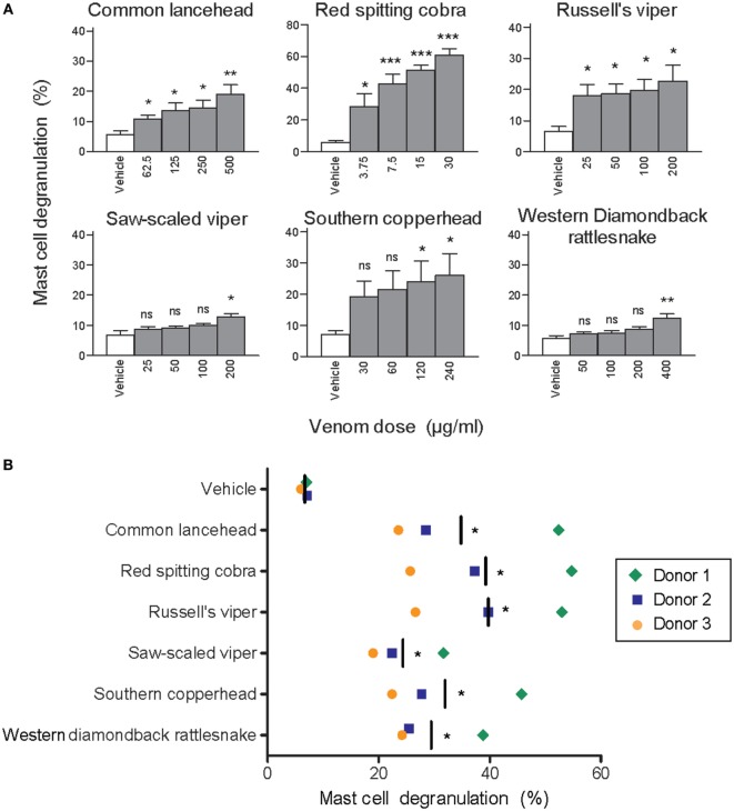
Figure 1 Whole venom from six different snakes induces degranulation of human mast cells. LAD2 MCs (A) or primary human skin mast cell (hsMCs) isolated from three healthy donors (B) were stimulated with venom from the six listed snake species. (B) Venom doses were as follows: common lancehead, saw-scaled viper, southern copperhead, red spitting cobra, Russell’s viper, and western diamondback rattlesnake. Negative controls (vehicle) were PBS only. Positive controls (not shown) were Ca ionomycin (1 µM) and caused an average of 55% degranulation in LAD2 (A), and 62, 36, and 8% degranulation in hsMCs from each donor (B). Data are pooled from four independent experiments and measurements were performed in triplicate. Student’s t-test compares venom to vehicle stimulation. *p < 0.05; **p < 0.01; ***p < 0.001.
Figure 1 Whole venom from six different snakes induces degranulation of human mast cells. LAD2 MCs (A) or primary human skin mast cell (hsMCs) isolated from three healthy donors (B) were stimulated with venom from the six listed snake species. (B) Venom doses were as follows: common lancehead, saw-scaled viper, southern copperhead, red spitting cobra, Russell’s viper, and western diamondback rattlesnake. Negative controls (vehicle) were PBS only. Positive controls (not shown) were Ca ionomycin (1 µM) and caused an average of 55% degranulation in LAD2 (A), and 62, 36, and 8% degranulation in hsMCs from each donor (B). Data are pooled from four independent experiments and measurements were performed in triplicate. Student’s t-test compares venom to vehicle stimulation. *p < 0.05; **p < 0.01; ***p < 0.001.CC BY · PubMed Central
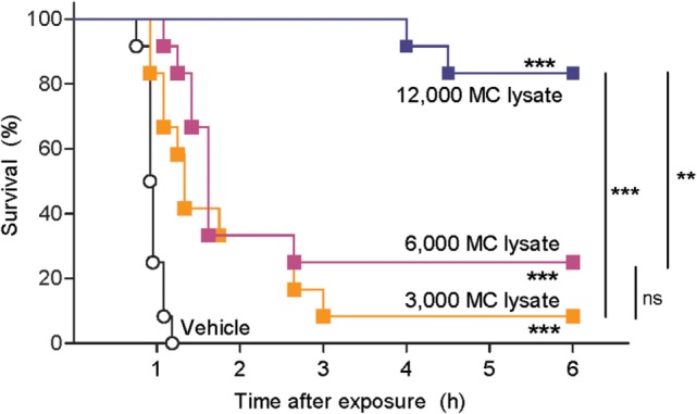
Figure 2 Lysate from purified human skin mast cells (hsMCs) protects zebrafish from toxicity of cobra venom. Zebrafish embryos aged 48 h post fertilization suspended in 100 µl of fish water received 625 ng red spitting cobra whole venom pre-treated with lysate of purified hsMCs (MC lysate) or PBS (vehicle). Lysate doses were 12-, 6-, or 3- thousand cells per fish. Survival was monitored at regular intervals for 6 h, with complete cessation of heartbeat as the predefined endpoint. Vehicle, lysate alone, or enzyme alone had no effect (data not shown). Data are pooled from three independent experiments, n = 12 fish per group. Kaplan–Meyer survival analysis, Log-rank (Mantel-Cox) test compares treatments to vehicle and to each other. **p < 0.01; ***p < 0.001.
Figure 2 Lysate from purified human skin mast cells (hsMCs) protects zebrafish from toxicity of cobra venom. Zebrafish embryos aged 48 h post fertilization suspended in 100 µl of fish water received 625 ng red spitting cobra whole venom pre-treated with lysate of purified hsMCs (MC lysate) or PBS (vehicle). Lysate doses were 12-, 6-, or 3- thousand cells per fish. Survival was monitored at regular intervals for 6 h, with complete cessation of heartbeat as the predefined endpoint. Vehicle, lysate alone, or enzyme alone had no effect (data not shown). Data are pooled from three independent experiments, n = 12 fish per group. Kaplan–Meyer survival analysis, Log-rank (Mantel-Cox) test compares treatments to vehicle and to each other. **p < 0.01; ***p < 0.001.CC BY · PubMed Central
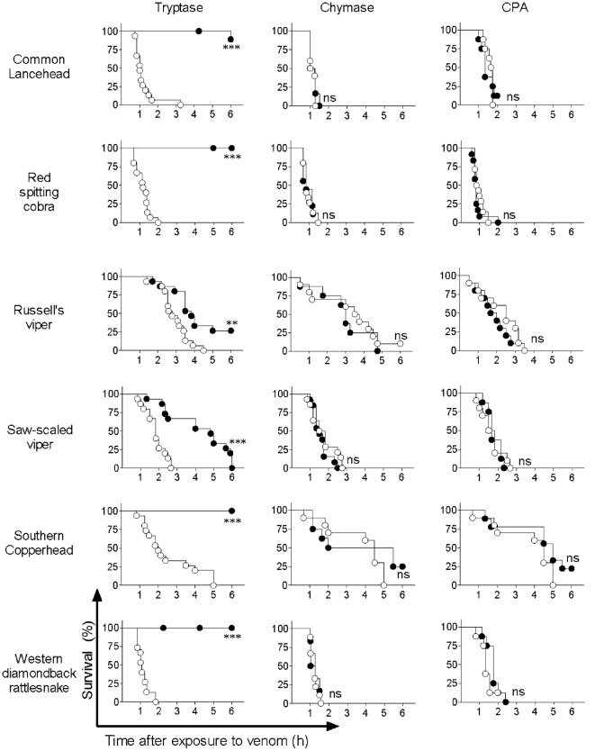
Figure 3 Purified human tryptase, but not chymase or carboxypeptidase A (CPA), detoxifies snake venoms. Venoms were incubated with 10 µg/ml human tryptase, chymase, or CPA. Treated and untreated venom were then administered to 48 h post fertilization zebrafish in 100 µl of water. Empty circles represent zebrafish receiving venom alone, and solid shapes represent fish receiving enzyme-treated venom. Vehicle or enzyme alone had no effect (data not shown). Data are pooled from three (tryptase) or two (chymase and CPA) independent experiments for n = 15 or 10 fish per group. Kaplan–Meyer survival analysis, Log-rank (Mantel-Cox) test compares treatments to vehicle. **p < 0.01; ***p < 0.001.
Figure 3 Purified human tryptase, but not chymase or carboxypeptidase A (CPA), detoxifies snake venoms. Venoms were incubated with 10 µg/ml human tryptase, chymase, or CPA. Treated and untreated venom were then administered to 48 h post fertilization zebrafish in 100 µl of water. Empty circles represent zebrafish receiving venom alone, and solid shapes represent fish receiving enzyme-treated venom. Vehicle or enzyme alone had no effect (data not shown). Data are pooled from three (tryptase) or two (chymase and CPA) independent experiments for n = 15 or 10 fish per group. Kaplan–Meyer survival analysis, Log-rank (Mantel-Cox) test compares treatments to vehicle. **p < 0.01; ***p < 0.001.CC BY · PubMed Central
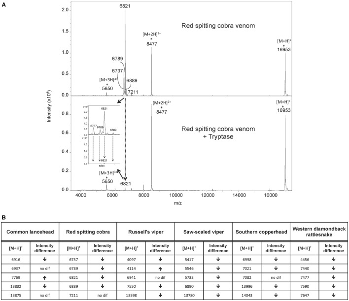
Figure 4 MALDI-TOF-MS analysis of venoms treated with purified human tryptase shows degradation of venom proteins. Samples were prepared at concentrations of 10 µg/µl venom and a ratio of 1000:1 venom to tryptase by mass (southern copperhead and common lancehead) or 0.1 µg/µl and a ratio of 10:1 venom to tryptase by mass (western diamondback rattlesnake, Russell’s viper, saw-scaled viper, and red spitting cobra). They were then incubated at 37°C for 30 min, and stored at −20°C. Control was prepared with venom and PBS alone. 17 µl of the venom sample (in PBS) was purified by reversed phase chromatography using C18 Zip TipsTM (Millipore). Mass spectra were acquired in positive ion reflectron mode within the m/z ratio range of m/z 3,500 to m/z 18,000. Panel (A) shows a representative MALDI-TOF-MS spectrum of red spitting cobra venom before and after incubation with purified human tryptase. Five signals (dashed arrows) over the acquired mass range were selected and their signal intensities normalized to the intensity of the doubly charged myoglobin signal (m/z 8,477). The relative signal intensity reduction of several peaks indicates protein degradation by tryptase. Spectra for additional venoms are provided in Figure 5. Comparisons of relative signal intensities for all six venoms are summarized in (B). Arrows indicate a reduced (↓) or increased (↑) relative signal intensity difference (Intens. dif.) of prominent peaks, or no difference (no dif.), with lower peaks indicating degradation of the polypeptide of the corresponding m/z ratio.
Figure 4 MALDI-TOF-MS analysis of venoms treated with purified human tryptase shows degradation of venom proteins. Samples were prepared at concentrations of 10 µg/µl venom and a ratio of 1000:1 venom to tryptase by mass (southern copperhead and common lancehead) or 0.1 µg/µl and a ratio of 10:1 venom to tryptase by mass (western diamondback rattlesnake, Russell’s viper, saw-scaled viper, and red spitting cobra). They were then incubated at 37°C for 30 min, and stored at −20°C. Control was prepared with venom and PBS alone. 17 µl of the venom sample (in PBS) was purified by reversed phase chromatography using C18 Zip TipsTM (Millipore). Mass spectra were acquired in positive ion reflectron mode within the m/z ratio range of m/z 3,500 to m/z 18,000. Panel (A) shows a representative MALDI-TOF-MS spectrum of red spitting cobra venom before and after incubation with purified human tryptase. Five signals (dashed arrows) over the acquired mass range were selected and their signal intensities normalized to the intensity of the doubly charged myoglobin signal (m/z 8,477). The relative signal intensity reduction of several peaks indicates protein degradation by tryptase. Spectra for additional venoms are provided in Figure 5. Comparisons of relative signal intensities for all six venoms are summarized in (B). Arrows indicate a reduced (↓) or increased (↑) relative signal intensity difference (Intens. dif.) of prominent peaks, or no difference (no dif.), with lower peaks indicating degradation of the polypeptide of the corresponding m/z ratio.CC BY · PubMed Central
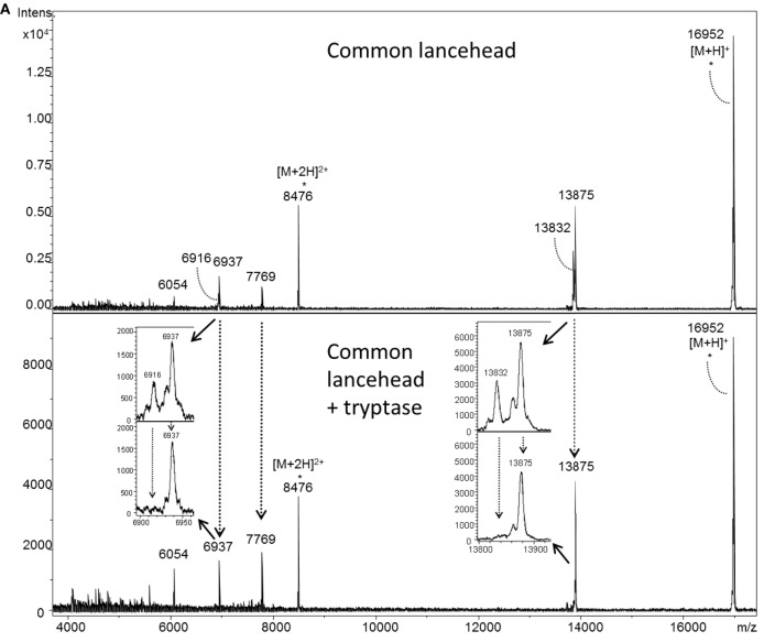
Figure 5 MALDI-TOF-MS analyses of whole venoms treated with purified human tryptase shows degradation of venom proteins. Venoms from five snake species [(A) common lancehead, (B) saw-scaled viper, (C) southern copperhead, (D) russel’s viper, (E), western diamondback rattlesnake] were treated with tryptase or vehicle control. Spectra were acquired within the mass range of m/z 3,500 to m/z 18,000. Samples contain equal amounts (0.5 µg) myoglobin as an internal reference (m/z 16,952 [M + H]+, m/z 8,476 [M + 2H]2+, m/z 5,651 [M + 3H]3+). Venom-derived signals (dashed arrows) distributed over the whole mass range were selected and normalized to the intensity of the doubly charged myoglobin signal (Figure 4B). The reduction in relative signal intensities of several peaks in tryptase-treated versus untreated venom indicates tryptase-mediated protein degradation.
Figure 5 MALDI-TOF-MS analyses of whole venoms treated with purified human tryptase shows degradation of venom proteins. Venoms from five snake species [(A) common lancehead, (B) saw-scaled viper, (C) southern copperhead, (D) russel’s viper, (E), western diamondback rattlesnake] were treated with tryptase or vehicle control. Spectra were acquired within the mass range of m/z 3,500 to m/z 18,000. Samples contain equal amounts (0.5 µg) myoglobin as an internal reference (m/z 16,952 [M + H]+, m/z 8,476 [M + 2H]2+, m/z 5,651 [M + 3H]3+). Venom-derived signals (dashed arrows) distributed over the whole mass range were selected and normalized to the intensity of the doubly charged myoglobin signal (Figure 4B). The reduction in relative signal intensities of several peaks in tryptase-treated versus untreated venom indicates tryptase-mediated protein degradation.CC BY · PubMed Central
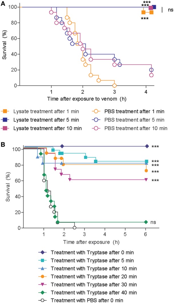
Figure 6 Skin mast cell lysate and purified human tryptase protects zebrafish from cobra venom when administered as a therapy. (A) 48 h post fertilization (hpf) zebrafish received 625 ng red spitting cobra whole venom in 100 µl each. 1, 5, or 10 min after addition of venom, zebrafish were treated with lysate from 12 thousand purified human skin mast cells per fish. Data are pooled from three independent experiments, with n = 4 zebrafish per group, each. (B) 48 hpf zebrafish received 625 ng red spitting cobra whole venom in 100 µl each. Between 0 and 40 min after addition of venom, zebrafish were treated with 10 µg/ml purified human tryptase. Data are pooled from six independent experiments with n = 5 embryos per group, each. Lysate, tryptase, or vehicle alone had no effect (data not shown). Kaplan–Meyer survival analysis, Log-rank (Mantel-Cox) test compares treatments to vehicle. ***p < 0.001.
Figure 6 Skin mast cell lysate and purified human tryptase protects zebrafish from cobra venom when administered as a therapy. (A) 48 h post fertilization (hpf) zebrafish received 625 ng red spitting cobra whole venom in 100 µl each. 1, 5, or 10 min after addition of venom, zebrafish were treated with lysate from 12 thousand purified human skin mast cells per fish. Data are pooled from three independent experiments, with n = 4 zebrafish per group, each. (B) 48 hpf zebrafish received 625 ng red spitting cobra whole venom in 100 µl each. Between 0 and 40 min after addition of venom, zebrafish were treated with 10 µg/ml purified human tryptase. Data are pooled from six independent experiments with n = 5 embryos per group, each. Lysate, tryptase, or vehicle alone had no effect (data not shown). Kaplan–Meyer survival analysis, Log-rank (Mantel-Cox) test compares treatments to vehicle. ***p < 0.001.CC BY · PubMed Central
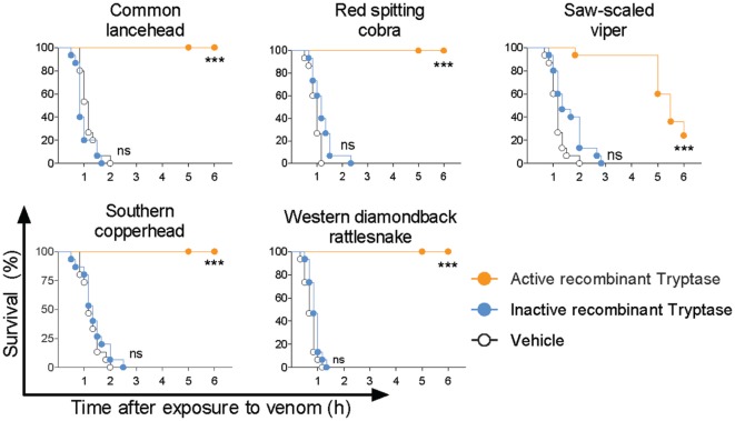
Figure 7 Active recombinant human tryptase β detoxifies snake venoms. Venoms were incubated with 10 µg/ml active human recombinant tryptase β and then administered to 48 h post fertilization zebrafish in 100 µl of water. Controls were inactive recombinant tryptase β and PBS (vehicle). Vehicle or enzymes alone had no effect (data not shown). Data are pooled from three to four independent experiments for n = 15–20 fish per group. Kaplan–Meyer survival analysis, Log-rank (Mantel-Cox) test compares treatments to vehicle. ***p < 0.001.
Figure 7 Active recombinant human tryptase β detoxifies snake venoms. Venoms were incubated with 10 µg/ml active human recombinant tryptase β and then administered to 48 h post fertilization zebrafish in 100 µl of water. Controls were inactive recombinant tryptase β and PBS (vehicle). Vehicle or enzymes alone had no effect (data not shown). Data are pooled from three to four independent experiments for n = 15–20 fish per group. Kaplan–Meyer survival analysis, Log-rank (Mantel-Cox) test compares treatments to vehicle. ***p < 0.001.CC BY · PubMed Central

Verification

Claims the paper makes

The claims and statistics of this paper haven’t been checked yet.

Run the check — free, no account needed. It reads the open-access text and checks the sources it cites.

A few free checks a day · papers already verified open instantly · results are public

Claims other papers make about this one

Each is checked against this paper’s own text: does it say what the citing paper says it does?

Self-check

Percentages, sums and reported differences re-computed from the paper’s own numbers.

The self-check needs the paper’s full text.

Citations

Cites

No references could be resolved yet.

Research works printed but not linked: 35

  1. Kasturiratne, 2008, The global burden of snakebite: a literature analysis and modelling based on regional estimates of envenoming and deaths, PLoS Med, 5, e218, 10.1371/journal.pmed.0050218 · DOI
  2. Chippaux, 1998, Snake-bites: appraisal of the global situation, Bull World Health Organ, 76, 515
  3. Warrell, 2010, Snake bite, Lancet, 375, 77, 10.1016/S0140-6736(09)61754-2 · DOI
  4. Williams, 2010, The global snake bite initiative: an antidote for snake bite, Lancet, 375, 89, 10.1016/S0140-6736(09)61159-4 · DOI
  5. Kasturiratne, 2017, The socio-economic burden of snakebite in Sri Lanka, PLoS Negl Trop Dis, 11, e0005647, 10.1371/journal.pntd.0005647 · DOI
  6. 2017, Snake-bite envenoming: a priority neglected tropical disease, Lancet, 390, 2, 10.1016/S0140-6736(17)31751-8 · DOI
  7. Williams, 2011, Ending the drought: new strategies for improving the flow of affordable, effective antivenoms in Asia and Africa, J Proteomics, 74, 1735, 10.1016/j.jprot.2011.05.027 · DOI
  8. de Silva, 2011, Low-dose adrenaline, promethazine, and hydrocortisone in the prevention of acute adverse reactions to antivenom following snakebite: a randomised, double-blind, placebo-controlled trial, PLoS Med, 8, e1000435, 10.1371/journal.pmed.1000435 · DOI
  9. Stone, 2013, Immune response to snake envenoming and treatment with antivenom; complement activation, cytokine production and mast cell degranulation, PLoS Negl Trop Dis, 7, e2326, 10.1371/journal.pntd.0002326 · DOI
  10. de Silva, 2016, Adverse reactions to snake antivenom, and their prevention and treatment, Br J Clin Pharmacol, 81, 446, 10.1111/bcp.12739 · DOI
  11. Vongphoumy, 2016, Prospective, consecutive case series of 158 snakebite patients treated at Savannakhet provincial hospital, Lao People’s Democratic Republic with high incidence of anaphylactic shock to horse derived F(ab′)2 antivenom, Toxicon, 117, 13, 10.1016/j.toxicon.2016.03.011 · DOI
  12. Metz, 2006, Mast cells can enhance resistance to snake and honeybee venoms, Science, 313, 526, 10.1126/science.1128877 · DOI · Paper page
  13. Schneider, 2007, Molecular mechanism of mast cell mediated innate defense against endothelin and snake venom sarafotoxin, J Exp Med, 204, 2629, 10.1084/jem.20071262 · DOI · Paper page
  14. Akahoshi, 2011, Mast cell chymase reduces the toxicity of Gila monster venom, Scorpion venom, and vasoactive intestinal polypeptide in mice, J Clin Invest, 121, 4180, 10.1172/JCI46139 · DOI · Paper page
  15. Kirshenbaum, 2003, Characterization of novel stem cell factor responsive human mast cell lines LAD 1 and 2 established from a patient with mast cell sarcoma/leukemia; activation following aggregation of FcepsilonRI or FcgammaRI, Leuk Res, 27, 677, 10.1016/S0145-2126(02)00343-0 · DOI
  16. Babina, 2001, Retinoic acid up-regulates myeloid ICAM-3 expression and function in a cell-specific fashion – evidence for retinoid signaling pathways in the mast cell lineage, J Leukoc Biol, 69, 361, 10.1189/jlb.69.3.361 · DOI
  17. Vukman, 2012, Mast cells cultured from IL-3-treated mice show impaired responses to bacterial antigen stimulation, Inflamm Res, 61, 79, 10.1007/s00011-011-0394-6 · DOI
  18. Bahri, 2018, Mast cell activation test in the diagnosis of allergic disease and anaphylaxis, J Allergy Clin Immunol, 10.1016/j.jaci.2018.01.043 · DOI
  19. Tsvilovskyy, 2018, Deletion of Orai2 augments endogenous CRAC currents and degranulation in mast cells leading to enhanced anaphylaxis, Cell Calcium, 71, 24, 10.1016/j.ceca.2017.11.004 · DOI
  20. Niles, 1998, Recombinant human mast cell tryptase beta: stable expression in Pichia pastoris and purification of fully active enzyme, Biotechnol Appl Biochem, 28, 125, 10.1111/j.1470-8744.1998.tb00522.x · DOI
  21. Schaschke, 2005, Affinity chromatography of tryptases: design, synthesis and characterization of a novel matrix-bound bivalent inhibitor, Chembiochem, 6, 95, 10.1002/cbic.200400217 · DOI
  22. Schaschke, 2001, Bivalent inhibition of human beta-tryptase, Chem Biol, 8, 313, 10.1016/S1074-5521(01)00011-4 · DOI
  23. Kolarich, 2012, Determination of site-specific glycan heterogeneity on glycoproteins, Nat Protoc, 7, 1285, 10.1038/nprot.2012.062 · DOI
  24. Seifert, 2009, AAPCC database characterization of native U.S. Venomous snake exposures, 2001-2005, Clin Toxicol (Phila), 47, 327, 10.1080/15563650902870277 · DOI
  25. Alirol, 2010, Snake bite in South Asia: a review, PLoS Negl Trop Dis, 4, e603, 10.1371/journal.pntd.0000603 · DOI
  26. Segura, 2010, Preclinical assessment of the neutralizing capacity of antivenoms produced in six Latin American countries against medically-relevant Bothrops snake venoms, Toxicon, 56, 980, 10.1016/j.toxicon.2010.07.001 · DOI
  27. Guhl, 2010, Mast cell lines HMC-1 and LAD2 in comparison with mature human skin mast cells – drastically reduced levels of tryptase and chymase in mast cell lines, Exp Dermatol, 19, 845, 10.1111/j.1600-0625.2010.01103.x · DOI
  28. 2010, WHO Guidelines for the Production, Control and Regulation of Snake Antivenom Immunoglobulins
  29. Metz, 2007, Mast cells in the promotion and limitation of chronic inflammation, Immunol Rev, 217, 304, 10.1111/j.1600-065X.2007.00520.x · DOI
  30. Du, 2002, Snake venom L-amino acid oxidases, Toxicon, 40, 659, 10.1016/S0041-0101(02)00157-5 · DOI
  31. Gutierrez, 2014, A multicomponent strategy to improve the availability of antivenom for treating snakebite envenoming, Bull World Health Organ, 92, 526, 10.2471/BLT.13.132431 · DOI
  32. Weber, 2003, Pattern analysis of human cutaneous mast cell populations by total body surface mapping, Br J Dermatol, 148, 224, 10.1046/j.1365-2133.2003.05090.x · DOI
  33. Sommerhoff, 1999, The structure of the human betaII-tryptase tetramer: fo(u)r better or worse, Proc Natl Acad Sci U S A, 96, 10984, 10.1073/pnas.96.20.10984 · DOI
  34. Gutierrez, 2000, Snake venom metalloproteinases: their role in the pathogenesis of local tissue damage, Biochimie, 82, 841, 10.1016/S0300-9084(00)01163-9 · DOI
  35. Sommerhoff, 2000, The human mast cell tryptase tetramer: a fascinating riddle solved by structure, Biochim Biophys Acta, 1477, 75, 10.1016/S0167-4838(99)00265-4 · DOI

Cited by

Sorted by citations

See all 30

Metadata

Authors
Anderson, E., Stavenhagen, K., Kolarich, D., Sommerhoff, C. P., Maurer, M., & Metz, M.
Venue
Frontiers in Immunology
Published
2018-07-09
Volume
9
ISSN
1664-3224
Cited by
30
References
0
Funders
  • Deutsche Forschungsgemeinschaft (ME 2668/3-2, ME 2668/2-1, SO 249/1-1)
  • Else Kröner-Fresenius-Stiftung
  • Max-Planck-Gesellschaft

About these checks

Registry, retraction, Cites and statistics results are computed, not judged, with one exception: a printed reference that no index lists is read once by a model before it counts as a citation error. Claims are extracted by a model and kept only when their quote appears verbatim in the text; each cited source is then checked, with a model’s help, for whether it says what the claim relies on. Those verdicts can be wrong — read the claim’s quote and the cited source before relying on one.

Statistics and arithmetic are recomputed, not judged.

Confidence bands are shown only for fields calibrated on our annotated test set. None is calibrated yet, so no band is shown.

What counts as a claim ›How we measure confidence ›How we check this ›

Are you an author, or do you see a problem with this page? Request a review

How to cite

Cite CoChat for the checks on this page, and the paper for its own findings.

This page
CoChat. (2026). Citation check: Human Mast Cell Tryptase Is a Potential Treatment for Snakebite Envenoming Across Multiple Snake Species. https://app.cochat.ai/papers/10.3389/fimmu.2018.01532
The paper
Anderson, E., Stavenhagen, K., Kolarich, D., Sommerhoff, C. P., Maurer, M., & Metz, M. (2018). Human Mast Cell Tryptase Is a Potential Treatment for Snakebite Envenoming Across Multiple Snake Species. Frontiers in Immunology. https://doi.org/10.3389/fimmu.2018.01532