

The DOI matches its Crossref record; 87 of the 87 references it cites were found in a registry or an index.
ccf-mtDNA as a Potential Link Between the Brain and Immune System in Neuro-Immunological Disorders
Open accessAbstract
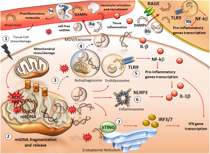
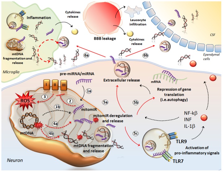
Fragments of mitochondrial DNA (mtDNA) are released outside the cell and they appear to persist in extracellular fluids as circulating, cell-free, mtDNA (ccf-mtDNA). When compared to nuclear DNA, such a double stranded mtDNA is more resistant to nuclease degradation. In fact, it is stable extracellularly where it can be detected in both plasma and cerebrospinal fluid (CSF), here acting as a potential biomarker in various disorders. In neurological diseases (Alzheimer's disease, Parkinson's disease and end-stage progressive Multiple Sclerosis), a decreased amount of CSF ccf-mtDNA is related with progressive cell dysfunction. This suggests an alteration in neuronal mtDNA levels (mtDNA replication, degradation and depletion) in vulnerable brain regions at early stages of neurodegeneration leading to reduced mtDNA release, which takes place before actual cell death occurs. On the other hand, elevated CSF ccf-mtDNA levels are reported in acute phases of relapsing-remitting Multiple Sclerosis (RRMS). This occurs during acute inflammation, which anticipates the neurodegenerative process. Thus, an increase in inflammatory cells in the affected regions is expected to add on mtDNA release into the CSF. In addition, similarly to bacterial DNA, the non-methylated CpG sites of mtDNA, which activate innate immunity and inflammation, are likely to participate in the molecular mechanisms of disease. Thus, ccf-mtDNA may represent a powerful biomarker for disease screening and prognosis at early stage, although its biological role may extend to generating the neurobiology of disease. The present manuscript discusses recent experimental findings in relationship with clinical evidence comparing neuro-immunological features of neurodegenerative disorders with frankly neuro-infectious diseases.
Overview
The claims and statistics of this paper haven’t been checked yet.
Run the check — free, no account needed. It reads the open-access text and checks the sources it cites.
Integrity signals
Registry
Cites
Statistics
Figures
Verification
Claims the paper makes
The claims and statistics of this paper haven’t been checked yet.
Run the check — free, no account needed. It reads the open-access text and checks the sources it cites.
Claims other papers make about this one
Each is checked against this paper’s own text: does it say what the citing paper says it does?
The source backs part of the claim, not all of it — or backs it for a different population, condition or comparison.
In the citing paper
“Assessing the presence of extracellular mitochondria or their fragments by measuring ccf-mtDNA levels could allow on-flight direct estimation of cellular stress to take preventive measures of cumulative damage196,199–206.”
From Key points for the development of antioxidant cocktails to prevent cellular stress and damage caused by reactive oxygen species (ROS) during manned space missions (2021) · cites this paper as [203]
Experimental — accuracy not yet measured
Self-check
Percentages, sums and reported differences re-computed from the paper’s own numbers.
The self-check needs the paper’s full text.
Citations
Cites
Delivery of siRNA to the mouse brain by systemic injection of targeted exosomes
Alvarez-Erviti, L., Seow, Y., Yin, H., Betts, C., Lakhal, S., & Wood, M. J. A. (2011) · Nature Biotechnology · doi:10.1038/nbt.1807 · Cited by 4,930
Circulating mitochondrial DAMPs cause inflammatory responses to injury
Zhang, Q., Raoof, M., Chen, Y., Sumi, Y., Sursal, T., Junger, W., Brohi, K., Itagaki, K., & Hauser, C. J. (2010) · Nature · doi:10.1038/nature08780 · Cited by 3,812
Bender, A., Krishnan, K. J., Morris, C. M., Taylor, G. A., Reeve, A. K., Perry, R. H., Jaros, E., Hersheson, J. S., Betts, J., Klopstock, T., et al. (2006) · Nature Genetics · doi:10.1038/ng1769 · Cited by 1,570
Healy, D. G., Falchi, M., O'Sullivan, S. S., Bonifati, V., Durr, A., Bressman, S., Brice, A., Aasly, J., Zabetian, C. P., Goldwurm, S., et al. (2008) · The Lancet Neurology · doi:10.1016/s1474-4422(08)70117-0 · Cited by 1,425
Mitochondria in innate immune responses
West, A. P., Shadel, G. S., & Ghosh, S. (2011) · Nature Reviews Immunology · doi:10.1038/nri2975 · Cited by 1,376
Zong, W., Rabinowitz, J. D., & White, E. (2016) · Molecular Cell · doi:10.1016/j.molcel.2016.02.011 · Cited by 1,320
Pathological mechanisms in progressive multiple sclerosis
Mahad, D., Trapp, B. D., & Lassmann, H. (2015) · The Lancet Neurology · doi:10.1016/s1474-4422(14)70256-x · Cited by 1,198
Catapult-like release of mitochondrial DNA by eosinophils contributes to antibacterial defense
Yousefi, S., Gold, J. A., Andina, N., Lee, J. J., Kelly, A. M., Kozlowski, E., Schmid, I., Straumann, A., Reichenbach, J., Gleich, G. J., et al. (2008) · Nature Medicine · doi:10.1038/nm.1855 · Cited by 967
Tapiola, T., Alafuzoff, I., Herukka, S.-K., Parkkinen, L., Hartikainen, P., Soininen, H., & Pirttilä, T. (2009) · Archives of Neurology · doi:10.1001/archneurol.2008.596 · Cited by 896
An unconventional role for miRNA: let-7 activates toll-like receptor 7 and causes neurodegeneration
Lehmann, S. M., Krüger, C., Park, B., Derkow, K., Rosenberger, K., Baumgart, J., Trimbuch, T., Eom, G., Hinz, M., Kaul, D., et al. (2012) · Nature Neuroscience · doi:10.1038/nn.3113 · Cited by 786
Neutrophil Extracellular Traps and Its Implications in Inflammation: An Overview
Delgado‐Rizo, V., Martinez-Guzman, M. A., Íñiguez-Gutiérrez, L., García-Orozco, A., Alvarado‐Navarro, A., & Fafutis‐Morris, M. (2017) · Frontiers in Immunology · doi:10.3389/fimmu.2017.00081 · Cited by 703
Astrocytes and Glioblastoma cells release exosomes carrying mtDNA
Guescini, M., Genedani, S., Stocchi, V., & Agnati, L. F. (2009) · Journal of Neural Transmission · doi:10.1007/s00702-009-0288-8 · Cited by 662
Impaired mitochondrial biogenesis contributes to mitochondrial dysfunction in Alzheimer’s disease
Sheng, B., Wang, X., Su, B., Lee, H., Casadesús, G., Perry, G. L., & Zhu, X. (2011) · Journal of Neurochemistry · doi:10.1111/j.1471-4159.2011.07581.x · Cited by 545
Localization of LRRK2 to membranous and vesicular structures in mammalian brain
Biskup, S., Moore, D. J., Celsi, F., Higashi, S., West, A. B., Andrabi, S. A., Kurkinen, K. M. A., Yu, S., Savitt, J. M., Waldvogel, H. J., et al. (2006) · Annals of Neurology · doi:10.1002/ana.21019 · Cited by 530
Mitochondrial dysfunction and mitophagy in Parkinson's: from familial to sporadic disease
Ryan, B. J., Hoek, S., Fon, E. A., & Wade-Martins, R. (2015) · Trends in Biochemical Sciences · doi:10.1016/j.tibs.2015.02.003 · Cited by 524
Is mitochondrial DNA content a potential biomarker of mitochondrial dysfunction?
Malik, A. N., & Czajka, A. (2012) · Mitochondrion · doi:10.1016/j.mito.2012.10.011 · Cited by 507
Endogenously oxidized mitochondrial DNA induces in vivo and in vitro inflammatory responses
Collins, L. V., Hajizadeh, S., Holme, E., Jonsson, I., & Tarkowski, A. (2004) · Journal of Leukocyte Biology · doi:10.1189/jlb.0703328 · Cited by 504
Hsieh, C.-H., Shaltouki, A., Gonzalez, A. E., da Cruz, A. B., Burbulla, L. F., Lawrence, E. S., Schüle, B., Krainc, D., Palmer, T. D., & Wang, X. (2016) · Cell stem cell · doi:10.1016/j.stem.2016.08.002 · Cited by 475
C. elegans neurons jettison protein aggregates and mitochondria under neurotoxic stress
Melentijevic, I., Toth, M. L., Arnold, M. L., Guasp, R. J., Harinath, G., Nguyen, K. C., Taub, D., Parker, J. A., Neri, C., Gabel, C. V., et al. (2017) · Nature · doi:10.1038/nature21362 · Cited by 454
Nakahira, K., Kyung, S.-Y., Rogers, A. J., Gazourian, L., Youn, S., Massaro, A. F., Quintana, C., Osorio, J. C., Wang, Z., Zhao, Y., et al. (2013) · PLoS Medicine · doi:10.1371/journal.pmed.1001577 · Cited by 445
Lukiw, W. J., Zhao, Y., & Cui, J. G. (2008) · Journal of Biological Chemistry · doi:10.1074/jbc.m805371200 · Cited by 441
MITOCHONDRIAL DNA IS RELEASED BY SHOCK AND ACTIVATES NEUTROPHILS VIA P38 MAP KINASE
Zhang, Q., Itagaki, K., & Hauser, C. J. (2010) · Shock · doi:10.1097/shk.0b013e3181cd8c08 · Cited by 361
STING-dependent cytosolic DNA sensing pathways
Barber, G. N. (2013) · Trends in Immunology · doi:10.1016/j.it.2013.10.010 · Cited by 341
Pickrell, A. M., Huang, C.-H., Kennedy, S. R., Ordureau, A., Sideris, D. P., Hoekstra, J. G., Harper, J. W., & Youle, R. J. (2015) · Neuron · doi:10.1016/j.neuron.2015.06.034 · Cited by 327
Wei, X., Shao, B., He, Z., Ye, T., Luo, M., Sang, Y., Liang, X., Wang, W., Luo, S., Yang, S., et al. (2015) · Cell Research · doi:10.1038/cr.2015.9 · Cited by 309
Schipler, A., & Iliakis, G. E. (2013) · Nucleic Acids Research · doi:10.1093/nar/gkt556 · Cited by 290
Mitochondrial dysfunction contributes to neurodegeneration in multiple sclerosis
Witte, M. E., Mahad, D., Lassmann, H., & van Horssen, J. (2013) · Trends in Molecular Medicine · doi:10.1016/j.molmed.2013.11.007 · Cited by 276
Defective mitochondrial DNA homeostasis in the substantia nigra in Parkinson disease
Dölle, C., Flønes, I. H., Nido, G. S., Miletić, H., Osuagwu, N., Kristoffersen, S., Lilleng, P. K., Larsen, J. P., Tysnes, O., Haugarvoll, K., et al. (2016) · Nature Communications · doi:10.1038/ncomms13548 · Cited by 271
Circulating MicroRNAs as biomarkers for disease staging in multiple sclerosis
Gandhi, R., Healy, B. C., Gholipour, T., Egorova, S., Musallam, A., Hussain, M. S., Nejad, P., Patel, B., Hei, H., Khoury, S. J., et al. (2013) · Annals of Neurology · doi:10.1002/ana.23880 · Cited by 261
MicroRNA regulation of autophagy
Frankel, L. B., & Lund, A. H. (2012) · Carcinogenesis · doi:10.1093/carcin/bgs266 · Cited by 261
Kataoka, N., Hioki, H., Kaneko, T., & Nakamura, K. (2014) · Cell Metabolism · doi:10.1016/j.cmet.2014.05.018 · Cited by 240
Early‐onset familial parkinsonism due to POLG mutations
Davidzon, G., Greene, P., Mancuso, M., Klos, K. J., Ahlskog, J. E., Hirano, M., & DiMauro, S. (2006) · Annals of Neurology · doi:10.1002/ana.20831 · Cited by 239
MicroRNAs: Roles in Regulating Neuroinflammation
Gaudet, A. D., Fonken, L. K., Watkins, L. R., Nelson, R. J., & Popovich, P. G. (2018) · The Neuroscientist · doi:10.1177/1073858417721150 · Cited by 236
Mitochondrial DNA Depletion in Respiratory Chain–Deficient P arkinson Disease Neurons
Grünewald, A., Rygiel, K. A., Hepplewhite, P. D., Morris, C. M., Picard, M. A., & Turnbull, D. M. (2015) · Annals of Neurology · doi:10.1002/ana.24571 · Cited by 229
Low cerebrospinal fluid concentration of mitochondrial DNA in preclinical Alzheimer disease
Podlesniy, P., Figueiro‐Silva, J., Llado, A., Antonell, A., Sanchez‐Valle, R., Alcolea, D., Lleo, A., Molinuevo, J. L., Serra, N., & Trullas, R. (2013) · Annals of Neurology · doi:10.1002/ana.23955 · Cited by 219
Cao, D., Li, L., & Chan, W. (2016) · International Journal of Molecular Sciences · doi:10.3390/ijms17060842 · Cited by 208
Haqqani, A. S., Delaney, C. E., Tremblay, T., Sodja, C., Sandhu, J. K., & Stanimirovic, D. B. (2013) · Fluids and Barriers of the CNS · doi:10.1186/2045-8118-10-4 · Cited by 200
Hajizadeh, S., DeGroot, J., TeKoppele, J. M., Tarkowski, A., & Collins, L. V. (2003) · Arthritis Res Ther · doi:10.1186/ar787 · Cited by 196
Oxidative Damage to RNA in Aging and Neurodegenerative Disorders
Nunomura, A., Moreira, P. I., Castellani, R. J., Lee, H., Zhu, X., Smith, M. A., & Perry, G. L. (2012) · Neurotoxicity Research · doi:10.1007/s12640-012-9331-x · Cited by 185
Advances in the understanding of mitochondrial DNA as a pathogenic factor in inflammatory diseases
Boyapati, R. K., Tamborska, A., Dorward, D. A., & Ho, G. (2017) · F1000Research · doi:10.12688/f1000research.10397.1 · Cited by 170
MitomiRs delineating the intracellular localization of microRNAs at mitochondria
Bandiera, S., Matégot, R., Girard, M., Jacques Demongeot, A., & Henrion‐Caude, A. (2013) · Free Radical Biology and Medicine · doi:10.1016/j.freeradbiomed.2013.06.013 · Cited by 166
Neurofilament light chain predicts disease activity in relapsing-remitting MS
Varhaug, K. N., Barro, C., Bjørnevik, K., Myhr, K., Torkildsen, Ø., Wergeland, S., Bindoff, L. A., Kühle, J., & Vedeler, C. A. (2017) · Neurology Neuroimmunology & Neuroinflammation · doi:10.1212/nxi.0000000000000422 · Cited by 164
Role of Mitochondrial DNA in Septic AKI via Toll-Like Receptor 9
Tsuji, N., Tsuji, T., Ohashi, N., Kato, A., Fujigaki, Y., & Yasuda, H. (2015) · Journal of the American Society of Nephrology · doi:10.1681/asn.2015040376 · Cited by 162
Coskun, P. E., Wyrembak, J., Derbereva, O., Melkonian, G., Doran, E. W., Lott, I. T., Head, E., Cotman, C. W., & Wallace, D. C. (2010) · Journal of Alzheimer s Disease · doi:10.3233/jad-2010-100351 · Cited by 161
The Role of microRNAs in Mitochondria: Small Players Acting Wide
Duarte, F. V., Palmeira, C. M., & Rolo, A. P. (2014) · Genes · doi:10.3390/genes5040865 · Cited by 150
Control of mitochondrial activity by miRNAs
Li, P., Jiao, J., Gao, G., & Prabhakar, B. S. (2011) · Journal of Cellular Biochemistry · doi:10.1002/jcb.24004 · Cited by 144
Nissanka, N., Bacman, S. R., Plastini, M. J., & Moraes, C. T. (2018) · Nature Communications · doi:10.1038/s41467-018-04895-1 · Cited by 139
TLR-2 and TLR-9 are sensors of apoptosis in a mouse model of doxorubicin-induced acute inflammation
Krysko, D. V., Kaczmarek, A., Krysko, O., Heyndrickx, L., Woznicki, J., Bogaert, P., Cauwels, A., Takahashi, N., Magez, S., Bachert, C., et al. (2011) · Cell Death and Differentiation · doi:10.1038/cdd.2011.4 · Cited by 129
Reduced cerebrospinal fluid mitochondrial DNA is a biomarker for early‐stage Parkinson's disease
Pyle, A., Brennan, R. R., Kurzawa‐Akanbi, M., Yarnall, A. J., Thouin, A., Mollenhauer, B., Burn, D. J., Chinnery, P. F., & Hudson, G. (2015) · Annals of Neurology · doi:10.1002/ana.24515 · Cited by 123
Rice, A. C., Keeney, P. M., Algarzae, N. K., Ladd, A. C., Thomas, R. R., & Bennett, J. P. (2014) · Journal of Alzheimer s Disease · doi:10.3233/jad-131715 · Cited by 117
Kageyama, Y., Kasahara, T., Kato, M., Sakai, S., Deguchi, Y., Tani, M., Kuroda, K., Hattori, K., Yoshida, S., Goto, Y., et al. (2017) · Journal of Affective Disorders · doi:10.1016/j.jad.2017.06.001 · Cited by 116
Mehra, N., Penning, M. T., Maas, J., van Daal, N., Giles, R. H., & Voest, E. E. (2007) · Clinical Cancer Research · doi:10.1158/1078-0432.ccr-06-1087 · Cited by 115
Carrizzo, A., Lenzi, P., Procaccini, C., Damato, A., Biagioni, F., Ambrosio, M., Amodio, G., Remondelli, P., Giudice, C. D., Izzo, R., et al. (2015) · Circulation · doi:10.1161/circulationaha.114.014822 · Cited by 106
Self-citationCossarizza, A., Pinti, M., Nasi, M., Gibellini, L., Manzini, S., Roat, E., Biasi, S. D., Bertoncelli, L., Montagna, J. P., Bisi, L., et al. (2011) · Mitochondrion · doi:10.1016/j.mito.2011.06.005 · Cited by 97
Plasma Mitochondrial DNA Concentrations after Trauma
Lam, N. Y. L., Rainer, T. H., Chiu, R. W. K., Joynt, G. M., & Lo, Y. M. D. (2004) · Clinical Chemistry · doi:10.1373/clinchem.2003.025783 · Cited by 97
Li, L., HANN, H.-W. L., Wan, S., Hann, R. S., Wang, C., Lai, Y., Ye, X., Evans, A. A., Myers, R. E., Ye, Z., et al. (2016) · Scientific Reports · doi:10.1038/srep23992 · Cited by 94
Mitochondrial quality control and neurological disease: an emerging connection
de Castro, I. P., Martins, L. M., & Tufi, R. (2010) · Expert Reviews in Molecular Medicine · doi:10.1017/s1462399410001456 · Cited by 92
Sursal, T., Stearns-Kurosawa, D. J., Itagaki, K., Oh, S., Sun, S., Kurosawa, S., & Hauser, C. J. (2012) · Shock · doi:10.1097/shk.0b013e318276f4ca · Cited by 91
Mitochondrial genome changes and neurodegenerative diseases
Pinto, M., & Moraes, C. T. (2013) · Biochimica et Biophysica Acta (BBA) - Molecular Basis of Disease · doi:10.1016/j.bbadis.2013.11.012 · Cited by 75
Hu, Q., Wood, C. R., Cimen, S., Venkatachalam, A. B., & Alwayn, I. P. J. (2015) · PLOS ONE · doi:10.1371/journal.pone.0140105 · Cited by 75
Circulating Cell-Free DNA Differentiates Severity of Inflammation
Frank, M. O. (2016) · Biological Research For Nursing · doi:10.1177/1099800416642571 · Cited by 70
Kataoka, H., Kono, H., Patel, Z., & Rock, K. L. (2014) · PLoS ONE · doi:10.1371/journal.pone.0104741 · Cited by 66
Nuclease resistant circular DNAs copurify with infectivity in scrapie and CJD
Manuelidis, L. (2010) · Journal of NeuroVirology · doi:10.1007/s13365-010-0007-0 · Cited by 58
Cell-free mitochondrial DNA in progressive multiple sclerosis
Lowes, H., Pyle, A., Duddy, M., & Hudson, G. (2018) · Mitochondrion · doi:10.1016/j.mito.2018.07.008 · Cited by 52
Keeney, P. M., & Bennett, J. P. (2010) · Molecular Neurodegeneration · doi:10.1186/1750-1326-5-21 · Cited by 51
Significance of Serum mtDNA Concentration in Lung Injury Induced by Hip Fracture
Gan, L., Chen, X., Sun, T., Li, Q., Zhang, R., Zhang, J., & Zhong, J. (2015) · Shock · doi:10.1097/shk.0000000000000366 · Cited by 50
Cerebrospinal fluid mitochondrial DNA in the Alzheimer's disease continuum
Cervera‐Carles, L., Alcolea, D. A., Estanga, A., Ecay‐Torres, M., Izagirre, A., Clerigué, M., García‐Sebastián, M., Villanúa, J., Escalas, C., Blesa, R., et al. (2016) · Neurobiology of Aging · doi:10.1016/j.neurobiolaging.2016.12.009 · Cited by 47
Mitochondrial DNA in CSF distinguishes LRRK2 from idiopathic Parkinson's disease
Podlesniy, P., Vilas, D., Taylor, P., Shaw, L. M. J., Tolosa, E., & Trullás, R. (2016) · Neurobiology of Disease · doi:10.1016/j.nbd.2016.05.019 · Cited by 45
Leurs, C. E., Podlesniy, P., Trullás, R., Balk, L. J., Steenwijk, M. D., Malekzadeh, A., Piehl, F., Uitdehaag, B. M. J., Killestein, J., van Horssen, J., et al. (2017) · Multiple Sclerosis Journal · doi:10.1177/1352458517699874 · Cited by 45
Ruffoli, R., Bartalucci, A., Frati, A., & Fornai, F. (2015) · Frontiers in Cellular Neuroscience · doi:10.3389/fncel.2015.00341 · Cited by 42
Self-citationNatale, G., Lenzi, P., Lazzeri, G., Falleni, A., Biagioni, F., Ryskalin, L., & Fornai, F. (2015) · Frontiers in Cellular Neuroscience · doi:10.3389/fncel.2015.00434 · Cited by 42
Self-citationFernandes, J., Michel, V., Camorlinga‐Ponce, M., Gómez, A., Maldonado‐Bernal, C., Reuse, H. D., Torres, J., & Touati, E. (2014) · Cancer Epidemiology Biomarkers & Prevention · doi:10.1158/1055-9965.epi-14-0471 · Cited by 39
Systematic analysis of microRNA fingerprints in thrombocythemic platelets using integrated platforms
Xu, X., Gnatenko, D. V., Ju, J., Hitchcock, I. S., Martin, D. W., Zhu, W., & Bahou, W. F. (2012) · Blood · doi:10.1182/blood-2012-02-411264 · Cited by 39
Mehta, S. R., Pérez‐Santiago, J., Hulgan, T., Day, T. R. C., Barnholtz‐Sloan, J. S., Gittleman, H. R., Letendre, S. L., Ellis, R. J., Heaton, R. K., Patton, S. M., et al. (2017) · Journal of Neuroinflammation · doi:10.1186/s12974-017-0848-z · Cited by 35
Arshad, O., Gadawska, I., Sattha, B., Côté, H. C. F., & Hsieh, A. Y. Y. (2018) · JAIDS Journal of Acquired Immune Deficiency Syndromes · doi:10.1097/qai.0000000000001650 · Cited by 33
Wnęk, M., Ressel, L., Ricci, E., Rodriguez-Martinez, C., Guerrero, J. C. V., Ismail, Z., Smith, C., Kipar, A., Sodeik, B., Chinnery, P. F., et al. (2016) · Acta Neuropathologica · doi:10.1007/s00401-016-1597-2 · Cited by 29
Evaluation of mitochondrial respiratory chain in the brain of rats after pneumococcal meningitis
Barichello, T., Savi, G. D., Simões, L. R., Generoso, J. S., Fraga, D. B., Bellettini, G., Daufenbach, J. F., Rezin, G. T., Scaini, G., & STRECK, E. L. (2010) · Brain Research Bulletin · doi:10.1016/j.brainresbull.2010.05.012 · Cited by 22
Neuron-Specific Mitochondrial DNA Deletion Levels in Sporadic Alzheimer´s Disease
Gerschütz, A., Heinsen, H., Grünblatt, E., Wagner, A., Bartl, J., Meißner, C., Fallgatter, A. J., Al‐Sarraj, S., Troakes, C., Ferrer, I., et al. (2013) · Current Alzheimer Research · doi:10.2174/15672050113106660166 · Cited by 18
Role of mitochondria in regulating microRNA activity and its relevance to the central nervous system
Wang, W.-X., & Springer, J. (2015) · Neural Regeneration Research · doi:10.4103/1673-5374.160061 · Cited by 18
Berezin, A. Е. (2015) · Angiology Open Access · doi:10.4172/2329-9495.1000161 · Cited by 17
Clinical implications and pathological associations of circulating mitochondrial DNA
Balogh, Z. J. (2016) · Frontiers in bioscience · doi:10.2741/4530 · Cited by 17
Brain diseases and tumorigenesis: The good and bad cops of pentraxin3
Fornai, F., Carrizzo, A., Ferrucci, M., Damato, A., Biagioni, F., Gaglione, A., Puca, A. A., & Vecchione, C. (2015) · The International Journal of Biochemistry & Cell Biology · doi:10.1016/j.biocel.2015.10.017 · Cited by 13
Self-citationGopinathan, U., Øvstebø, R., Brusletto, B. S., Olstad, O. K., Kierulf, P., Brandtzaeg, P., & Berg, J. P. (2017) · BMC Immunology · doi:10.1186/s12865-017-0229-5 · Cited by 9
Mechanical ventilation and Streptococcus pneumoniae pneumonia alter mitochondrial homeostasis
Blot, M., Pauchard, L.-A., Dunn-Siegrist, I., Donze, J., Malnuit, S., Rebaud, C., Croisier, D., Piroth, L., Pugin, J., & Charles, P. (2018) · Scientific Reports · doi:10.1038/s41598-018-30226-x · Cited by 8
Circulating Mitochondrial DNA Is Elevated in Patients with Immediate Primary Graft Dysfunction
Ibrahim, M., Strah, H. M., Huang, H. J., Krupnick, A. S., Kreisel, D., Hachem, R. R., Trulock, E. P., Alouch, A., & Gelman, A. E. (2013) · The Journal of Heart and Lung Transplantation · doi:10.1016/j.healun.2013.01.885 · Cited by 6
A biomarker and a target for progressive MS
Kingwell, K. (2009) · Nature Reviews Drug Discovery · doi:10.1038/nrd3006 · Cited by 4
Penetrance of LRRK2 mutations in familial Parkinson disease
(2009) · Nature Reviews Neurology · doi:10.1038/ncpneuro1026 · Cited by 2
Cited by
Sorted by citations
Lin, M., Liu, N., Qin, Z., & Wang, Y. (2022) · Acta Pharmacologica Sinica · doi:10.1038/s41401-022-00879-6 · Cited by 350
Nanoparticulate cell-free DNA scavenger for treating inflammatory bone loss in periodontitis
Huang, H., Pan, W., Wang, Y., Kim, H. S., Shao, D., Huang, B., Ho, T., Lao, Y., Quek, C. H., Shi, J., et al. (2022) · Nature Communications · doi:10.1038/s41467-022-33492-6 · Cited by 153
GDF15 and Cardiac Cells: Current Concepts and New Insights
Rochette, L., Dogon, G., Zeller, M., Cottin, Y., & Vergely, C. (2021) · International Journal of Molecular Sciences · doi:10.3390/ijms22168889 · Cited by 158
Mechanisms of mitochondrial DNA escape and its relationship with different metabolic diseases
Pérez-Treviño, P., Velasquez-Esparza, M. M., & Garcı́a, N. (2020) · Biochimica et Biophysica Acta (BBA) - Molecular Basis of Disease · doi:10.1016/j.bbadis.2020.165761 · Cited by 134
Zhao, Y., Liu, B., Xu, L., Yu, S., Fu, J., Wang, J., Yan, X., & Su, J. (2021) · Antioxidants · doi:10.3390/antiox10121917 · Cited by 117
Metadata
- Authors
- Gambardella, S., Limanaqi, F., Ferese, R., Biagioni, F., Campopiano, R., Centonze, D., & Fornai, F.
- Affiliations
- Istituto Neurologico Mediterraneo (IT)
- University of Pisa (IT)
- University of Rome Tor Vergata (IT)
- Corresponding author
- Gambardella, Stefano
- ORCID
- Venue
- Frontiers in Immunology
- Publisher
- Frontiers Media SA
- Published
- 2019-05-09
- Volume
- 10
- Pages
- 1064-1064
- ISSN
- 1664-3224 (ISSN-L)
- PMC
- PMC6520662
- OpenAlex
- W2944647162
- Cited by
- 141
- References
- 87
- Citations by year
- 2026: 12 · 2025: 22 · 2024: 22 · 2023: 27 · 2022: 22 · 2021: 21 · 2020: 13 · 2019: 1
- FWCI
- 1.26
- Type
- journal article
- Language
- en
- Open access
- gold
- doi.org
- Licence
- Locations
- Frontiers in Immunology (publishedVersion, cc-by)
- PubMed Central (publishedVersion, cc-by)
- PubMed (publishedVersion)
- Archivio Istituzionale della Ricerca (Universita Degli Studi Di Milano) (submittedVersion, other-oa)
- CINECA IRIS Institutial research information system (University of Pisa) (submittedVersion)
- Cineca Institutional Research Information System (Tor Vergata University) (submittedVersion, cc-by)
- DOAJ (DOAJ: Directory of Open Access Journals) (submittedVersion, cc-by-sa)
- CINECA IRIS Institutional Research information system (University of Urbino) (submittedVersion)
- Funders
- Ministero della Salute
- Topics
- Life Sciences › Biochemistry, Genetics and Molecular Biology › Molecular Biology › Mitochondrial Function and Pathology
- Life Sciences › Neuroscience › Neurology › Neuroinflammation and Neurodegeneration Mechanisms
- Health Sciences › Medicine › Clinical Neurology › Multiple Sclerosis Research Studies
- Keywords
- circulating cell-free mitochondrial DNA, cerebrospinal fluid biomarkers, neuroinflammation, neurodegeneration, Alzheimer's disease, Parkinson's disease, multiple sclerosis, relapsing-remitting multiple sclerosis, mitochondrial DNA release, CpG sites, innate immunity, central nervous system infection
- MeSH terms
- Cell-Free Nucleic Acids, Animals, Brain, Disease Susceptibility, DNA, Mitochondrial, Gene Expression Regulation, Humans, Immune System, Mitochondria, Nervous System Diseases, Neuroimmunomodulation, Signal Transduction, Biomarkers, Neurodegenerative Diseases, MicroRNAs, Exosomes
About these checks
Registry, retraction, Cites and statistics results are computed, not judged, with one exception: a printed reference that no index lists is read once by a model before it counts as a citation error. Claims are extracted by a model and kept only when their quote appears verbatim in the text; each cited source is then checked, with a model’s help, for whether it says what the claim relies on. Those verdicts can be wrong — read the claim’s quote and the cited source before relying on one.
Statistics and arithmetic are recomputed, not judged.
Confidence bands are shown only for fields calibrated on our annotated test set. None is calibrated yet, so no band is shown.
What counts as a claim ›How we measure confidence ›How we check this ›
Are you an author, or do you see a problem with this page? Request a review
