

The DOI matches its Crossref record; 445 of the 446 references it cites were found in a registry or an index, and 1 are not checked yet.
Immunomodulatory Role of the Extracellular Matrix Within the Liver Disease Microenvironment
Open accessAbstract
Chronic liver disease when accompanied by underlying fibrosis, is characterised by an accumulation of extracellular matrix (ECM) proteins and chronic inflammation. Although traditionally considered as a passive and largely architectural structure, the ECM is now being recognised as a source of potent DAMPs with immune-active peptides and domains. In parallel, the ECM anchors a range of cytokines, chemokines and growth factors, all of which are capable of modulating the immune responses. A growing body of evidence shows that ECM proteins themselves are capable of interacting both directly, via a wide range of receptors including integrins and TLRs, and indirectly with immune cells. ECM deposition and remodelling during fibrosis can result in release or formation of ECM-DAMPs, which can drive immune cellular infiltration and sterile inflammation. Both the ECM and immune response are interlinked and integral in driving fibrosis, although their precise interactions in the context of chronic liver disease are poorly understood. This review aims to shed light on the ECM-immune interactions underlying liver disease, in particular fibrosis of different aetiologies, by describing the main ECM intact proteins and ECM-DAMPs with known immune regulatory activity. We also discuss how tissue engineering could provide new opportunities to explore ECM-immune cell interactions in bioreactor-based dynamic 3D models containing intact decellularized ECM scaffolds. A better understanding of these interactions would aid in the identification of new antifibrotic and immunomodulatory targets for treatment and biomarkers in liver disease.
Overview
The claims and statistics of this paper haven’t been checked yet.
Run the check — free, no account needed. It reads the open-access text and checks the sources it cites.
Integrity signals
Registry
Cites
Statistics
Figures
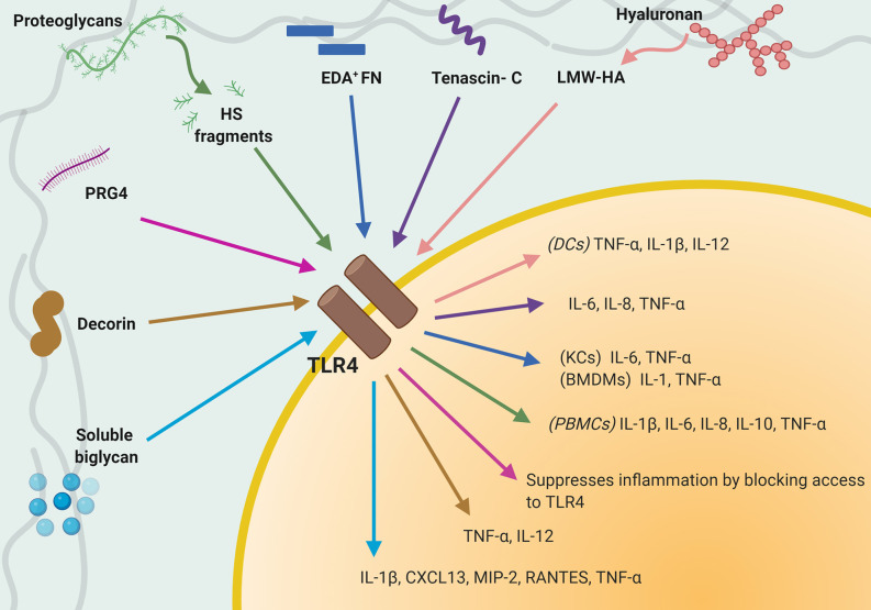
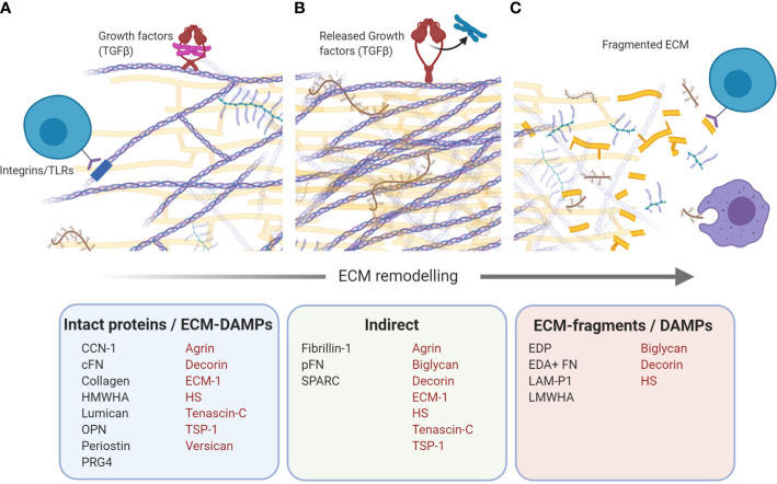
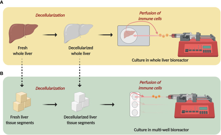
Verification
Claims the paper makes
The claims and statistics of this paper haven’t been checked yet.
Run the check — free, no account needed. It reads the open-access text and checks the sources it cites.
Claims other papers make about this one
Each is checked against this paper’s own text: does it say what the citing paper says it does?
The source backs part of the claim, not all of it — or backs it for a different population, condition or comparison.
In the citing paper
“In contrast to HMWHAs, low molecular weight hyaluronic acids (LMWHAs) are released during ECM damage or degradation, which act as DAMPs and drive inflammatory responses such as enhancing immune cells chemotaxis, DCs maturation, and M1 polarization of macrophages via the same receptors (CD44 and TLRs) [63, 100].”
From Immunogenicity of decellularized extracellular matrix scaffolds: a bottleneck in tissue engineering and regenerative medicine (2023) · cites this paper as [100]
Experimental — accuracy not yet measuredThe source backs part of the claim, not all of it — or backs it for a different population, condition or comparison.
In the citing paper
“Activation of these receptors accompanies a decrease in DC maturation and promotion in the polarization of macrophages toward M2 phenotype besides differentiation of T cells into regulatory phenotype and induction of apoptosis in activated T cells.”
From Immunogenicity of decellularized extracellular matrix scaffolds: a bottleneck in tissue engineering and regenerative medicine (2023) · cites this paper as [100]
Experimental — accuracy not yet measuredThe source backs part of the claim, not all of it — or backs it for a different population, condition or comparison.
In the citing paper
“Immunomodulatory traits of dECM scaffolds are largely attributed to the ability of their collagens to trigger leukocyte-associated immunoglobulin-like receptors-1 (LAIR-1) of immune cells and consequently downregulation of immune cell activity [100].”
From Immunogenicity of decellularized extracellular matrix scaffolds: a bottleneck in tissue engineering and regenerative medicine (2023) · cites this paper as [100]
Experimental — accuracy not yet measuredThe source backs part of the claim, not all of it — or backs it for a different population, condition or comparison.
In the citing paper
“In another example, high molecular weight hyaluronic acids (HMWHAs), well-known GAGs of dECM, exert anti-inflammatory effects by impeding antigen-antibody interaction and triggering toll-like receptors (TLRs), a subgroup of PRRs, and CD44 receptors of innate and adaptive immune cells.”
From Immunogenicity of decellularized extracellular matrix scaffolds: a bottleneck in tissue engineering and regenerative medicine (2023) · cites this paper as [100]
Experimental — accuracy not yet measuredThe cited source says what the claim attributes to it.
In the citing paper
“ECM is a reservoir for the cryptic particles that activate or suppress inflammation and orchestrates innate and adaptive immune responses [100, 101].”
From Immunogenicity of decellularized extracellular matrix scaffolds: a bottleneck in tissue engineering and regenerative medicine (2023) · cites this paper as [100]
The check wasn’t confident either way, so it gives no verdict — never disputed.
In the citing paper
“For instance, harsh decellularization procedures may unveil certain cryptic helical and terminal antigenic sites of collagen, prompting the initiation of antibody production against the ECM [73].”
From Liver tissue engineering using decellularized scaffolds: Current progress, challenges, and opportunities (2024) · cites this paper as [73]
Experimental — accuracy not yet measured
Self-check
Percentages, sums and reported differences re-computed from the paper’s own numbers.
The self-check needs the paper’s full text.
Citations
Cites
Items 401–446 of 446
Kohan, M., Muro, A. F., Bader, R., & Berkman, N. (2010) · Journal of Allergy and Clinical Immunology · doi:10.1016/j.jaci.2010.10.021 · Cited by 32
Anatomy, pathophysiology and basic mechanisms of disease
Crawford, J. M., & Burt, A. D. (2011) · Elsevier eBooks · doi:10.1016/b978-0-7020-3398-8.00001-5 · Cited by 32
GPR56 in Cancer Progression: Current Status and Future Perspective
Yang, L., & Xu, L. (2012) · Future Oncology · doi:10.2217/fon.12.27 · Cited by 31
Gillis, S. E., & Nagy, L. E. (1997) · PubMed · Cited by 31
Targeting early changes in the synovial microenvironment: a new class of immunomodulatory therapy?
Aungier, S. R., Cartwright, A. J., Schwenzer, A., Marshall, J. L., Dyson, M. R., Slavny, P., Parthiban, K., Karatt-Vellatt, A., Sahbudin, I., Culbert, E. J., et al. (2018) · Annals of the Rheumatic Diseases · doi:10.1136/annrheumdis-2018-214294 · Cited by 31
Self-citationYANG, Z. K., Zhang, H., Niu, Y., Zhang, W., Zhu, L., Li, X., Lü, S., Fan, J., Li, X., Ning, G., et al. (2016) · Scientific Reports · doi:10.1038/srep37886 · Cited by 30
Gillis, S. E., & Nagy, L. E. (1997) · Alcoholism Clinical and Experimental Research · doi:10.1111/j.1530-0277.1997.tb03849.x · Cited by 30
Differential roles of osteopontin/Eta-1 in early and late lpr disease
Weber, G. F., & Cantor, H. (2001) · Clinical & Experimental Immunology · doi:10.1046/j.1365-2249.2001.01702.x · Cited by 30
Su, P., Chen, S., Zheng, Y., Zhou, H., Yan, C., Yu, F., Zhang, Y., He, L., Zhang, Y., Wang, Y., et al. (2016) · The Journal of Immunology · doi:10.4049/jimmunol.1502457 · Cited by 30
Nahon, J. E., Hoekstra, M., van Harmelen, V., Rensen, P. C. N., van Dijk, K. W., Kooijman, S., & Eck, M. V. (2018) · Biochimica et Biophysica Acta (BBA) - Molecular Basis of Disease · doi:10.1016/j.bbadis.2018.11.009 · Cited by 29
Maimaitiyiming, H., Zhou, Q. T., & Wang, S. (2016) · PLoS ONE · doi:10.1371/journal.pone.0156144 · Cited by 29
Doddapattar, P., Jain, M. K., Dhanesha, N., Lentz, S. R., & Chauhan, A. K. (2018) · Arteriosclerosis Thrombosis and Vascular Biology · doi:10.1161/atvbaha.117.310345 · Cited by 29
Hirayama, C., Suzuki, H., Takada, A., Fujisawa, K., Tanikawa, K., & Igarashi, S. (1996) · Journal of Gastroenterology · doi:10.1007/bf02389524 · Cited by 28
Tanaka, H., El-Karef, A. A., Kaito, M., Kinoshita, N., Fujita, N., Horiike, S., Watanabe, S., Yoshida, T., & Adachi, Y. (2006) · Liver International · doi:10.1111/j.1478-3231.2005.01229.x · Cited by 28
Agrin Binds BMP2, BMP4 and TGFβ1
Bányai, L., Sonderegger, P., & Patthy, L. (2010) · PLoS ONE · doi:10.1371/journal.pone.0010758 · Cited by 27
Repair in avascular tissues: fibrosis in the transparent structures of the eye and thrombospondin 1.
Hiscott, P. S., Armstrong, D., Batterbury, M., & Kaye, S. B. (1999) · PubMed · doi:10.14670/hh-14.1309 · Cited by 26
Yilmaz, Y. N., Öztürk, O., Alahdab, Y. Ö., Şenateş, E., Çolak, Y., Doğanay, H. L., Coşkunpınar, E. M., Oltulu, Y. M., Eren, F., Atuğ, Ö., et al. (2012) · Digestive and Liver Disease · doi:10.1016/j.dld.2012.08.017 · Cited by 26
Serum tenascin levels in chronic liver disease
Yamauchi, M., Mizuhara, Y., Maezawa, Y., & Toda, G. (1994) · Liver International · doi:10.1111/j.1600-0676.1994.tb00064.x · Cited by 26
Yuting, Y., Lifeng, F., & Qiwei, H. (2018) · Biochemical and Biophysical Research Communications · doi:10.1016/j.bbrc.2018.12.006 · Cited by 25
Liver fibrogenesis: un update on established and emerging basic concepts
Novo, E., Bocca, C., Foglia, B., Protopapa, F., Maggiora, M., Parola, M., & Cannito, S. (2020) · Archives of Biochemistry and Biophysics · doi:10.1016/j.abb.2020.108445 · Cited by 24
Ethanol Feeding Potentiates the Pro-Inflammatory Response of Kupffer Cells to Cellular Fibronectin
Aziz-Seible, R. S., Lee, S. M. L., Kharbanda, K. K., McVicker, B. L., & Casey, C. A. (2011) · Alcoholism Clinical and Experimental Research · doi:10.1111/j.1530-0277.2010.01389.x · Cited by 23
CCN proteins in normal and injured liver
Weiskirchen, R. (2010) · Frontiers in bioscience · doi:10.2741/3832 · Cited by 23
Hepatic elastin content is predictive of adverse outcome in advanced fibrotic liver disease
Kendall, T. J., Dolman, G. E., Duff, C. M., Paish, E. C., Zaitoun, A. M., Irving, W. L., Fallowfield, J. A., & Guha, I. N. (2018) · Histopathology · doi:10.1111/his.13499 · Cited by 23
Urinary assays for desmosine and hydroxylysylpyridinoline in the detection of cirrhosis
Afdhal, N. H., Keaveny, A. P., Cohen, S. A., Nunes, D., Maldonado, N., O’Brien, M. J., & Stone, P. J. (1997) · Journal of Hepatology · doi:10.1016/s0168-8278(97)80142-0 · Cited by 22
Expression of LAIR-1 (CD305) on Human Blood Monocytes as a Marker of Hepatic Cirrhosis Progression
Martinez‐Esparza, M., Ruiz‐Alcaraz, A. J., Carmona‐Martínez, V., Fernández-Fernández, M. D., Antón, G., Muñoz‐Tornero, M., Lencina, M., Pagán, I., de la Peña, J., & García–Peñarrubia, P. (2019) · Journal of Immunology Research · doi:10.1155/2019/2974753 · Cited by 22
Li, Y., Wu, S., Xiong, S., & Ouyang, G. (2014) · Journal of Hepatology · doi:10.1016/j.jhep.2014.10.005 · Cited by 20
The dual role of osteopontin in acetaminophen hepatotoxicity
He, C.-Y., Liang, B., Fan, X., Cao, L., Chen, R., Guo, Y., & Zhao, J. (2012) · Acta Pharmacologica Sinica · doi:10.1038/aps.2012.47 · Cited by 18
Interferon-gamma, an Anti-fibrogenic Cytokine which Binds to Heparan Sulfate
Lortat‐Jacob, H., Esterre, P., & Grimaud, J.-A. (1994) · Pathology - Research and Practice · doi:10.1016/s0344-0338(11)80996-9 · Cited by 18
Elastosis in fibrotic and cirrhotic processes of the liver.
LIBAN, E., & Ungar, H. (1959) · PubMed · Cited by 18
Hennig, E. E., Mikuła, M., Goryca, K., Paziewska, A., Ledwon, J. K., Nesteruk, M., Woszczyński, M., Walewska‐Zielecka, B., Pyśniak, K., & Ostrowski, J. (2014) · Journal of Cellular and Molecular Medicine · doi:10.1111/jcmm.12328 · Cited by 17
Xuan, J., Chen, Y., Zhu, L., Guo, Y., Deng, L., Zheng, Y., Wang, Z., Wang, Z., & Ao, M. (2017) · Oncotarget · doi:10.18632/oncotarget.21358 · Cited by 16
Clinical Consequences of Infection in Cirrhosis: Organ Failures and Acute‐on‐Chronic Liver Failure
Wong, F. S.-H. (2019) · Clinical Liver Disease · doi:10.1002/cld.813 · Cited by 15
Intrahepatic gene expression profiles in chronic hepatitis B and autoimmune liver disease
Furuta, K., Sato, S., Yamauchi, T., Ozawa, T., Harada, M., & Kakumu, S. (2008) · Journal of Gastroenterology · doi:10.1007/s00535-008-2237-y · Cited by 15
Kobayashi, T., Kanno, K., Nguyen, P. T., Sugiyama, A., Kawahara, A., Otani, Y., Kishikawa, N., Ito, M., & Tazuma, S. (2020) · Journal of Gastroenterology and Hepatology · doi:10.1111/jgh.15088 · Cited by 14
Tang, T., Thompson, J. C., Wilson, P. G., Nelson, C., Williams, K. J., & Tannock, L. R. (2012) · Connective Tissue Research · doi:10.3109/03008207.2012.715700 · Cited by 13
Mustonen, A., Salvén, A., Kärjä, V. J., Rilla, K. J., Matilainen, J., & Nieminen, P. (2019) · Glycobiology · doi:10.1093/glycob/cwz002 · Cited by 13
Ceci, L., Francis, H., Zhou, T., Giang, T., Yang, Z., Meng, F., Wu, N., Kennedy, L. L., Kyritsi, K., Meadows, V., et al. (2020) · American Journal Of Pathology · doi:10.1016/j.ajpath.2020.07.007 · Cited by 13
Correction: Cellular Senescence in Livers from Children with End Stage Liver Disease
Gutiérrez‐Reyes, G., de León, M. D. C. G., Varela‐Fascinetto, G., Valencia, P., Tamayo, R. P., Rosado, C. G., Farfán-Labonne, B. E., Rochilin, N. M., Martínez-García, R., Aguirre-Valádez, J., et al. (2010) · PLoS ONE · doi:10.1371/annotation/6082f3f8-2b92-42a2-8d6f-b9210d2f25bf · Cited by 12
Aziz-Seible, R. S. (2011) · World Journal of Hepatology · doi:10.4254/wjh.v3.i2.45 · Cited by 11
Elastin content in human fibrotic and cirrhotic liver.
Kanta, J., Velebný, V., Mergancová, J., Ettlerová, E., & Chlumská, A. (1990) · PubMed · Cited by 10
Analysis of Plasma Tenascin-C in Post-HCV Cirrhosis: A Prospective Study
Benbow, J. H., Elam, A. D., Bossi, K. L., Massengill, D. L., Brandon‐Warner, E., Anderson, W. E., Culberson, C. R., Russo, M. W., deLemos, A. S., & Schrum, L. W. (2018) · Digestive Diseases and Sciences · doi:10.1007/s10620-017-4860-z · Cited by 10
Suppression of hepatic dysfunction in tenascin-X-deficient mice fed a high-fat diet
Yamaguchi, S., Kawakami, K., Satoh, K., Fukunaga, N., Akama, K., & Matsumoto, K.-I. (2017) · Molecular Medicine Reports · doi:10.3892/mmr.2017.7052 · Cited by 8
Use of a three-dimensional humanized liver model for the study of viral gene vectors
Wagner, A., Röhrs, V., Dehne, E., Hiller, T., Kedzierski, R., Fechner, H., Lauster, R., & Kurreck, J. (2015) · Journal of Biotechnology · doi:10.1016/j.jbiotec.2015.08.012 · Cited by 7
Cells Responsible for Extracellular Matrix Production in the Liver
Ueno, T., Sata, M., & Tanikawa, K. (2003) · Elsevier eBooks · doi:10.1016/b978-012525251-5/50007-5 · Cited by 6
Liver: Anatomy, Microscopic Structure, and Cell Types
Kanel, G. C. (2008) · Textbook of Gastroenterology · doi:10.1002/9781444303254.ch79 · Cited by 3
The TNF receptor family member CD27 signals to Jun N-terminal kinase via Traf-2
Eugster (1998) · Pure Amsterdam UMC · doi:10.1002/(sici)1521-4141(199807)28:07 · Cited by 2
Cited by
Sorted by citations
Fibroblasts: Origins, definitions, and functions in health and disease
Plikus, M. V., Wang, X., Sinha, S., Forte, E., Thompson, S. M., Herzog, E. L., Driskell, R. R., Rosenthal, N. A., Biernaskie, J. A., & Horsley, V. (2021) · Cell · doi:10.1016/j.cell.2021.06.024 · Cited by 1,247
The extracellular matrix and the immune system: A mutually dependent relationship
Sutherland, T. E., Dyer, D. P., & Allen, J. E. (2023) · Science · doi:10.1126/science.abp8964 · Cited by 519
Kasravi, M., Ahmadi, A., Babajani, A., Mazloomnejad, R., Hatamnejad, M. R., Shariatzadeh, S., Bahrami, S., & Niknejad, H. (2023) · Biomaterials Research · doi:10.1186/s40824-023-00348-z · Cited by 316
Bhattacharya, M., & Ramachandran, P. (2023) · Nature Immunology · doi:10.1038/s41590-023-01551-9 · Cited by 148
The Role of Epithelial Damage in the Pulmonary Immune Response
Burgoyne, R. A., Fisher, A. J., & Borthwick, L. A. (2021) · Cells · doi:10.3390/cells10102763 · Cited by 153
Metadata
- Authors
- McQuitty, C. E., Williams, R., Chokshi, S., & Urbani, L.
- Affiliations
- King's College London (GB)
- Corresponding author
- Urbani, Luca
- Venue
- Frontiers in Immunology
- Publisher
- Frontiers Media SA
- Published
- 2020-11-11
- Volume
- 11
- Pages
- 574276-574276
- ISSN
- 1664-3224 (ISSN-L)
- PMC
- PMC7686550
- OpenAlex
- W3098031338
- Cited by
- 146
- References
- 446
- Citations by year
- 2026: 23 · 2025: 22 · 2024: 34 · 2023: 31 · 2022: 19 · 2021: 17
- FWCI
- 2.23
- Type
- journal article
- Language
- en
- Open access
- gold
- doi.org
- Licence
- Locations
- Frontiers in Immunology (publishedVersion, cc-by)
- PubMed Central (publishedVersion, cc-by)
- PubMed (publishedVersion)
- DOAJ (DOAJ: Directory of Open Access Journals) (submittedVersion, cc-by-sa)
- Topics
- Health Sciences › Medicine › Hepatology › Liver physiology and pathology
- Health Sciences › Medicine › Hepatology › Liver Diseases and Immunity
- Health Sciences › Medicine › Surgery › Tissue Engineering and Regenerative Medicine
- Keywords
- extracellular matrix, chronic liver disease, liver fibrosis, damage-associated molecular patterns, sterile inflammation, integrins, TLRs, immune cell infiltration, tissue engineering, biomarkers
- MeSH terms
- Anti-Inflammatory Agents, Extracellular Matrix, Humans, Inflammation, Liver, Liver Cirrhosis, Liver Diseases, Cytokines, Extracellular Matrix Proteins, Immunomodulation
About these checks
Registry, retraction, Cites and statistics results are computed, not judged, with one exception: a printed reference that no index lists is read once by a model before it counts as a citation error. Claims are extracted by a model and kept only when their quote appears verbatim in the text; each cited source is then checked, with a model’s help, for whether it says what the claim relies on. Those verdicts can be wrong — read the claim’s quote and the cited source before relying on one.
Statistics and arithmetic are recomputed, not judged.
Confidence bands are shown only for fields calibrated on our annotated test set. None is calibrated yet, so no band is shown.
What counts as a claim ›How we measure confidence ›How we check this ›
Are you an author, or do you see a problem with this page? Request a review
