

The DOI matches its Crossref record, with a small difference in title or year.
Quick and wide-range taxonomical repertoire establishment of the cystic fibrosis lung microbiota by tandem mass spectrometry on sputum samples
Abstract
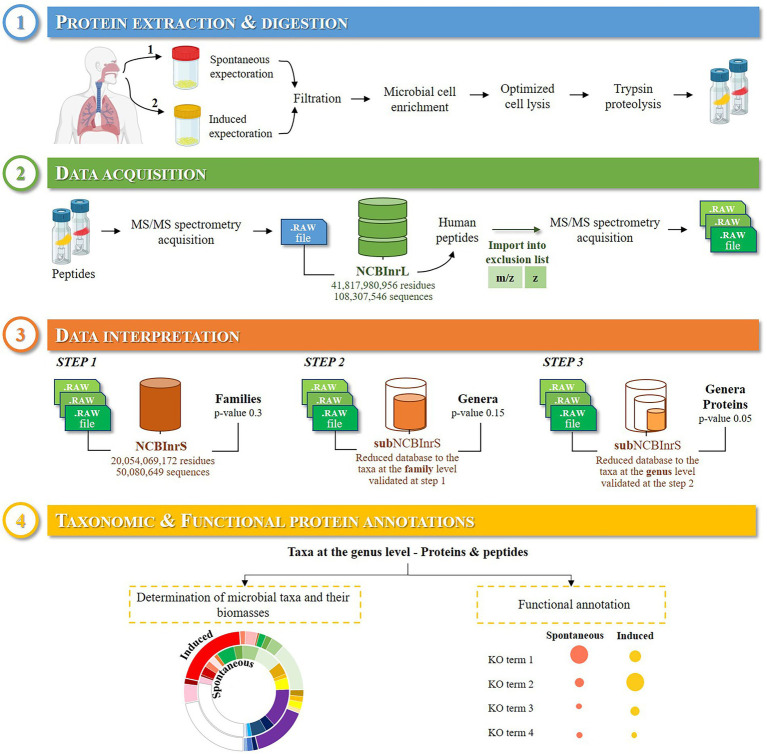
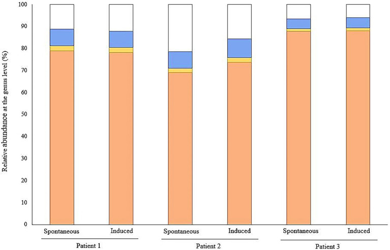
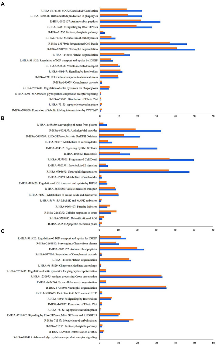
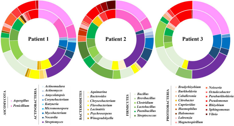
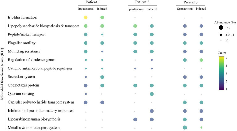
Microorganisms proteotyping by tandem mass spectrometry has been recently shown as a powerful methodology to identify the wide-range taxonomy and biomass of microbiota. Sputum is the recommended specimen for routine microbiological monitoring of Cystic Fibrosis (CF) patients but has been rarely submitted to tandem mass spectrometry-based proteotyping. In this study, we compared the microbial components of spontaneous and induced sputum samples from three cystic fibrosis patients. Although the presence of microbial proteins is much lower than host proteins, we report that the microbiota’s components present in the samples can be identified, as well as host biomarkers and functional insights into the microbiota. No significant difference was found in microorganism abundance between paired spontaneous and induced sputum samples. Microbial proteins linked to resistance, iron uptake, and biofilm-forming ability were observed in sputa independently of the sampling method. This unbiased and enlarged view of the CF microbiome could be highly complementary to culture and relevant for the clinical management of CF patients by improving knowledge about the host-pathogen dynamics and CF pathophysiology.
Overview
The claims and statistics of this paper haven’t been checked yet.
Run the check — free, no account needed. It reads the open-access text and checks the sources it cites.
Integrity signals
Registry
Cites
Statistics
Figures
Verification
Claims the paper makes
The claims and statistics of this paper haven’t been checked yet.
Run the check — free, no account needed. It reads the open-access text and checks the sources it cites.
Claims other papers make about this one
Each is checked against this paper’s own text: does it say what the citing paper says it does?
The cited source says what the claim attributes to it.
In the citing paper
“Proteotyping of organisms based on tandem mass spectrometry data has been successfully used to identify bacteria [6, 7], and more complex medical microbiota [8, 9] or environmental microbiota [10].”
From Metaproteomic analysis of King Ghezo tomb wall (Abomey, Benin) confirms 19th century voodoo sacrifices (2024) · cites this paper as [9]
Self-check
Percentages, sums and reported differences re-computed from the paper’s own numbers.
The self-check needs the paper’s full text.
Citations
Cites
No references could be resolved yet.
Research works printed but not linked: 44
- Aakko, 2020, Data-independent acquisition mass spectrometry in Metaproteomics of gut microbiota-implementation and computational analysis, J. Proteome Res., 19, 432, 10.1021/acs.jproteome.9b00606 · DOI
- Acosta, 2018, Sputum microbiota is predictive of long-term clinical outcomes in young adults with cystic fibrosis, Thorax, 73, 1016, 10.1136/thoraxjnl-2018-211510 · DOI
- Bassignani, 2021, Benefits of iterative searches of large databases to interpret large human gut Metaproteomic data sets, J. Proteome Res., 20, 1522, 10.1021/acs.jproteome.0c00669 · DOI
- Bevivino, 2019, Deciphering the ecology of cystic fibrosis bacterial communities: towards systems-level integration, Trends Mol. Med., 25, 1110, 10.1016/j.molmed.2019.07.008 · DOI
- Blau, 2014, Induced sputum compared to bronchoalveolar lavage in young, non-expectorating cystic fibrosis children, J. Cyst. Fibros., 13, 106, 10.1016/j.jcf.2013.05.013 · DOI
- Bodas, 2019, Adapting Proteostasis and autophagy for controlling the pathogenesis of cystic fibrosis lung disease, Front. Pharmacol., 10, 20, 10.3389/fphar.2019.00020 · DOI
- Christie-Oleza, 2012, Proteomic insights into the lifestyle of an environmentally relevant marine bacterium, ISME J., 6, 124, 10.1038/ismej.2011.86 · DOI
- Coburn, 2015, Lung microbiota across age and disease stage in cystic fibrosis, Sci. Rep., 5, 10241, 10.1038/srep10241 · DOI
- Cuthbertson, 2020, Lung function and microbiota diversity in cystic fibrosis, Microbiome, 8, 45, 10.1186/s40168-020-00810-3 · DOI
- Dubourg, 2015, Correlation between sputum and bronchoalveolar lavage fluid cultures, J. Clin. Microbiol., 53, 994, 10.1128/jcm.02918-14 · DOI
- Feigelman, 2017, Sputum DNA sequencing in cystic fibrosis: non-invasive access to the lung microbiome and to pathogen details, Microbiome, 5, 20, 10.1186/s40168-017-0234-1 · DOI
- Foster, 2017, Metacoder: an R package for visualization and manipulation of community taxonomic diversity data, PLoS Comput. Biol., e1005404, 10.1371/journal.pcbi.1005404 · DOI
- Françoise, 2020, The microbiome in cystic fibrosis pulmonary disease, 536, 10.3390/genes11050536 · DOI
- Graf, 2021, An innovative protocol for metaproteomic analyses of microbial pathogens in cystic fibrosis sputum, Front. Cell. Infect. Microbiol., 11, 724569, 10.3389/fcimb.2021.724569 · DOI
- Grenga, 2019, Pathogen proteotyping: a rapidly developing application of mass spectrometry to address clinical concerns, Clin. Mass Spectrom., 14, 9, 10.1016/j.clinms.2019.04.004 · DOI
- Grenga, 2022, Taxonomical and functional changes in COVID-19 faecal microbiome could be related to SARS-CoV-2 faecal load, Environ. Microbiol., 10.1111/1462-2920.16028 · DOI
- Hahn, 2016, Different next generation sequencing platforms produce different microbial profiles and diversity in cystic fibrosis sputum, J. Microbiol. Methods, 130, 95, 10.1016/j.mimet.2016.09.002 · DOI
- Hardouin, 2021, Metaproteomics to decipher CF host-microbiota interactions: overview, challenges and future perspectives, Genes, 12, 892, 10.3390/genes12060892 · DOI
- Hayoun, 2019, Evaluation of sample preparation methods for fast proteotyping of microorganisms by tandem mass spectrometry, Front. Microbiol., 10, 1985, 10.3389/fmicb.2019.01985 · DOI
- Héry-Arnaud, 2019, The lung and gut microbiome: what has to be taken into consideration for cystic fibrosis?, J. Cyst. Fibros., 18, 13, 10.1016/j.jcf.2018.11.003 · DOI
- Hogan, 2016, Analysis of lung microbiota in bronchoalveolar lavage, protected brush and sputum samples from subjects with mild-to-moderate cystic fibrosis lung disease, PLoS One, 11, e0149998, 10.1371/journal.pone.0149998 · DOI
- Jagtap, 2018, BAL fluid metaproteome in acute respiratory failure, Am. J. Respir. Cell Mol. Biol., 59, 648, 10.1165/rcmb.2018-0068LE · DOI
- Jouffret, 2021, Increasing the power of interpretation for soil metaproteomics data, Microbiome, 9, 195, 10.1186/s40168-021-01139-1 · DOI · Paper page
- Kanehisa, 2016, BlastKOALA and GhostKOALA: KEGG tools for functional characterization of genome and metagenome sequences, J. Mol. Biol., 428, 726, 10.1016/j.jmb.2015.11.006 · DOI
- Laube, 2006, Antimicrobial peptides in the airway, Curr. Top. Microbiol. Immunol., 306, 153, 10.1007/3-540-29916-5_6 · DOI
- Lecaille, 2016, Antimicrobial proteins and peptides in human lung diseases: a friend and foe partnership with host proteases, Biochimie, 122, 151, 10.1016/j.biochi.2015.08.014 · DOI
- Lim, 2013, Metagenomics and metatranscriptomics: windows on CF-associated viral and microbial communities, J. Cyst. Fibros., 12, 154, 10.1016/j.jcf.2012.07.009 · DOI
- Mappa, 2021, Assessing the ratio of bacillus spores and vegetative cells by shotgun proteomics, Environ. Sci. Pollut. Res. Int., 28, 25107, 10.1007/s11356-018-3341-z · DOI
- Marchandin, 2012, Atypical Bacteria in the CF Airways: Diversity, Clinical Consequences, Emergence and Adaptation
- Menetrey, 2021, Achromobacter xylosoxidans and Stenotrophomonas maltophilia: emerging pathogens well-armed for life in the cystic fibrosis patients' lung, Genes, 12, 610, 10.3390/genes12050610 · DOI
- Pattison, 2017, Proteomic profile of cystic fibrosis sputum cells in adults chronically infected with Pseudomonas aeruginosa, Eur. Respir. J., 50, 1601569, 10.1183/13993003.01569-2016 · DOI
- Perez-Riverol, 2022, The PRIDE database resources in 2022: a hub for mass spectrometry-based proteomics evidences, Nucleic Acids Res., 50, D543, 10.1093/nar/gkab1038 · DOI · Paper page
- Pible, 2020, Estimating relative biomasses of organisms in microbiota using “phylopeptidomics”, Microbiome, 8, 30, 10.1186/s40168-020-00797-x · DOI
- Quinn, 2014, Biogeochemical forces shape the composition and physiology of polymicrobial communities in the cystic fibrosis lung, mBio, 5, e00956, 10.1128/mBio.00956-13 · DOI · Paper page
- Rubiano-Labrador, 2014, Proteogenomic insights into salt tolerance by a halotolerant alpha-proteobacterium isolated from an Andean saline spring, J. Proteome, 97, 36, 10.1016/j.jprot.2013.05.020 · DOI
- Soleti, 2013, Apoptotic process in cystic fibrosis cells, Apoptosis, 18, 1029, 10.1007/s10495-013-0874-y · DOI
- Suarez, 2013, Ribosomal proteins as biomarkers for bacterial identification by mass spectrometry in the clinical microbiology laboratory, J. Microbiol. Methods, 94, 390, 10.1016/j.mimet.2013.07.021 · DOI
- Tyrrell, 2016, Iron acquisition in the cystic fibrosis lung and potential for novel therapeutic strategies, Microbiology, 162, 191, 10.1099/mic.0.000220 · DOI · Paper page
- Van Den Bossche, The Metaproteomics initiative: a coordinated approach for propelling the functional characterization of microbiomes, Microbiome, 9, 243, 10.1186/s40168-021-01176-w · DOI · Paper page
- Van Den Bossche, Critical assessment of MetaProteome investigation (CAMPI): a multi-laboratory comparison of established workflows, Nat. Commun., 12, 7305, 10.1038/s41467-021-27542-8 · DOI
- van der Gast, 2011, Partitioning core and satellite taxa from within cystic fibrosis lung bacterial communities, ISME J., 5, 780, 10.1038/ismej.2010.175 · DOI
- Wang, 2020, Metaproteomics: a strategy to study the taxonomy and functionality of the gut microbiota, J. Proteome, 219, 103737, 10.1016/j.jprot.2020.103737 · DOI
- Zhao, 2020, Autophagy, an important therapeutic target for pulmonary fibrosis diseases, Clin. Chim. Acta, 502, 139, 10.1016/j.cca.2019.12.016 · DOI · Paper page
- Zhou, 2019, Metascape provides a biologist-oriented resource for the analysis of systems-level datasets, Nat. Commun., 10, 1523, 10.1038/s41467-019-09234-6 · DOI · Paper page
Cited by
Sorted by citations
Charlier, P., Bourdin, V., N'Dah, D., Kielbasa, M., Pible, O., & Armengaud, J. (2024) · PROTEOMICS · doi:10.1002/pmic.202400048 · Cited by 8
Self-citation
Metadata
- Authors
- Hardouin, P., Pible, O., Marchandin, H., Culotta, K., Armengaud, J., Chiron, R., & Grenga, L.
- Venue
- Frontiers in Microbiology
- Publisher
- Frontiers Media SA
- Published
- 2022-10-13
- Volume
- 13
- ISSN
- 1664-302X
- PMC
- PMC9608366
- Cited by
- 11
- References
- 0
About these checks
Registry, retraction, Cites and statistics results are computed, not judged, with one exception: a printed reference that no index lists is read once by a model before it counts as a citation error. Claims are extracted by a model and kept only when their quote appears verbatim in the text; each cited source is then checked, with a model’s help, for whether it says what the claim relies on. Those verdicts can be wrong — read the claim’s quote and the cited source before relying on one.
Statistics and arithmetic are recomputed, not judged.
Confidence bands are shown only for fields calibrated on our annotated test set. None is calibrated yet, so no band is shown.
What counts as a claim ›How we measure confidence ›How we check this ›
Are you an author, or do you see a problem with this page? Request a review
How to cite
Cite CoChat for the checks on this page, and the paper for its own findings.
- This page
- CoChat. (2026). Citation check: Quick and wide-range taxonomical repertoire establishment of the cystic fibrosis lung microbiota by tandem mass spectrometry on sputum samples. https://app.cochat.ai/papers/10.3389/fmicb.2022.975883
- The paper
- Hardouin, P., Pible, O., Marchandin, H., Culotta, K., Armengaud, J., Chiron, R., & Grenga, L. (2022). Quick and wide-range taxonomical repertoire establishment of the cystic fibrosis lung microbiota by tandem mass spectrometry on sputum samples. Frontiers in Microbiology. https://doi.org/10.3389/fmicb.2022.975883
