

The DOI matches its Crossref record; 63 of the 63 references it cites were found in a registry or an index.
Microglia Morphological Categorization in a Rat Model of Neuroinflammation by Hierarchical Cluster and Principal Components Analysis
Open accessAbstract
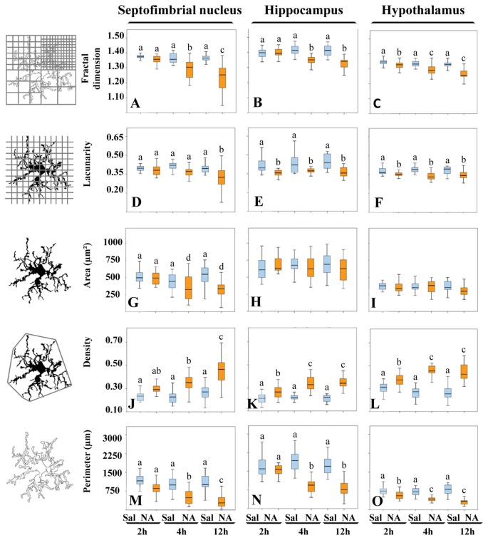
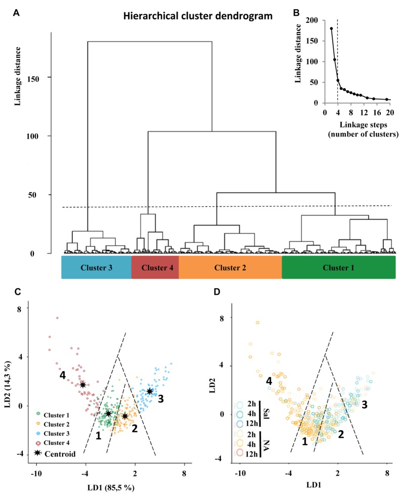
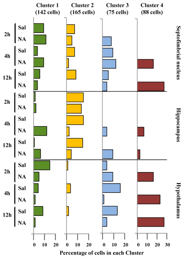
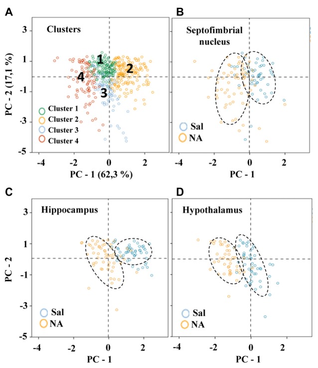
It is known that microglia morphology and function are closely related, but only few studies have subjectively described different morphological subtypes. To address this issue, morphological parameters of microglial cells were analyzed in a rat model of aseptic neuroinflammation. After the injection of a single dose of the enzyme neuraminidase within the lateral ventricle an acute inflammatory process occurs. Sections from NA-injected animals and sham controls were immunolabeled with the microglial marker IBA1, which highlights ramifications and features of the cell shape. Using images obtained by section scanning, individual microglial cells were sampled from various regions (septofimbrial nucleus, hippocampus and hypothalamus) at different times post-injection (2, 4 and 12 hours). Each cell yielded a set of 15 morphological parameters by means of image analysis software. Five initial parameters (including fractal measures) were statistically different in cells from NA-injected rats (most of them IL-1β positive, i.e. M1-state) compared to those from control animals (none of them IL-1β positive, i.e. surveillant state). However, additional multimodal parameters were revealed more suitable for hierarchical cluster analysis. This method pointed out the classification of microglia population in four clusters. Furthermore, a linear discriminant analysis suggested three specific parameters to objectively classify any microglia by a decision tree. In addition, a principal components analysis revealed two extra valuable variables that allowed to further classifying microglia in a total of eight sub-clusters or types. The spatio-temporal distribution of these different morphotypes in our rat inflammation model allowed to relate specific morphotypes with microglial activation status and brain location. An objective method for microglia classification based on morphological parameters is proposed.
Overview
The claims and statistics of this paper haven’t been checked yet.
Run the check — free, no account needed. It reads the open-access text and checks the sources it cites.
Integrity signals
Registry
Cites
Statistics
Figures
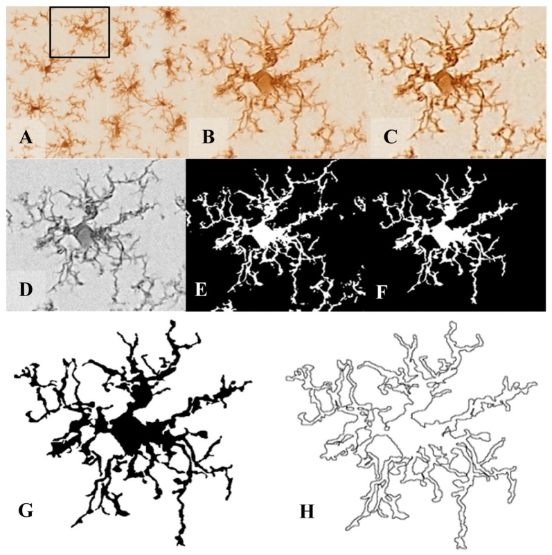
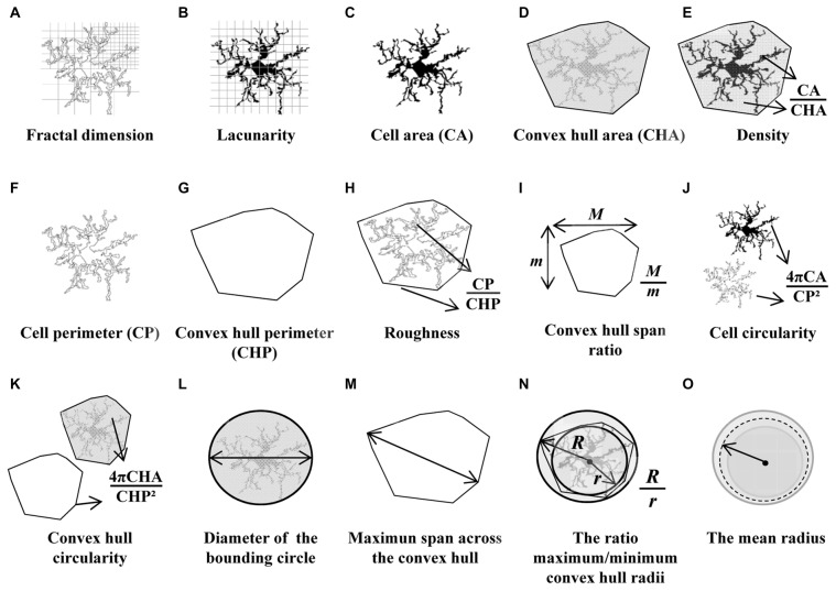
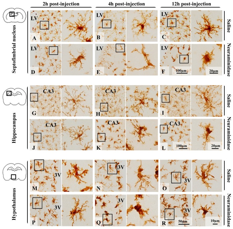
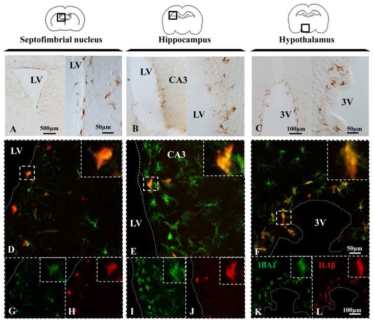
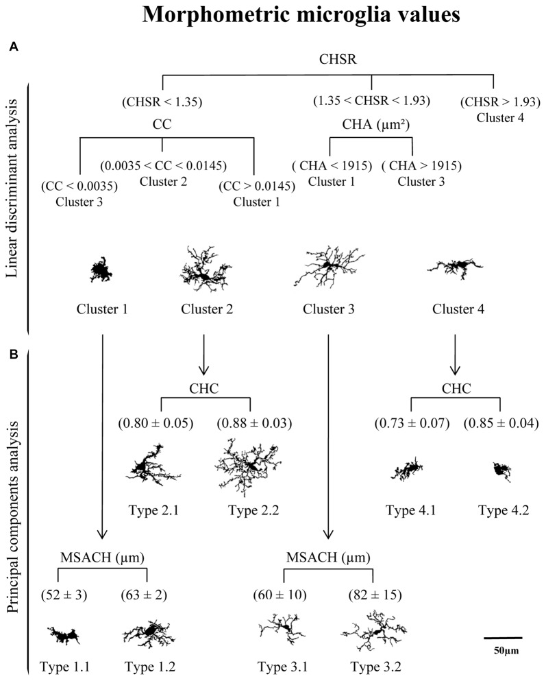
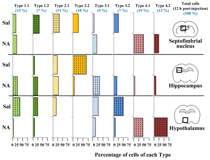
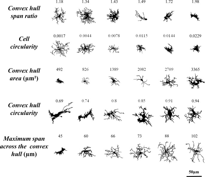
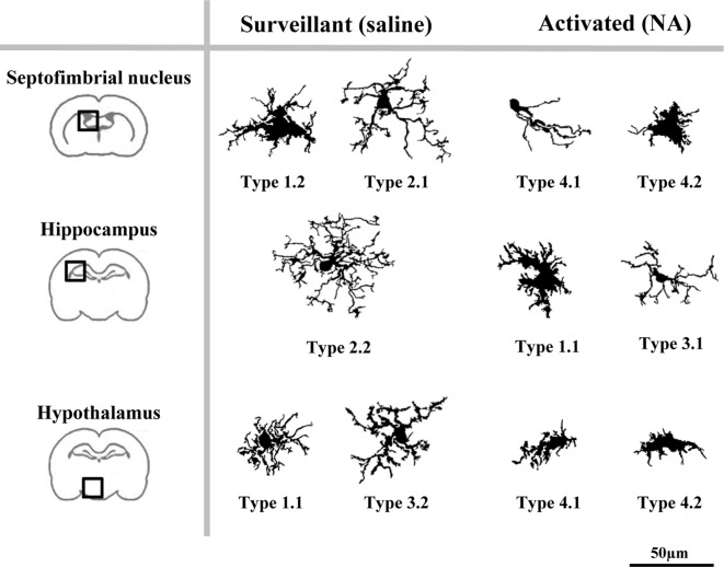
Verification
Claims the paper makes
The claims and statistics of this paper haven’t been checked yet.
Run the check — free, no account needed. It reads the open-access text and checks the sources it cites.
Claims other papers make about this one
Each is checked against this paper’s own text: does it say what the citing paper says it does?
The cited source says what the claim attributes to it.
In the citing paper
“We found that, upon LPS/ATP treatment, iMG infused into our tubular organoids displayed a lower complexity in patterns as well as a spherical shape, exemplified by lower fractal dimensions, lower lacunarity, and higher convex-hull circularity (Figure 6c).56”
From Tubular human brain organoids to model microglia-mediated neuroinflammation (2021) · cites this paper as [56]
Self-check
Percentages, sums and reported differences re-computed from the paper’s own numbers.
The self-check needs the paper’s full text.
Citations
Cites
The Rat Brain in Stereotaxic Coordinates
Paxinos, G., & Watson, C. (1983) · Elsevier eBooks · doi:10.1016/c2009-0-63235-9 · Cited by 28,585
The Fractal Geometry of Nature
Mandelbrot, B. B., & WHEELER, J. A. (1983) · American Journal of Physics · doi:10.1119/1.13295 · Cited by 21,964
Hierarchical Grouping to Optimize an Objective Function
Ward, J. H. (1963) · Journal of the American Statistical Association · doi:10.1080/01621459.1963.10500845 · Cited by 21,341
THE USE OF MULTIPLE MEASUREMENTS IN TAXONOMIC PROBLEMS
FISHER, R. A. (1936) · Annals of Eugenics · doi:10.1111/j.1469-1809.1936.tb02137.x · Cited by 15,227
Resting microglial cells are highly dynamic surveillants of brain parenchyma in vivo
Nimmerjahn, A., Kirchhoff, F., & Helmchen, F. (2005) · Science · doi:10.1126/science.1110647 · Cited by 5,827
A dendrite method for cluster analysis
Caliński, T., & Harabasz, J. (1974) · Communications in Statistics · doi:10.1080/03610927408827101 · Cited by 4,480
Synaptic Pruning by Microglia Is Necessary for Normal Brain Development
Paolicelli, R. C., Bolasco, G., Pagani, F., Maggi, L., Scianni, M., Panzanelli, P., Giustetto, M., Ferreira, T. A., Guiducci, E., Dumas, L., et al. (2011) · Science · doi:10.1126/science.1202529 · Cited by 4,244
Microglia Sculpt Postnatal Neural Circuits in an Activity and Complement-Dependent Manner
Schafer, D. P., Lehrman, E. K., Kautzman, A. G., Koyama, R., Mardinly, A. R., Yamasaki, R., Ransohoff, R. M., Greenberg, M. E., Barres, B. A., & Stevens, B. (2012) · Neuron · doi:10.1016/j.neuron.2012.03.026 · Cited by 4,089
Kettenmann, H., Hanisch, U.-K., Noda, M., & Verkhratsky, A. (2011) · Physiological Reviews · doi:10.1152/physrev.00011.2010 · Cited by 3,547
Hierarchical Grouping to Optimize an Objective Function
Ward, J. H. (1963) · Journal of the American Statistical Association · doi:10.2307/2282967 · Cited by 3,294
Thorndike, R. L. (1953) · Psychometrika · doi:10.1007/bf02289263 · Cited by 2,264
Microglial M1/M2 polarization and metabolic states
Orihuela, R., McPherson, C. A., & Harry, G. J. (2016) · British Journal of Pharmacology · doi:10.1111/bph.13139 · Cited by 2,070
Heterogeneity in the distribution and morphology of microglia in the normal adult mouse brain
Lawson, L. J., Perry, V. H., Dri, P., & Gordon, S. (1990) · Neuroscience · doi:10.1016/0306-4522(90)90229-w · Cited by 2,045
Modulation of macrophage phenotype by cell shape
McWhorter, F. Y., Wang, T., Nguyen, P., Chung, T., & Liu, W. F. (2013) · Proceedings of the National Academy of Sciences · doi:10.1073/pnas.1308887110 · Cited by 1,482
Microglia: new roles for the synaptic stripper
Kettenmann, H., Kirchhoff, F., & Verkhratsky, A. (2013) · Neuron · doi:10.1016/j.neuron.2012.12.023 · Cited by 1,173
Streit, W. J., Walter, S. A., & Pennell, N. A. (1999) · Progress in Neurobiology · doi:10.1016/s0301-0082(98)00069-0 · Cited by 1,096
Reciprocal Regulation between Resting Microglial Dynamics and Neuronal Activity In Vivo
Li, Y., Du, X.-F., Liu, C., Wen, Z., & Du, J. (2012) · Developmental Cell · doi:10.1016/j.devcel.2012.10.027 · Cited by 590
Janus-faced microglia: beneficial and detrimental consequences of microglial phagocytosis
Sierra, A., Abiega, O., Shahraz, A., & Neumann, H. (2013) · Frontiers in Cellular Neuroscience · doi:10.3389/fncel.2013.00006 · Cited by 574
Quantitating the subtleties of microglial morphology with fractal analysis
Karperien, A., Ahammer, H., & Jelinek, H. F. (2013) · Frontiers in Cellular Neuroscience · doi:10.3389/fncel.2013.00003 · Cited by 541
Morrison, H. W., & Filosa, J. A. (2013) · Journal of Neuroinflammation · doi:10.1186/1742-2094-10-4 · Cited by 506
Cellular forms and functions of brain microglia
Davis, E. J., Foster, T. D., & Thomas, W. E. (1994) · Brain Research Bulletin · doi:10.1016/0361-9230(94)90189-9 · Cited by 359
Microglial Dynamics and Role in the Healthy and Diseased Brain: A Paradigm of Functional Plasticity
Gomez-Nicola, D., & Perry, V. H. (2015) · The Neuroscientist · doi:10.1177/1073858414530512 · Cited by 340
Hinwood, M., Tynan, R. J., Charnley, J. L., Beynon, S. B., Day, T. A., & Walker, F. R. (2012) · Cerebral Cortex · doi:10.1093/cercor/bhs151 · Cited by 336
Morphometric characterization of microglial phenotypes in human cerebral cortex
Torres-Platas, S. G., Comeau, S., Rachalski, A., Bo, G. D., Cruceanu, C., Turecki, G., Giros, B., & Mechawar, N. (2014) · Journal of Neuroinflammation · doi:10.1186/1742-2094-11-12 · Cited by 329
Homeostatic and injury‐induced microglia behavior in the aging brain
Hefendehl, J. K., Neher, J. J., Sühs, R. B., Kohsaka, S., Skodras, A., & Jucker, M. (2014) · Aging Cell · doi:10.1111/acel.12149 · Cited by 319
Sierra, A., de Castro, F., del Río‐Hortega, J., Rafael Iglesias‐Rozas, J., Garrosa, M., & Kettenmann, H. (2016) · Glia · doi:10.1002/glia.23046 · Cited by 266
Olah, M., Biber, K. P. H., Vinet, J., & Boddeke, H. W. G. M. (2011) · CNS & Neurological Disorders - Drug Targets · doi:10.2174/187152711794488575 · Cited by 260
Walker, F. R., Beynon, S. B., Jones, K., Zhao, Z., Kongsui, R., Cairns, M. J., & Nilsson, M. (2014) · Brain Behavior and Immunity · doi:10.1016/j.bbi.2013.12.010 · Cited by 237
Kozlowski, C., & Weimer, R. M. (2012) · PLoS ONE · doi:10.1371/journal.pone.0031814 · Cited by 218
Use of Fractal Theory in Neuroscience: Methods, Advantages, and Potential Problems
Fernández, E., & Jelinek, H. F. (2001) · Methods · doi:10.1006/meth.2001.1201 · Cited by 217
Hovens, I. B., Nyakas, C. J., & Schoemaker, R. (2014) · Neuroimmunology and Neuroinflammation · doi:10.4103/2347-8659.139719 · Cited by 186
PPAR ? agonists stimulate oligodendrocyte differentiation in tissue culture
Saluja, I., Granneman, J. G., & Skoff, R. P. (2001) · Glia · doi:10.1002/1098-1136(200103)33:3 · Cited by 186
Neurotransmitter signaling in the pathophysiology of microglia
Domercq, M., Vázquez‐Villoldo, N., & Matute, C. (2013) · Frontiers in Cellular Neuroscience · doi:10.3389/fncel.2013.00049 · Cited by 174
Microglia and synapse interactions: fine tuning neural circuits and candidate molecules
Miyamoto, A., Wake, H., Moorhouse, A. J., & Nabekura, J. (2013) · Frontiers in Cellular Neuroscience · doi:10.3389/fncel.2013.00070 · Cited by 147
Zanier, E. R., Fumagalli, S., Perego, C., Pischiutta, F., & De Simoni, M.-G. (2015) · Intensive Care Medicine Experimental · doi:10.1186/s40635-015-0039-0 · Cited by 147
Diverse microglial motility behaviors during clearance of dead cells in hippocampal slices
Petersen, M. A., & Dailey, M. E. (2004) · Glia · doi:10.1002/glia.10362 · Cited by 139
Microglial control of neuronal activity
Béchade, C., Cantaut-Belarif, Y., & Bessis, A. (2013) · Frontiers in Cellular Neuroscience · doi:10.3389/fncel.2013.00032 · Cited by 134
Increased Morphological Diversity of Microglia in the Activated Hypothalamic Supraoptic Nucleus
Ayoub, A. E., & Salm, A. K. (2003) · Journal of Neuroscience · doi:10.1523/jneurosci.23-21-07759.2003 · Cited by 122
Baron, R., Babcock, A. A., Nemirovsky, A., Finsen, B., & Monsonego, A. (2014) · Aging Cell · doi:10.1111/acel.12210 · Cited by 118
Kongsui, R., Beynon, S. B., Johnson, S. J., & Walker, F. R. (2014) · Journal of Neuroinflammation · doi:10.1186/s12974-014-0182-7 · Cited by 115
Morphological and Phagocytic Profile of Microglia in the Developing Rat Cerebellum
Perez‐Pouchoulen, M., VanRyzin, J. W., & McCarthy, M. M. (2015) · eNeuro · doi:10.1523/eneuro.0036-15.2015 · Cited by 100
Glenn, J. A., Ward, S. A., Stone, C. R., Booth, P. L., & Thomas, W. E. (1992) · PubMed · Cited by 89
Sołtys, Z., Orzylowska, O., Zaremba, M., Orłowski, D., Piechota, M., Fiedorowicz, A., Janeczko, K. J., & Oderfeld-Nowak, B. (2005) · Journal of Neuroscience Methods · doi:10.1016/j.jneumeth.2005.01.009 · Cited by 82
Emerging roles for telomerase in neuronal development and apoptosis
Mattson, M. P., & Klapper, W. (2001) · Journal of Neuroscience Research · doi:10.1002/1097-4547(20010101)63:1 · Cited by 76
Early morphofunctional plasticity of microglia in response to acute lipopolysaccharide
Madore, C., Joffre, C., Delpech, J.-C., Smedt, V. D., Aubert, A., Coste, L., Layé, S., & Nadjar, A. (2013) · Brain Behavior and Immunity · doi:10.1016/j.bbi.2013.08.008 · Cited by 70
The use of cluster analysis for cell typing
Schweitzer, L., & Renehan, W. E. (1997) · Brain Research Protocols · doi:10.1016/s1385-299x(96)00014-1 · Cited by 65
Yamada, J., & Jinno, S. (2012) · The Journal of Comparative Neurology · doi:10.1002/cne.23228 · Cited by 58
Grondona, J. M., Pérez‐Martín, M., Cifuentes, M., Pérez, J., Jimenez, A. J., Pérez-Fígares, J. M., & Fernández‐Llebrez, P. (1996) · Journal of Neuropathology & Experimental Neurology · doi:10.1097/00005072-199609000-00007 · Cited by 54
Self-citationGómez-Roldán, M. D. C., Pérez‐Martín, M., Capilla‐González, V., Cifuentes, M., Pérez, J., García‐Verdugo, J. M., & Fernández‐Llebrez, P. (2008) · The Journal of Comparative Neurology · doi:10.1002/cne.21618 · Cited by 45
Self-citationEyo, U. B., Miner, S. A., Weiner, J. A., & Dailey, M. E. (2015) · Brain Behavior and Immunity · doi:10.1016/j.bbi.2015.11.009 · Cited by 39
Ohgomori, T., Yamada, J., Takeuchi, H., Kadomatsu, K., & Jinno, S. (2016) · European Journal of Neuroscience · doi:10.1111/ejn.13227 · Cited by 38
Reviewing lacunarity analysis and classification of microglia in neuroscience
Karperien, A., Jelinek, H. F., & Milošević, N. T. (2011) · Charles Sturt University Research Output (CRO) · Cited by 36
Kongsui, R., Johnson, S. J., Graham, B. A., Nilsson, M., & Walker, F. R. (2015) · Neuroscience · doi:10.1016/j.neuroscience.2015.09.061 · Cited by 31
Amadio, S., Ninno, A. D., Montilli, C., Businaro, L., Gerardino, A., & Volonté, C. (2013) · BMC Neuroscience · doi:10.1186/1471-2202-14-121 · Cited by 30
BOX-COUNTING ANALYSIS OF MICROGLIA FORM IN SCHIZOPHRENIA, ALZHEIMER'S DISEASE AND AFFECTIVE DISORDER
Karperien, A., Jelinek, H. F., & Buchan, A. M. (2008) · Fractals · doi:10.1142/s0218348x08003880 · Cited by 30
de Sousa, A. A., dos Reis, R. R., de Lima, C. M., Augusto‐Oliveira, M., Fernandes, T. N., Gomes, G. F., Diniz, D. G., Magalhães, N. G. D. M., Diniz, C. G., Sosthenes, M. C. K., et al. (2015) · European Journal of Neuroscience · doi:10.1111/ejn.12951 · Cited by 28
Neuroinflammation Induced by Intracerebroventricular Injection of Microbial Neuraminidase
Granados‐Durán, P., López-Ávalos, M. D., Grondona, J. M., Gómez-Roldán, M. D. C., Cifuentes, M., Pérez‐Martín, M., Álvarez, M., de Fonseca, F. R., & Fernández‐Llebrez, P. (2015) · Frontiers in Medicine · doi:10.3389/fmed.2015.00014 · Cited by 24
Self-citationKapoor, K., Bhandare, A., Nedoboy, P. E., Mohammed, S., Farnham, M. M. J., & Pilowsky, P. M. (2016) · Neuroscience · doi:10.1016/j.neuroscience.2016.04.044 · Cited by 23
Filho, C. S., de Lima, C. M., Fôro, C. A. R., Augusto‐Oliveira, M., Magalhães, N. G. D. M., Diniz, C. G., Diniz, D. G., Vasconcelos, P. F. D. C., & Diniz, C. W. P. (2014) · Journal of Chemical Neuroanatomy · doi:10.1016/j.jchemneu.2014.10.001 · Cited by 21
Rommy von Bernhardi, M., & Nicholls, J. G. (1999) · Journal of Experimental Biology · doi:10.1242/jeb.202.6.723 · Cited by 14
Lacunarity Analysis and Classification of Microglia in Neuroscience
Karperien, A., Jelinek, H. F., & Milošević, N. T. (2011) · doi:10.13140/2.1.3576.6082 · Cited by 8
Diniz, D. G., Silva, G. O., Naves, T. B., Fernandes, T. N., Araújo, S. C., Diniz, J. A. P., Farias, L. H. S., Sosthenes, M. C. K., Diniz, C. G., Anthony, D. C., et al. (2016) · Frontiers in Neuroanatomy · doi:10.3389/fnana.2016.00023 · Cited by 7
Erratum: Neurotransmitter signaling in the pathophysiology of microglia
Domercq, M., Vázquez‐Villoldo, N., & Matute, C. (2013) · Frontiers in Cellular Neuroscience · doi:10.3389/fncel.2013.00107 · Cited by 6
Research works printed but not linked: 4
- Río-Hortega, El “tercer elemento de los centros nerviosos”. I. La microglía en estado normal, Boletín Soc. Española Biol., 8, 67
- Río-Hortega, El “tercer elemento de los centros nerviosos”. II. Intervención de la microglía en los procesos patológicos (células en bastoncito y cuerpos granuloadiposos), Boletín Soc. Española Biol., 8, 91
- Río-Hortega, El “tercer elemento de los centros nerviosos”. III. Naturaleza probable de la microglía, Boletín Soc. Española Biol., 8, 108
- Río-Hortega, El “tercer elemento de los centros nerviosos”. IV. Poder fagocitario y movilidad de la microglía, Boletín Soc. Española Biol., 8, 154
Cited by
Sorted by citations
Young, K., & Morrison, H. W. (2018) · Journal of Visualized Experiments · doi:10.3791/57648 · Cited by 680
Microglial TREM2/DAP12 signaling: a double-edged sword in neural diseases
Konishi, H., & Kiyama, H. (2018) · Frontiers in Cellular Neuroscience · doi:10.3389/fncel.2018.00206 · Cited by 300
Goshi, N., Morgan, R. K., Lein, P. J., & Şeker, E. (2020) · Journal of Neuroinflammation · doi:10.1186/s12974-020-01819-z · Cited by 248
Neuroinflammation and protein pathology in Parkinson’s disease dementia
Kouli, A., Camacho, M., Allinson, K. S. J., & Williams‐Gray, C. H. (2020) · Acta Neuropathologica Communications · doi:10.1186/s40478-020-01083-5 · Cited by 196
Classification of Microglial Morphological Phenotypes Using Machine Learning
Leyh, J., Paeschke, S., Mages, B., Michalski, D., Nowicki, M., Bechmann, I., & Winter, K. (2021) · Frontiers in Cellular Neuroscience · doi:10.3389/fncel.2021.701673 · Cited by 196
Metadata
- Authors
- Fernández-Arjona, M. D. M., Grondona, J. M., Granados-Durán, P., Fernández-Llebrez, P., & López-Ávalos, M. D.
- Affiliations
- Instituto de Investigación Biomédica de Málaga (ES)
- Corresponding author
- López-Ávalos, María D.
- Venue
- Frontiers in Cellular Neuroscience
- Publisher
- Frontiers Media SA
- Published
- 2017-08-08
- Volume
- 11
- Pages
- 235-235
- ISSN
- 1662-5102 (ISSN-L)
- PMC
- PMC5550745
- OpenAlex
- W2742827847
- Cited by
- 417
- References
- 61
- Citations by year
- 2026: 26 · 2025: 47 · 2024: 52 · 2023: 46 · 2022: 72 · 2021: 81 · 2020: 56 · 2019: 20 · 2018: 16
- FWCI
- 12.53
- Type
- journal article
- Language
- en
- Open access
- gold
- doi.org
- Licence
- cc-by
- Locations
- Frontiers in Cellular Neuroscience (publishedVersion, cc-by)
- Europe PMC (PubMed Central) (publishedVersion, cc-by)
- PubMed Central (publishedVersion, cc-by)
- PubMed (publishedVersion)
- RISalud Institutional Health Repository of Andalucía (submittedVersion, cc-by)
- DOAJ (DOAJ: Directory of Open Access Journals) (submittedVersion, cc-by-sa)
- Funders
- Consejería de Economía, Innovación, Ciencia y Empleo, Junta de Andalucía (P11-CVI-07637)
- Topics
- Life Sciences › Neuroscience › Neurology › Neuroinflammation and Neurodegeneration Mechanisms
- Life Sciences › Biochemistry, Genetics and Molecular Biology › Biophysics › Cell Image Analysis Techniques
- Life Sciences › Neuroscience › Cellular and Molecular Neuroscience › Neuroscience and Neuropharmacology Research
- Keywords
- microglial morphology, Iba1, fractal analysis, IL-1β, microglial activation states, hippocampus, hypothalamus, rat model
About these checks
Registry, retraction, Cites and statistics results are computed, not judged, with one exception: a printed reference that no index lists is read once by a model before it counts as a citation error. Claims are extracted by a model and kept only when their quote appears verbatim in the text; each cited source is then checked, with a model’s help, for whether it says what the claim relies on. Those verdicts can be wrong — read the claim’s quote and the cited source before relying on one.
Statistics and arithmetic are recomputed, not judged.
Confidence bands are shown only for fields calibrated on our annotated test set. None is calibrated yet, so no band is shown.
What counts as a claim ›How we measure confidence ›How we check this ›
Are you an author, or do you see a problem with this page? Request a review
