

The DOI matches its Crossref record; 134 of the 134 references it cites were found in a registry or an index.
Glia Crosstalk in Neuroinflammatory Diseases
Open accessAbstract
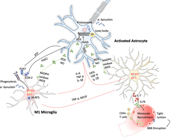
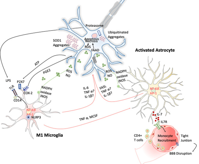
Neuroinflammation constitutes a fundamental cellular process to signal the loss of brain homeostasis. Glial cells play a central role in orchestrating these neuroinflammation processes in both deleterious and beneficial ways. These cellular responses depend on their intercellular interactions with neurons, astrocytes, the blood-brain barrier (BBB), and infiltrated T cells in the central nervous system (CNS). However, this intercellular crosstalk seems to be activated by specific stimuli for each different neurological scenario. This review summarizes key studies linking neuroinflammation with certain neurodegenerative diseases such as Alzheimer disease (AD), Parkinson disease (PD), and amyotrophic lateral sclerosis (ALS) and for the development of better therapeutic strategies based on immunomodulation.
Overview
The claims and statistics of this paper haven’t been checked yet.
Run the check — free, no account needed. It reads the open-access text and checks the sources it cites.
Integrity signals
Registry
Cites
Statistics
Figures
Verification
Claims the paper makes
The claims and statistics of this paper haven’t been checked yet.
Run the check — free, no account needed. It reads the open-access text and checks the sources it cites.
Claims other papers make about this one
Each is checked against this paper’s own text: does it say what the citing paper says it does?
The cited source says what the claim attributes to it.
In the citing paper
“Gliosis is consistent with the presence of neuroinflammation [46].”
From Pharmacological modulation of TSPO in microglia/macrophages and neurons in a chronic neurodegenerative model of prion disease (2023) · cites this paper as [46]
Self-check
Percentages, sums and reported differences re-computed from the paper’s own numbers.
The self-check needs the paper’s full text.
Citations
Cites
Staging of brain pathology related to sporadic Parkinson’s disease
Braak, H., Tredici, K. D., Rüb, U., de Vos, R. A. I., Jansen Steur, E. N. H., & Braak, E. (2003) · Neurobiology of Aging · doi:10.1016/s0197-4580(02)00065-9 · Cited by 10,777
The role of pattern-recognition receptors in innate immunity: update on Toll-like receptors
Kawai, T., & Akira, S. (2010) · Nature Immunology · doi:10.1038/ni.1863 · Cited by 9,304
Polazzi, E., Mengoni, I., Caprini, M., Peña‐Altamira, E., Kurtys, E., & Monti, B. (2012) · Neurosignals · doi:10.1159/000337115 · Cited by 9,023
Neurotoxic reactive astrocytes are induced by activated microglia
Liddelow, S. A., Guttenplan, K. A., Clarke, L. E., Bennett, F. C., Bohlen, C. J., Schirmer, L., Bennett, M. L., Münch, A. E., Chung, W.-S., Peterson, T. C., et al. (2017) · Nature · doi:10.1038/nature21029 · Cited by 7,971
Astrocytes: Biology and pathology
Sofroniew, M. V., & Vinters, H. V. (2010) · Acta Neuropathologica · doi:10.1007/s00401-009-0619-8 · Cited by 5,196
Fate Mapping Analysis Reveals That Adult Microglia Derive from Primitive Macrophages
Ginhoux, F., Greter, M., Leboeuf, M., Nandi, S., See, P., Gokhan, S., Mehler, M. F., Conway, S. J., Ng, L. G., Stanley, E. R., et al. (2010) · Science · doi:10.1126/science.1194637 · Cited by 5,175
Meta-Analysis of 74,046 Individuals Identifies 11 New Susceptibility Loci for Alzheimer’s Disease
(EADI), E. A. D. I., Lambert, J.-C., (GERAD), G. A. E. R. I. A. D., Alzheimer's Disease Genetic Consortium (ADGC), (CHARGE), C. F. H. A. A. R. I. G. E., Ibrahim-Verbaas, C. A., Harold, D., Naj, A. C., Sims, R., Bellenguez, C., et al. (2013) · Nature Genetics · doi:10.1038/ng.2802 · Cited by 4,336
Synaptic Pruning by Microglia Is Necessary for Normal Brain Development
Paolicelli, R. C., Bolasco, G., Pagani, F., Maggi, L., Scianni, M., Panzanelli, P., Giustetto, M., Ferreira, T. A., Guiducci, E., Dumas, L., et al. (2011) · Science · doi:10.1126/science.1202529 · Cited by 4,244
Kettenmann, H., Hanisch, U.-K., Noda, M., & Verkhratsky, A. (2011) · Physiological Reviews · doi:10.1152/physrev.00011.2010 · Cited by 3,547
Bauernfeind, F. G., Horvath, G. L., Stutz, A., Alnemri, E. S., MacDonald, K. L., Speert, D. P., Fernandes‐Alnemri, T., Wu, J., Monks, B. G., Fitzgerald, K. A., et al. (2009) · The Journal of Immunology · doi:10.4049/jimmunol.0901363 · Cited by 3,087
Cited by
Sorted by citations
Alzheimer’s disease and its treatment by different approaches: A review
Srivastava, S., Ahmad, R., & Khare, S. K. (2021) · European Journal of Medicinal Chemistry · doi:10.1016/j.ejmech.2021.113320 · Cited by 506
Long-Term Respiratory and Neurological Sequelae of COVID-19
Wang, F., Kream, R. M., & Stefano, G. B. (2020) · Medical Science Monitor · doi:10.12659/msm.928996 · Cited by 441
Houldsworth, A. (2023) · Brain Communications · doi:10.1093/braincomms/fcad356 · Cited by 455
Microglia-Astrocyte Communication in Alzheimer’s Disease
Wu, Y., & Eisel, U. L. M. (2023) · Journal of Alzheimer s Disease · doi:10.3233/jad-230199 · Cited by 125
Xia, Q., Zhan, G., Mao, M., Zhao, Y., & Li, X. (2022) · Experimental & Molecular Medicine · doi:10.1038/s12276-022-00734-y · Cited by 112
Metadata
- Authors
- Bernaus, A., Blanco, S., & Sevilla, A.
- Affiliations
- Universitat de Barcelona (ES)
- Consejo Superior de Investigaciones Científicas (ES)
- Corresponding author
- Sevilla, Ana
- Venue
- Frontiers in Cellular Neuroscience
- Publisher
- Frontiers Media SA
- Published
- 2020-07-29
- Volume
- 14
- Pages
- 209-209
- ISSN
- 1662-5102 (ISSN-L)
- PMC
- PMC7403442
- OpenAlex
- W3046216659
- Cited by
- 157
- References
- 134
- Citations by year
- 2026: 16 · 2025: 20 · 2024: 39 · 2023: 29 · 2022: 32 · 2021: 20 · 2020: 1
- FWCI
- 7.94
- Type
- journal article
- Language
- en
- Open access
- gold
- doi.org
- Licence
- Locations
- Frontiers in Cellular Neuroscience (publishedVersion, cc-by)
- PubMed Central (publishedVersion, cc-by)
- PubMed (publishedVersion)
- DIGITAL.CSIC (Spanish National Research Council (CSIC)) (submittedVersion, cc-by)
- DOAJ (DOAJ: Directory of Open Access Journals) (submittedVersion, cc-by-sa)
- Funders
- Ministerio de Ciencia y Tecnología (RYC-2016-19962)
- Ministerio de Ciencia, Innovación y Universidades (PGC2018-098626-B100)
- Merck Company Foundation
- Fundación Científica Asociación Española Contra el Cáncer
- Topics
- Life Sciences › Neuroscience › Neurology › Neuroinflammation and Neurodegeneration Mechanisms
- Life Sciences › Neuroscience › Cellular and Molecular Neuroscience › Neuroscience and Neuropharmacology Research
- Life Sciences › Neuroscience › Neurology › Neurological Disease Mechanisms and Treatments
- Keywords
- neuroinflammation, astrocytes, blood-brain barrier, infiltrating T cells, neurodegenerative diseases, Alzheimer's disease, Parkinson's disease, amyotrophic lateral sclerosis, immunomodulation, neuron-glia interactions
About these checks
Registry, retraction, Cites and statistics results are computed, not judged, with one exception: a printed reference that no index lists is read once by a model before it counts as a citation error. Claims are extracted by a model and kept only when their quote appears verbatim in the text; each cited source is then checked, with a model’s help, for whether it says what the claim relies on. Those verdicts can be wrong — read the claim’s quote and the cited source before relying on one.
Statistics and arithmetic are recomputed, not judged.
Confidence bands are shown only for fields calibrated on our annotated test set. None is calibrated yet, so no band is shown.
What counts as a claim ›How we measure confidence ›How we check this ›
Are you an author, or do you see a problem with this page? Request a review
