

The DOI matches its Crossref record; 101 of the 101 references it cites were found in a registry or an index.
Neural masses and fields in dynamic causal modeling
Open accessAbstract
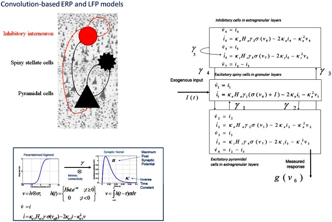
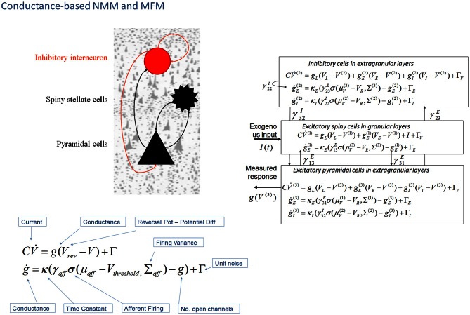
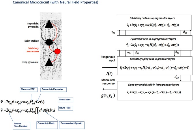
Dynamic causal modeling (DCM) provides a framework for the analysis of effective connectivity among neuronal subpopulations that subtend invasive (electrocorticograms and local field potentials) and non-invasive (electroencephalography and magnetoencephalography) electrophysiological responses. This paper reviews the suite of neuronal population models including neural masses, fields and conductance-based models that are used in DCM. These models are expressed in terms of sets of differential equations that allow one to model the synaptic underpinnings of connectivity. We describe early developments using neural mass models, where convolution-based dynamics are used to generate responses in laminar-specific populations of excitatory and inhibitory cells. We show that these models, though resting on only two simple transforms, can recapitulate the characteristics of both evoked and spectral responses observed empirically. Using an identical neuronal architecture, we show that a set of conductance based models-that consider the dynamics of specific ion-channels-present a richer space of responses; owing to non-linear interactions between conductances and membrane potentials. We propose that conductance-based models may be more appropriate when spectra present with multiple resonances. Finally, we outline a third class of models, where each neuronal subpopulation is treated as a field; in other words, as a manifold on the cortical surface. By explicitly accounting for the spatial propagation of cortical activity through partial differential equations (PDEs), we show that the topology of connectivity-through local lateral interactions among cortical layers-may be inferred, even in the absence of spatially resolved data. We also show that these models allow for a detailed analysis of structure-function relationships in the cortex. Our review highlights the relationship among these models and how the hypothesis asked of empirical data suggests an appropriate model class.
Overview
The claims and statistics of this paper haven’t been checked yet.
Run the check — free, no account needed. It reads the open-access text and checks the sources it cites.
Integrity signals
Registry
Cites
Statistics
Figures
Verification
Claims the paper makes
The claims and statistics of this paper haven’t been checked yet.
Run the check — free, no account needed. It reads the open-access text and checks the sources it cites.
Claims other papers make about this one
Each is checked against this paper’s own text: does it say what the citing paper says it does?
The source backs part of the claim, not all of it — or backs it for a different population, condition or comparison.
In the citing paper
“By adjusting a few key parameters (like the gain of neuronal populations that carry error signals), these models can simulate how precision (confidence) is encoded and modulated in the brain (21, 22).”
From Predictive coding and neurocomputational psychiatry: a mechanistic framework for understanding mental disorders (2026) · cites this paper as [21]
Experimental — accuracy not yet measured
Self-check
Percentages, sums and reported differences re-computed from the paper’s own numbers.
The self-check needs the paper’s full text.
Citations
Cites
Distributed hierarchical processing in the primate cerebral cortex
Felleman, D. J., & Van Essen, D. C. (1991) · Cerebral Cortex · doi:10.1093/cercor/1.1.1 · Cited by 7,783
Friston, K. J., Harrison, L., & Penny, W. (2003) · NeuroImage · doi:10.1016/s1053-8119(03)00202-7 · Cited by 4,583
Self-citationA theory of cortical responses.
Friston, K. (2005) · Philosophical Transactions of the Royal Society B: Biological Sciences · doi:10.1098/rstb.2005.1622 · Cited by 4,578
Self-citationExcitatory and Inhibitory Interactions in Localized Populations of Model Neurons
Wilson, H. R., & Cowan, J. D. (1972) · Biophysical Journal · doi:10.1016/s0006-3495(72)86068-5 · Cited by 3,579
Preface: Cerebral Cortex Has Come of Age
Goldman, P. S., & RAKIC, P. T. (1991) · Cerebral Cortex · doi:10.1093/cercor/1.1.1-a · Cited by 3,537
Canonical Microcircuits for Predictive Coding
Bastos, A. M., Usrey, W. M., Adams, R. A., Mangun, G. R., Fries, P., & Friston, K. J. (2012) · Neuron · doi:10.1016/j.neuron.2012.10.038 · Cited by 2,421 · cited 8×
“The canonical microcircuit or “CMC” models a refinement of the Jansen and Rit convolution models that explicitly accommodates the neuronal sources of forward and backward connections in cortical hierarchies (Bastos et al., 2012). These are distinct superficial and deep pyramidal cell populations, respectively that, cr…”
BackgroundMethodInfluentialSelf-citationVoltage oscillations in the barnacle giant muscle fiber
Morris, C., & Lecar, H. (1981) · Biophysical Journal · doi:10.1016/s0006-3495(81)84782-0 · Cited by 2,302
Electric Fields of the Brain: The Neurophysics of EEG
Nunez, P. L., & Harth, E. M. (1982) · Physics Today · doi:10.1063/1.2915137 · Cited by 2,257
Box, G. E. P. (1976) · Journal of the American Statistical Association · doi:10.1080/01621459.1976.10480949 · Cited by 2,114
Baillet, S., Mosher, J., & Leahy, R. (2001) · IEEE Signal Processing Magazine · doi:10.1109/79.962275 · Cited by 1,846
The free-energy principle: a rough guide to the brain?
Friston, K. (2009) · Trends in Cognitive Sciences · doi:10.1016/j.tics.2009.04.005 · Cited by 1,831
Self-citationA mathematical theory of the functional dynamics of cortical and thalamic nervous tissue
Wilson, H. R., & Cowan, J. D. (1973) · Kybernetik · doi:10.1007/bf00288786 · Cited by 1,823
Whittington, M. A., Traub, R. D., & Jefferys, J. G. R. (1995) · Nature · doi:10.1038/373612a0 · Cited by 1,613
Electric Fields of the Brain: The Neurophysics of EEG
Fermaglich, J. (1982) · JAMA · doi:10.1001/jama.1982.03320380071046 · Cited by 1,557
Mass action in the nervous system
Freeman, W. J. (1975) · Cited by 1,480
The mismatch negativity: a review of underlying mechanisms
Garrido, M. I., Kilner, J. M., Stephan, K. E., & Friston, K. J. (2009) · Clinical Neurophysiology · doi:10.1016/j.clinph.2008.11.029 · Cited by 1,441 · cited 1×
“Early applications of this model led to a series of EEG-based investigations into oddball effects by Garrido et al. (2007a, 2008, 2009).”
BackgroundSelf-citationNonlinear dynamical analysis of EEG and MEG: Review of an emerging field
Stam, C. J. (2005) · Clinical Neurophysiology · doi:10.1016/j.clinph.2005.06.011 · Cited by 1,410
Jansen, B. H., & Rit, V. G. (1995) · Biological Cybernetics · doi:10.1007/bf00199471 · Cited by 1,159
The dynamic brain: from spiking neurons to neural masses and cortical fields
Deco, G., Jirsa, V. K., Robinson, P. A., Breakspear, M., & Friston, K. (2008) · PLoS Computational Biology · doi:10.1371/journal.pcbi.1000092 · Cited by 1,084
Self-citationComparing dynamic causal models.
Penny, W. D., Stephan, K. E., Mechelli, A., & Friston, K. J. (2004) · NeuroImage · doi:10.1016/j.neuroimage.2004.03.026 · Cited by 848
Self-citationSimulation of chaotic EEG patterns with a dynamic model of the olfactory system
Freeman, W. J. (1987) · Biological Cybernetics · doi:10.1007/bf00317988 · Cited by 838
Regional and cellular fractionation of working memory
Goldman‐Rakic, P. S. (1996) · Proceedings of the National Academy of Sciences · doi:10.1073/pnas.93.24.13473 · Cited by 826
A functional microcircuit for cat visual cortex
Douglas, R. J., & Martin, K. A. (1991) · The Journal of Physiology · doi:10.1113/jphysiol.1991.sp018733 · Cited by 736
A neural mass model for MEG/EEG:
Olivier David, A., & Friston, K. J. (2003) · NeuroImage · doi:10.1016/j.neuroimage.2003.07.015 · Cited by 705
Self-citationOngoing Cortical Activity at Rest: Criticality, Multistability, and Ghost Attractors
Deco, G., & Jirsa, V. (2012) · Journal of Neuroscience · doi:10.1523/jneurosci.2523-11.2012 · Cited by 704
Very Slow Activity Fluctuations in Monkey Visual Cortex: Implications for Functional Brain Imaging
Leopold, D. A. (2003) · Cerebral Cortex · doi:10.1093/cercor/13.4.422 · Cited by 675
Memory‐based or afferent processes in mismatch negativity (MMN): A review of the evidence
Näätänen, R., Jacobsen, T., & Winkler, I. (2005) · Psychophysiology · doi:10.1111/j.1469-8986.2005.00256.x · Cited by 657
Dopamine-Mediated Stabilization of Delay-Period Activity in a Network Model of Prefrontal Cortex
Durstewitz, D., Seamans, J. K., & B, T. J. S. A. (2000) · Journal of Neurophysiology · doi:10.1152/jn.2000.83.3.1733 · Cited by 624
Dynamic causal modeling of evoked responses in EEG and MEG
David, O., Kiebel, S. J., Harrison, L. M., Mattout, J., Kilner, J. M., & Friston, K. J. (2006) · NeuroImage · doi:10.1016/j.neuroimage.2005.10.045 · Cited by 620
Self-citationBrunel, N., & Wang, X.-J. (2001) · Journal of Computational Neuroscience · doi:10.1023/a:1011204814320 · Cited by 620
Laminar differences in gamma and alpha coherence in the ventral stream
Buffalo, E. A., Fries, P., Landman, R., Buschman, T. J., & Desimone, R. (2011) · Proceedings of the National Academy of Sciences · doi:10.1073/pnas.1011284108 · Cited by 606
Durstewitz, D., & Seamans, J. K. (2008) · Biological Psychiatry · doi:10.1016/j.biopsych.2008.05.015 · Cited by 586
EEG and MEG Data Analysis in SPM8
Vladimir Litvak, A., Mattout, J., Kiebel, S. J., Phillips, C. L. M., Henson, R. N., Kilner, J. M., Barnes, G. R., Oostenveld, R., Jean Daunizeau, A., Flandin, G., et al. (2011) · Computational Intelligence and Neuroscience · doi:10.1155/2011/852961 · Cited by 586
Self-citationMuthukumaraswamy, S. D., Edden, R. A. E., Jones, D. K., Swettenham, J. B., & Singh, K. D. (2009) · Proceedings of the National Academy of Sciences · doi:10.1073/pnas.0900728106 · Cited by 581
Marreiros, A. C., Stephan, K. E., & Friston, K. J. (2010) · Scholarpedia · doi:10.4249/scholarpedia.9568 · Cited by 564
Self-citationBrunel, N., & Wang, X. (2014) · Journal of Computational Neuroscience · doi:10.1007/s10827-014-0506-8 · Cited by 538
Generative Models of Cortical Oscillations: Neurobiological Implications of the Kuramoto Model
Breakspear, M. J., Heitmann, S., & Daffertshofer, A. (2010) · Frontiers in Human Neuroscience · doi:10.3389/fnhum.2010.00190 · Cited by 531
A Universal Model for Spike-Frequency Adaptation
Benda, J., & Herz, A. V. M. (2003) · Neural Computation · doi:10.1162/089976603322385063 · Cited by 524
Ashburner, J. (2011) · NeuroImage · doi:10.1016/j.neuroimage.2011.10.025 · Cited by 522
Preserved Feedforward But Impaired Top-Down Processes in the Vegetative State
Boly, M., Garrido, M. I., Gosseries, O., Bruno, M., Boveroux, P., Schnakers, C., Massimini, M., Vladimir Litvak, A., Laureys, S., & Friston, K. J. (2011) · Science · doi:10.1126/science.1202043 · Cited by 508
Self-citationField Theory of Electromagnetic Brain Activity
Jirsa, V., & Haken, H. (1996) · Physical Review Letters · doi:10.1103/physrevlett.77.960 · Cited by 487
Breakspear, M. J., Roberts, J. A., Terry, J. R., Rodrigues, S., Mahant, N., & Robinson, P. A. (2005) · Cerebral Cortex · doi:10.1093/cercor/bhj072 · Cited by 463
The functional anatomy of the MMN: a DCM study of the roving paradigm
Garrido, M. I., Friston, K. J., Kiebel, S. J., Stephan, K. E., Baldeweg, T., & Kilner, J. M. (2008) · NeuroImage · doi:10.1016/j.neuroimage.2008.05.018 · Cited by 458
Self-citationRelevance of nonlinear lumped-parameter models in the analysis of depth-EEG epileptic signals
Wendling, F., Bellanger, J.-J., Bartoloméi, F., & Chauvel, P. (2000) · Biological Cybernetics · doi:10.1007/s004220000160 · Cited by 410
Mass action in the nervous system
Walter, D. O. (1976) · Electroencephalography and Clinical Neurophysiology · doi:10.1016/0013-4694(76)90110-3 · Cited by 377
Prediction of electroencephalographic spectra from neurophysiology
Robinson, P. A., RENNIE, C. J., Wright, J. J., Bahramali, H., Gordon, E., & ROWE, D. L. (2001) · Physical review. E, Statistical physics, plasmas, fluids, and related interdisciplinary topics · doi:10.1103/physreve.63.021903 · Cited by 366
Box, G. E. P. (1976) · Journal of the American Statistical Association · doi:10.2307/2286841 · Cited by 366
Edden, R. A. E., Muthukumaraswamy, S. D., Freeman, T. C. A., & Singh, K. D. (2009) · Journal of Neuroscience · doi:10.1523/jneurosci.4426-09.2009 · Cited by 360
The surface area of human V1 predicts the subjective experience of object size
Schwarzkopf, D. S., Song, C., & Rees, G. E. (2010) · Nature Neuroscience · doi:10.1038/nn.2706 · Cited by 358
The brain wave equation: a model for the EEG
Nunez, P. L. (1974) · Mathematical Biosciences · doi:10.1016/0025-5564(74)90020-0 · Cited by 338
Dynamic causal modelling: A critical review of the biophysical and statistical foundations
Daunizeau, J., David, O., & Stephan, K. E. (2011) · NeuroImage · doi:10.1016/j.neuroimage.2009.11.062 · Cited by 331
A quantitative description of NMDA receptor-channel kinetic behavior
Jahr, C. E., & Stevens, C. F. (1990) · Journal of Neuroscience · doi:10.1523/jneurosci.10-06-01830.1990 · Cited by 327
Connectivity Changes Underlying Spectral EEG Changes during Propofol-Induced Loss of Consciousness
Boly, M., Moran, R., Murphy, M., Boveroux, P., Bruno, M., Noirhomme, Q., Ledoux, D., Bonhomme, V. L., Brichant, J.-F., Tononi, G., et al. (2012) · Journal of Neuroscience · doi:10.1523/jneurosci.3769-11.2012 · Cited by 326
Self-citationDynamic causal modeling for EEG and MEG
Kiebel, S., Garrido, M., Moran, R., Chen, C.-C., & Friston, K. J. (2009) · Human Brain Mapping · doi:10.1002/hbm.20775 · Cited by 318
Self-citationEvoked brain responses are generated by feedback loops
Garrido, M. I., Kilner, J. M., Kiebel, S. J., & Friston, K. J. (2007) · Proceedings of the National Academy of Sciences · doi:10.1073/pnas.0706274105 · Cited by 296
Self-citationDynamics of the brain at global and microscopic scales: Neural networks and the EEG
Wright, J. J., & Liley, D. T. J. (1996) · Behavioral and Brain Sciences · doi:10.1017/s0140525x00042679 · Cited by 274
Maier (2010) · Frontiers in System Neuroscience · doi:10.3389/fnsys.2010.00031 · Cited by 266
Simulation of emission spectra from nonuniform reactive laser-induced plasmas
Hermann, J., Lorusso, A., Perrone, A., Strafella, F., Dutouquet, C., & Torralba, B. (2015) · Physical Review E · doi:10.1103/physreve.92.053103 · Cited by 257
Propagation of electrical signals along giant nerve fibres
Hodgkin, A. L., & Huxley, A. F. (1952) · Proceedings of the Royal Society B Biological Sciences · doi:10.1098/rspb.1952.0054 · Cited by 256
A derivation of a macroscopic field theory of the brain from the quasi-microscopic neural dynamics
Jirsa, V., & Haken, H. (1997) · Physica D Nonlinear Phenomena · doi:10.1016/s0167-2789(96)00166-2 · Cited by 236
Dynamic causal modelling of evoked responses in EEG/MEG with lead field parameterization
Kiebel, S. J., David, O., & Friston, K. J. (2006) · NeuroImage · doi:10.1016/j.neuroimage.2005.12.055 · Cited by 233
Self-citationDynamic causal modelling of evoked potentials: A reproducibility study
Garrido, M. I., Kilner, J. M., Kiebel, S. J., Stephan, K. E., & Friston, K. J. (2007) · NeuroImage · doi:10.1016/j.neuroimage.2007.03.014 · Cited by 233
Self-citationA neural mass model of spectral responses in electrophysiology
Moran, R., Kiebel, S. J., Stephan, K. E., Reilly, R. B., Jean Daunizeau, A., & Friston, K. J. (2007) · NeuroImage · doi:10.1016/j.neuroimage.2007.05.032 · Cited by 214
Self-citationBifurcation Analysis of Jansen's Neural Mass Model
Grimbert, F., & Faugeras, O. D. (2006) · Neural Computation · doi:10.1162/neco.2006.18.12.3052 · Cited by 196
da Silva, F. L., Pijn, J. P. M., & Boeijinga, P. H. (1989) · Brain Topography · doi:10.1007/bf01128839 · Cited by 191
Steyn‐Ross, M. L., Steyn‐Ross, D. A., Sleigh, J. W., & Liley, D. T. J. (1999) · Physical review. E, Statistical physics, plasmas, fluids, and related interdisciplinary topics · doi:10.1103/physreve.60.7299 · Cited by 176
Population dynamics: Variance and the sigmoid activation function
Marreiros, A. C., Jean Daunizeau, A., Kiebel, S. J., & Friston, K. J. (2008) · NeuroImage · doi:10.1016/j.neuroimage.2008.04.239 · Cited by 175
Self-citationDopamine Enhances EPSCs in Layer II–III Pyramidal Neurons in Rat Prefrontal Cortex
Gonzalez-Islas, C. E., & Hablitz, J. J. (2003) · Journal of Neuroscience · doi:10.1523/jneurosci.23-03-00867.2003 · Cited by 173
Large-scale neural dynamics: Simple and complex
Coombes, S. (2010) · NeuroImage · doi:10.1016/j.neuroimage.2010.01.045 · Cited by 163
Gorelova, N. A., & Yang, C. R. (2000) · Journal of Neurophysiology · doi:10.1152/jn.2000.84.1.75 · Cited by 162
Mechanisms of Cortical Electrical Activity and Emergence of Gamma Rhythm
RENNIE, C. J., Wright, J. J., & Robinson, P. A. (2000) · Journal of Theoretical Biology · doi:10.1006/jtbi.2000.2040 · Cited by 154
Electric fields of the brain the neurophysics of EEG
Nunez, P. L., & Srinivasan, R. (2006) · Cited by 151
Nonlinear EEG analysis based on a neural mass model
Valdes-Sosa, P. A., Jiménez, J. C., Riera, J. J., Biscay, R., & Ozaki, T. (1999) · Biological Cybernetics · doi:10.1007/s004220050572 · Cited by 141
Jansen, B. H., & Rit, V. G. (1995) · Biological Cybernetics · doi:10.1007/s004220050191 · Cited by 140
An in vivo assay of synaptic function mediating human cognition
Moran, R. J., Symmonds, M., Stephan, K. E., Friston, K. J., & Dolan, R. J. (2011) · Current Biology · doi:10.1016/j.cub.2011.06.053 · Cited by 137
Self-citationDCM for complex-valued data: Cross-spectra, coherence and phase-delays
Friston, K. J., Bastos, A. M., Vladimir Litvak, A., Stephan, K. E., Fries, P., & Moran, R. (2011) · NeuroImage · doi:10.1016/j.neuroimage.2011.07.048 · Cited by 135
Self-citationBayesian estimation of synaptic physiology from the spectral responses of neural masses
Moran, R. J., Stephan, K. E., Kiebel, S. J., Rombach, N., O'Connor, W. T., Murphy, K. J., Reilly, R. B., & Friston, K. J. (2008) · NeuroImage · doi:10.1016/j.neuroimage.2008.01.025 · Cited by 126
Self-citationNonlinear phase desynchronization in human electroencephalographic data
Breakspear, M. J. (2002) · Human Brain Mapping · doi:10.1002/hbm.10011 · Cited by 117
Modeling ketamine effects on synaptic plasticity during the mismatch negativity
Schmidt, A., Diaconescu, A. O., Kometer, M., Friston, K. J., Stephan, K. E., & Vollenweider, F. X. (2013) · Cerebral Cortex · doi:10.1093/cercor/bhs238 · Cited by 109
Self-citationDynamic causal modeling with neural fields
Pinotsis, D. A., Moran, R., & Friston, K. J. (2011) · NeuroImage · doi:10.1016/j.neuroimage.2011.08.020 · Cited by 106
Self-citationPopulation dynamics under the Laplace assumption
Marreiros, A. C., Kiebel, S. J., Jean Daunizeau, A., Harrison, L. M., & Friston, K. J. (2008) · NeuroImage · doi:10.1016/j.neuroimage.2008.10.008 · Cited by 85
Self-citationMarten, F., Rodrigues, S., Suffczyński, P., Richardson, M. P., & Terry, J. R. (2009) · Physical Review E · doi:10.1103/physreve.79.021911 · Cited by 82
Parallelism and Functional Specialization in Human Visual CortexPreprint
Zeki, S. M. (1990) · Cold Spring Harbor Symposia on Quantitative Biology · doi:10.1101/sqb.1990.055.01.062 · Cited by 82
(2004) · Elsevier eBooks · doi:10.1016/b978-012264841-0/50054-8 · Cited by 81
Nevado‐Holgado, A. J., Marten, F., Richardson, M. P., & Terry, J. R. (2011) · NeuroImage · doi:10.1016/j.neuroimage.2011.08.111 · Cited by 80
Consistent spectral predictors for dynamic causal models of steady-state responses
Moran, R. J., Stephan, K. E., Dolan, R. J., & Friston, K. J. (2011) · NeuroImage · doi:10.1016/j.neuroimage.2011.01.012 · Cited by 75
Self-citationMathematical neuroscience: from neurons to circuits to systems
Gutkin, B. S., Pinto, D. J., & Ermentrout, B. (2003) · Journal of Physiology-Paris · doi:10.1016/j.jphysparis.2003.09.005 · Cited by 73
Topographic Organization of Nonlinear Interdependence in Multichannel Human EEG
Breakspear, M. J., & Terry, J. R. (2002) · NeuroImage · doi:10.1006/nimg.2002.1106 · Cited by 70
Schwarzkopf, D. S., Robertson, D. J., Song, C., Barnes, G. R., & Rees, G. E. (2012) · Journal of Neuroscience · doi:10.1523/jneurosci.4771-11.2012 · Cited by 65
Dynamic causal modelling of lateral interactions in the visual cortex
Pinotsis, D. A., Schwarzkopf, D. S., Vladimir Litvak, A., Rees, G. E., Barnes, G. R., & Friston, K. J. (2012) · NeuroImage · doi:10.1016/j.neuroimage.2012.10.078 · Cited by 64
Self-citationCampo, P., Garrido, M. I., Moran, R., Maestú, F., García‐Morales, I., Gil‐Nagel, A., del Pozo, F., Dolan, R. J., & Friston, K. J. (2011) · Cerebral Cortex · doi:10.1093/cercor/bhr201 · Cited by 64
Self-citationPatchy propagators, brain dynamics, and the generation of spatially structured gamma oscillations
Robinson, P. A. (2006) · Physical Review E · doi:10.1103/physreve.73.041904 · Cited by 62
Dynamic Finite Size Effects in Spiking Neural Networks
Buice, M. A., & Chow, C. C. (2013) · PLoS Computational Biology · doi:10.1371/journal.pcbi.1002872 · Cited by 61
Dynamical Reconnection and Stability Constraints on Cortical Network Architecture
Robinson, P. A., Henderson, J. A., Matar, E., Riley, P., & Gray, R. T. (2009) · Physical Review Letters · doi:10.1103/physrevlett.103.108104 · Cited by 60
Anatomical connectivity and the resting state activity of large cortical networks
Pinotsis, D. A., Hansen, E. C. A., Friston, K. J., & Jirsa, V. (2012) · NeuroImage · doi:10.1016/j.neuroimage.2012.10.016 · Cited by 56
Self-citationA dynamic causal model study of neuronal population dynamics
Marreiros, A. C., Kiebel, S. J., & Friston, K. J. (2010) · NeuroImage · doi:10.1016/j.neuroimage.2010.01.098 · Cited by 53
Self-citationPacemaker-Induced Coherence in Cortical Networks
Destexhe, A., & BABLOYANTZ, A. (1991) · Neural Computation · doi:10.1162/neco.1991.3.2.145 · Cited by 52
Comparing dynamic causal models
Penny, W. D. (2004) · NeuroImage · doi:10.1016/s1053-8119(04)00164-8 · Cited by 50
Stability of small-world networks of neural populations
Gray, R. T., Fung, C. K. C., & Robinson, P. A. (2008) · Neurocomputing · doi:10.1016/j.neucom.2008.09.006 · Cited by 30
Neural fields, spectral responses and lateral connections
Pinotsis, D. A., & Friston, K. J. (2010) · NeuroImage · doi:10.1016/j.neuroimage.2010.11.081 · Cited by 28
Self-citationSynaptic Scaling Stabilizes Persistent Activity Driven by Asynchronous Neurotransmitter Release
Volman, V., & Gerkin, R. C. (2011) · Neural Computation · doi:10.1162/neco_a_00098 · Cited by 12
Cited by
Sorted by citations
Great Expectations: Is there Evidence for Predictive Coding in Auditory Cortex?
Heilbron, M., & Chait, M. (2018) · Neuroscience · doi:10.1016/j.neuroscience.2017.07.061 · Cited by 428
A guide to group effective connectivity analysis, part 1: First level analysis with DCM for fMRI
Zeidman, P., Jafarian, A., Corbin, N., Seghier, M. L., Razi, A., Price, C. J., & Friston, K. J. (2019) · NeuroImage · doi:10.1016/j.neuroimage.2019.06.031 · Cited by 369
Self-citationA systematic framework for functional connectivity measures
Wang, H., Bénar, C., Quilichini, P., Friston, K. J., Jirsa, V., & Bernard, C. (2014) · Frontiers in Neuroscience · doi:10.3389/fnins.2014.00405 · Cited by 342
Self-citationBeyond the Connectome: The Dynome
Kopell, N., Gritton, H. J., Whittington, M. A., & Kramer, M. (2014) · Neuron · doi:10.1016/j.neuron.2014.08.016 · Cited by 331
Dynamic causal modelling revisited
Friston, K. J., Preller, K. H., Mathys, C., Cagnan, H., Heinzle, J., Razi, A., & Zeidman, P. (2019) · NeuroImage · doi:10.1016/j.neuroimage.2017.02.045 · Cited by 271
Self-citation
Metadata
- Authors
- Moran, R., Pinotsis, D. A., & Friston, K.
- Affiliations
- Wellcome Centre for Human Neuroimaging (GB)
- Corresponding author
- Moran, Rosalyn
- Venue
- Frontiers in Computational Neuroscience
- Publisher
- Frontiers Media SA
- Published
- 2013
- Volume
- 7
- Pages
- 57-57
- ISSN
- 1662-5188 (ISSN-L)
- PMC
- PMC3664834
- OpenAlex
- W2142039948
- Cited by
- 255
- References
- 100
- Citations by year
- 2026: 14 · 2025: 19 · 2024: 19 · 2023: 26 · 2022: 19 · 2021: 29 · 2020: 28 · 2019: 16 · 2018: 16 · 2017: 17 · 2016: 18 · 2015: 16 · 2014: 15 · 2013: 3
- FWCI
- 9.51
- Type
- journal article
- Language
- en
- Open access
- gold
- doi.org
- Licence
- cc-by
- Locations
- Frontiers in Computational Neuroscience (publishedVersion, cc-by)
- Europe PMC (PubMed Central) (publishedVersion, cc-by)
- PubMed Central (publishedVersion, cc-by)
- PubMed (publishedVersion)
- Research Portal (King's College London) (publishedVersion, other-oa)
- City Research Online (City University London) (acceptedVersion)
- DOAJ (DOAJ: Directory of Open Access Journals) (submittedVersion, cc-by-sa)
- UCL Discovery (University College London) (submittedVersion)
- Bristol Research (University of Bristol) (submittedVersion)
- VTechWorks (Virginia Tech) (submittedVersion, cc-by)
- Funders
- Wellcome Trust (091593, 088130)
- Topics
- Life Sciences › Neuroscience › Cognitive Neuroscience › Neural dynamics and brain function
- Life Sciences › Neuroscience › Cognitive Neuroscience › Functional Brain Connectivity Studies
- Health Sciences › Medicine › Radiology, Nuclear Medicine and Imaging › Advanced Neuroimaging Techniques and Applications
- Keywords
- dynamic causal modeling, neural mass model, neural field models, conductance-based models, effective connectivity, ion channel dynamics, multiple resonances, structure-function relationship, EEG, magnetoencephalography, local field potentials, electrocorticography
About these checks
Registry, retraction, Cites and statistics results are computed, not judged, with one exception: a printed reference that no index lists is read once by a model before it counts as a citation error. Claims are extracted by a model and kept only when their quote appears verbatim in the text; each cited source is then checked, with a model’s help, for whether it says what the claim relies on. Those verdicts can be wrong — read the claim’s quote and the cited source before relying on one.
Statistics and arithmetic are recomputed, not judged.
Confidence bands are shown only for fields calibrated on our annotated test set. None is calibrated yet, so no band is shown.
What counts as a claim ›How we measure confidence ›How we check this ›
Are you an author, or do you see a problem with this page? Request a review
