

The DOI matches its Crossref record; 71 of the 73 references it cites were found in a registry or an index.
Activation of the hypothalamic-pituitary-adrenal stress axis induces cellular oxidative stress
Open accessAbstract
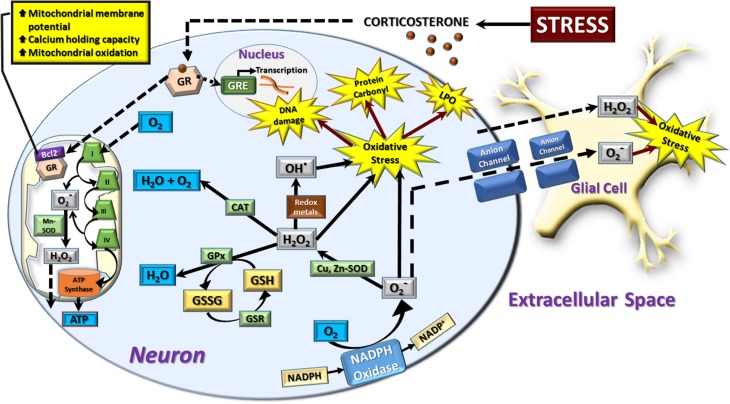
Glucocorticoids released from the adrenal gland in response to stress-induced activation of the hypothalamic-pituitary-adrenal (HPA) axis induce activity in the cellular reduction-oxidation (redox) system. The redox system is a ubiquitous chemical mechanism allowing the transfer of electrons between donor/acceptors and target molecules during oxidative phosphorylation while simultaneously maintaining the overall cellular environment in a reduced state. The objective of this review is to present an overview of the current literature discussing the link between HPA axis-derived glucocorticoids and increased oxidative stress, particularly focussing on the redox changes observed in the hippocampus following glucocorticoid exposure.
Overview
The claims and statistics of this paper haven’t been checked yet.
Run the check — free, no account needed. It reads the open-access text and checks the sources it cites.
Integrity signals
Registry
Cites
Statistics
Figures
Verification
Claims the paper makes
The claims and statistics of this paper haven’t been checked yet.
Run the check — free, no account needed. It reads the open-access text and checks the sources it cites.
Claims other papers make about this one
Each is checked against this paper’s own text: does it say what the citing paper says it does?
The source backs part of the claim, not all of it — or backs it for a different population, condition or comparison.
In the citing paper
“Chronic prenatal stress produces affective abnormalities together with HPA axis hyperactivity and epigenetic disruptions, illustrating the interplay between inflammation, endocrine function, and chromatin regulation in stress vulnerability (Possamai-Della et al., 2023; Glover et al., 2009; Spiers et al., 2014).”
From Histone modifications and depression: epigenetic mechanisms, therapeutic targets, and translational outlook (2026) · cites this paper as Spiers et al., 2014
Experimental — accuracy not yet measured
Self-check
Percentages, sums and reported differences re-computed from the paper’s own numbers.
The self-check needs the paper’s full text.
Citations
Cites
Free radicals in biology and medicine
Gutteridge, J. M. C. (1991) · General Pharmacology The Vascular System · doi:10.1016/0306-3623(91)90478-o · Cited by 17,178
Free radicals and antioxidants in normal physiological functions and human disease
Valko, M., Leibfritz, D., Moncol, J., Cronin, M. T. D., Mazur, M., & Telser, J. (2007) · The International Journal of Biochemistry & Cell Biology · doi:10.1016/j.biocel.2006.07.001 · Cited by 14,269
Oxidants, antioxidants, and the degenerative diseases of aging.
Ames, B. N., Shigenaga, M. K., & Hagen, T. M. (1993) · Proceedings of the National Academy of Sciences · doi:10.1073/pnas.90.17.7915 · Cited by 5,958
Oxidative stress: oxidants and antioxidants
Sies, H. (1997) · Experimental Physiology · doi:10.1113/expphysiol.1997.sp004024 · Cited by 4,577
Itoh, K., Wakabayashi, N., Katoh, Y., Ishii, T., Igarashi, K., Engel, J. D., & Yamamoto, M. (1999) · Genes & Development · doi:10.1101/gad.13.1.76 · Cited by 3,634
Mitochondrial free radical generation, oxidative stress, and aging
Cadenas, E., & Davies, K. J. A. (2000) · Free Radical Biology and Medicine · doi:10.1016/s0891-5849(00)00317-8 · Cited by 3,010
Nordberg, J., & Arnér, E. S. J. (2001) · Free Radical Biology and Medicine · doi:10.1016/s0891-5849(01)00724-9 · Cited by 2,720
Brain corticosteroid receptor balance in health and disease
de Kloet, E. R., Vreugdenhil, E., Oitzl, M. S., & Joëls, M. (1998) · Endocrine Reviews · doi:10.1210/edrv.19.3.0331 · Cited by 2,614
Brain corticosteroid receptor balance in health and disease
de Kloet, E. R. (1998) · Endocrine Reviews · doi:10.1210/er.19.3.269 · Cited by 2,614
Two receptor systems for corticosterone in rat brain: Microdistribution and differential occupation
REUL, J. M. H. M., & KLOET, E. R. D. (1985) · Endocrinology · doi:10.1210/endo-117-6-2505 · Cited by 2,580
Research works printed but not linked: 3
- Barycki, 2007, Antioxidant Molecules and Redox Cofactors, Redox Biochemistry
- Fridovich, 2007, Antioxidant Enzymes, Redox Biochemistry
- Rovira, 2007, Redox Regulation of Physiological Processes, Redox Biochemistry
Cited by
Sorted by citations
Items 201–290 of 290
Lv, L.-Z., Wang, Z.-D., Ren, J.-J., Li, L.-H., Liu, X.-B., Li, J.-L., Zhu, H., Jiang, B., Han, Y.-P., Zhou, X.-M., et al. (2026) · Frontiers in Genetics · doi:10.3389/fgene.2025.1722453 · Cited by 7
Lim, S., Jeon, J.-H., Lim, B., Jang, M., Kim, D., Kim, C. H., & Kim, J. (2023) · Journal of Animal Science · doi:10.1093/jas/skad336 · Cited by 5
Câmara, A. B., & Brandão, I. A. (2025) · Journal of Psychiatric Research · doi:10.1016/j.jpsychires.2025.02.012 · Cited by 4
Depletion of MGO or Its Derivatives Ameliorate CUMS-Induced Neuroinflammation
Liu, B., Dong, K., Zhao, Y. F., Wang, X., Sun, Z., Xie, F., & Qian, L. (2025) · Cells · doi:10.3390/cells14060397 · Cited by 4
Elliot‐Portal, E., Arias‐Reyes, C., Laouafa, S., Tam, R., Kinkead, R., & Soliz, J. (2021) · Frontiers in Physiology · doi:10.3389/fphys.2021.701344 · Cited by 4
Navarro, B. F., Aguayo, A. A. C., Gómez, Y. L. T., Alfaro, M. C., & Torner, L. (2024) · Antioxidants · doi:10.3390/antiox13070802 · Cited by 4
Beaven, E. A., Colthorpe, K. L., Spiers, J. G., Chen, H. C., Lavidis, N. A., & Albrecht, J. (2016) · Journal of Basic and Clinical Physiology and Pharmacology · doi:10.1515/jbcpp-2016-0006 · Cited by 4
Self-citationExperimental Comparative Study of potential anxiolytic effect of Vitamin C and Buspirone in rats
Soliman, G. F., Khattab, A. A., & Habil, M. R. (2018) · Functional Foods in Health and Disease · doi:10.31989/ffhd.v8i2.365 · Cited by 4
Womersley, J. S., Spies, G., Tromp, G. C., Seedat, S., & Hemmings, S. M. J. (2021) · Journal of NeuroVirology · doi:10.1007/s13365-021-01009-4 · Cited by 4
Cao, J.-J., Yin, C.-L., Li, X., Sha, X.-J., Li, L., Sun, C.-Y., & Zhang, L. (2025) · World Journal of Psychiatry · doi:10.5498/wjp.v15.i8.103302 · Cited by 3
Roslan, M. S., Mazlishah, M. S. H., Mamat, N., Aziz, N. A. A., Hashim, N. H., & Mohamad‐Fauzi, N. (2025) · Small Ruminant Research · doi:10.1016/j.smallrumres.2025.107645 · Cited by 3
Zhao, D., Shi, Y., Long, X., Tan, Q., Yang, J., & Li, H. (2025) · PLoS ONE · doi:10.1371/journal.pone.0322853 · Cited by 3
Possible Adaptation of the Adrenal Gland Hsp72 Expression to Hypoxic Stress
Akın, Ş., Baştuğ, M., Çolak, R., Fıçıcılar, H., Saglam, B. S., Koşar, Ş. N., & Demirel, H. A. (2021) · High Altitude Medicine & Biology · doi:10.1089/ham.2021.0025 · Cited by 3
The involvement of oxidative stress in non-Hodgkin’s lymphomas; a review of the literature
Corbeanu, R., & Găman, A. M. (2022) · Journal of Mind and Medical Sciences · doi:10.22543/7674.91.p115 · Cited by 3
Dey, R., & Bishayi, B. (2022) · Inflammopharmacology · doi:10.1007/s10787-022-01012-z · Cited by 3
Oxidative Stress-Induced Male Infertility: Role of Antioxidants in Cellular Defense Mechanisms
Vazquez, J. F. S., Rosas, I. M., Villar, L., Macias, L. B. L., Dominguez, L. B. R., Kesari, K. K., Martínez, E. M., León, E. B., & Roychoudhury, S. (2022) · Advances in experimental medicine and biology · doi:10.1007/978-3-031-12966-7_16 · Cited by 3
Brammer-Robbins, E., Nouri, M., Griffin, E. K., Aristizabal‐Henao, J. J., Denslow, N. D., Bowden, J. A., Larkin, I. V., & Martyniuk, C. J. (2023) · General and Comparative Endocrinology · doi:10.1016/j.ygcen.2023.114250 · Cited by 3
Tomoda, F., Koike, T., Nitta, A., Kurosaki, H., Sugimori, H., Oh-hara, M., & Kinugawa, K. (2023) · Journal of Hypertension · doi:10.1097/hjh.0000000000003507 · Cited by 3
The Effects of Feed Additives on Farm Animals Under Heat Stress Conditions
Sakkas, P. (2023) · Springer eBooks · doi:10.1007/978-3-031-42855-5_11 · Cited by 4
Muthuvel, A., Sulekha, S. M. P., & Reddy, G. (2020) · Pharmacognosy Magazine · doi:10.4103/pm.pm_517_19 · Cited by 3
Telomeres and Early Life Stress
Ridout, K. K., Ridout, S. J., Goonan, K., Tyrka, A. R., & Price, L. H. (2017) · Elsevier eBooks · doi:10.1016/b978-0-12-802175-0.00018-8 · Cited by 3
Aghajani, M., Mahdavi, M. R. V., Najafabadi, M. K., Ghazanfari, T., Moradi, F., Golchoobian, R., Askari, H., Sanadgol, N., & Moghaddam, E. K. (2017) · NeuroImmunoModulation · doi:10.1159/000480732 · Cited by 4
Badmus, O. O., Anoka, N. A., Ologe, M. O., & Olatunji, L. A. (2020) · Environmental Toxicology and Pharmacology · doi:10.1016/j.etap.2020.103423 · Cited by 3
Treatment of a Woman With Glycyrrhiza glabra for Acute Sinusitis: A Case Report
Martin, B. R., Reshamwala, G., & Short, M. (2018) · Journal of Chiropractic Medicine · doi:10.1016/j.jcm.2018.04.005 · Cited by 4
Fidelis, D. E. D. S., Fernandes, M. S. D. S., Ramos, T. L., Silva, C. B. G., Pereira, A. R., Nóbrega, C. G. D. O., de Souza, V. M. O., Yağın, F. H., Aygün, Y., Lagranha, C. J., et al. (2025) · Frontiers in Physiology · doi:10.3389/fphys.2025.1626477 · Cited by 4
Lomiwes, D. D., Kanon, A. P., Ha, B., Cooney, J. M., Scholey, A. B., Lyall, K. A., Jensen, D. J. A., & Hurst, R. D. (2025) · International Journal of Molecular Sciences · doi:10.3390/ijms262311568 · Cited by 2
Kim, K., Chae, Y., Kim, Y.-J., Zee, S., Park, S.-H., & Park, C.-B. (2026) · Environmental Toxicology and Pharmacology · doi:10.1016/j.etap.2026.104942 · Cited by 2
Mansour, D. A., Hassan, A. A., & Baraka, S. M. (2026) · Tissue and Cell · doi:10.1016/j.tice.2026.103559 · Cited by 5
Peluso, A., Lamichhane, D. K., VanDerslice, J. A., Logan, J., Kapadia, A. A., Trafton, J. A., & Bakian, A. V. (2026) · American Journal of Epidemiology · doi:10.1093/aje/kwag099 · Cited by 2
From Stress to Neurodegeneration: A New Look at the Pathogenesis of Parkinson’s Disease
Kazanskaya, R. B., Tsytsarev, V., Volnova, A. B., Gainetdinov, R. R., & Lopachev, A. V. (2026) · Biomedicines · doi:10.3390/biomedicines14051130 · Cited by 2
Sunday-Jimmy, P., Fialkowski, R. J., Bush, B. J., Dijkstra, P. D., & Wong, R. Y. (2026) · Scientific Reports · doi:10.1038/s41598-026-55780-7 · Cited by 2
Down-regulating the stress axis: Living in the present while preparing for the future
Viblanc, V. A., Pardonnet, S., Tamian, A., McCaw, L. K., Dobson, F. S., & Boonstra, R. (2024) · General and Comparative Endocrinology · doi:10.1016/j.ygcen.2024.114541 · Cited by 2
Weggen, J. B., Darling, A. M., Autler, A. S., Hogwood, A. C., Decker, K., Richardson, J. W., Tuzzolo, G., & Garten, R. S. (2024) · Experimental Physiology · doi:10.1113/ep091762 · Cited by 2
Martino, P., Perez‐Alarcón, M., Deconinck, L., Raedt, R. D., Vanderhasselt, M., Kozusznik, M. W., Kooy, R. F., Hidalgo, V., Venero, C., Salvador, A., et al. (2025) · Psychoneuroendocrinology · doi:10.1016/j.psyneuen.2025.107358 · Cited by 2
Lichtner, K. E., Dziubek, J. K., Joseph, N. A., Chapman, S. E., Chace, T. J., Sun, D., Bitzer, Z. T., Stier, A., Mauck, R. A., Jones, P. L., et al. (2025) · Journal of Experimental Zoology Part A Ecological and Integrative Physiology · doi:10.1002/jez.2917 · Cited by 2
Dunacka, J., Grembecka, B., & Wrona, D. (2025) · Cells · doi:10.3390/cells14151189 · Cited by 3
Lys, O. B., Reheda, M. S., Sementsiv, N. G., Regeda-Furdychko, M. M., & Reheda, S. M. (2021) · ScienceRise Pharmaceutical Science · doi:10.15587/2519-4852.2021.238320 · Cited by 3
Toh, P., Seale, L. A., Berry, M. J., & Torres, D. J. (2023) · Frontiers in Molecular Neuroscience · doi:10.3389/fnmol.2023.1115993 · Cited by 2
Keeping the lights on: a new role for an old drug to support cone survival in Retinitis Pigmentosa
Napoli, D., Marco, B. D., Salamone, G., Orsini, N., Mazziotti, R. M., & Strettoi, E. (2025) · Progress in Retinal and Eye Research · doi:10.1016/j.preteyeres.2025.101403 · Cited by 2
Jang, J., Roh, J., Kim, G. S., Park, J., Hwang, G. J., Park, B., Lee, H., Takahashi, S., Hong, Y., Ko, S., et al. (2025) · ACS Omega · doi:10.1021/acsomega.5c03784 · Cited by 1
Stocker, A., Giangreco, B., Revet, A., Alameda, L., Bui, É., Bürgin, D., Clemens, V., Cortese, S., Schechter, D. S., Schmid, M., et al. (2025) · Neuroscience & Biobehavioral Reviews · doi:10.1016/j.neubiorev.2025.106393 · Cited by 1
Huang, X., Gao, Z., Gao, P., Ji, H., Xu, L., Feng, D., Fan, W., & Song, S. (2025) · Animal Reproduction Science · doi:10.1016/j.anireprosci.2025.108026 · Cited by 1
Zeni, A. L. B., Dalmagro, A. P., Junges, L. H., Cavichioli, N., & Sasse, O. R. (2024) · Planta Medica · doi:10.1055/a-2404-3294 · Cited by 1
Vitamin C Ameliorates the Antifertility Effects of Inhaled Marijuana in Rats
Ephraim, U. N., & Godwin, U. U. (2019) · Science Journal of Public Health · doi:10.11648/j.sjph.20190705.12 · Cited by 1
Management of ovarian functions by melatonin
Haldar, C., & Chowdhury, J. P. (2021) · Journal of Reproductive Healthcare and Medicine · doi:10.25259/jrhm_39_2020 · Cited by 1
Divyashree, S., & Yajurvedi, H. N. (2017) · International Journal of Pharma and Bio Sciences · doi:10.22376/ijpbs.2017.8.1.p283-290 · Cited by 1
Cellular resilience and baboon aging
Adekunbi, D. A., Nathanielsz, P. W., & Salmon, A. B. (2021) · Aging · doi:10.18632/aging.203728 · Cited by 1
Lu, J., Liu, G., Wang, Z., Cao, J., Chen, Y., & Dong, Y. (2020) · doi:10.21203/rs.2.20359/v1 · Cited by 1
Kubow, S. J. (2015) · Clinical Research Open Access · doi:10.16966/2469-6714.103
Peñagarikano, O. (2016) · Science Translational Medicine · doi:10.1126/scitranslmed.aal3703
You, L. (2018) · The Atrium (University of Guelph)
Effect of stress induced changes in neuropeptide Y on antioxidant status.
Zainab, S., Khan, U. A., Munir, T. A., Ilahi, A., Khan, A. S., & Javed, A. (2020) · The Professional Medical Journal · doi:10.29309/tpmj/2020.27.05.3506
CORTISOL HORMONE VARIATIONS DUE TO SOUND STRESS: PLEASANT AND UNPLEASANT SOUND
Velayudhan, N., & Andhare, V. (2020) · INTERNATIONAL JOURNAL OF SCIENTIFIC RESEARCH · doi:10.36106/ijsr/6831882
Arjadi, F., Gumilas, N. S. A., Harini, I. M., Samodro, P., & Andromeda, A. (2021) · Jurnal kedokteran dan kesehatan Indonesia · doi:10.20885/jkki.vol12.iss2.art3
Diabetes and Depression: A Bidirectional Phenomenon
Modak, S., Ghosh, M., Chakraborty, M., Chutia, D., & Bhuyan, N. R. (2021) · International Journal of Pharmaceutical Sciences Review and Research · doi:10.47583/ijpsrr.2021.v70i02.005
Ashoub, A. H., Abdel‐Naby, D. H., Safar, M. M., El‐Ghazaly, M. A., & Kenawy, S. A. (2022) · Egyptian Journal of Radiation Sciences and Applications · doi:10.21608/ejrsa.2022.113062.1126
Telomeres and Early-Life Stress
Mayer, S. E., Conklin, Q. A., Ridout, S. J., & Ridout, K. K. (2020) · Elsevier eBooks · doi:10.1016/b978-0-12-813156-5.00027-3
Ogbuagu, N. E., Ayo, J. O., Aluwong, T., & Akor-Dewu, M. B. (2022) · Comparative Clinical Pathology · doi:10.1007/s00580-022-03342-4
Investigation of oxidative stress parameters and hormone levels in men with suspected infertility
Afşin, M. (2022) · Journal of Bionic Memory · doi:10.53545/jbm.2022.19
Sizer, E., Çobanoğlu, T., & Kaplan, İ. (2023) · Journal of Surgery and Medicine · doi:10.28982/josam.7856
da Rosa, J. P., Sandrini, I. G., Della, T. P., Aguiar‐Geraldo, J. M., Machado-Laureano, M. L., Zugno, A. I., Quevedo, J., & Valvassori, S. S. (2024) · Behavioural Brain Research · doi:10.1016/j.bbr.2024.115008
AnIn VitroMale Germ Cell Assay and Its Application for Detecting Phase-specificity of Genotoxins
Habas, K., Brinkworth, M. H., & Anderson, D. C. (2024) · Royal Society of Chemistry eBooks · doi:10.1039/bk9781837670192-00123
Jang, J., Roh, J., Kim, G. S., Park, J., Hwang, G. J., Park, B., Lee, H., Takahashi, S., Hong, Y., Ko, S., et al. (2025) · Journal of Natural Products · doi:10.1021/acs.jnatprod.4c01201
RetractedNurkhalis, N., Jamil, K. F., Imran, I., & Yamin, M. (2025) · Research Square · doi:10.21203/rs.3.rs-6388485/v1
Cao, W., Chen, S., Sun, Q., & Zhou, X. (2025) · BMC Gastroenterology · doi:10.1186/s12876-025-03889-w
Berro, L. F., Albuquerque, I. C., Rubio, D. A. V., dos Santos, L. A. F. V., Retamoso, V. R., Pesarico, A. P., Manfredini, V., & Piccoli, J. D. C. E. (2025) · Stress and Health · doi:10.1002/smi.70073
Faramarzifar, H., Nazari, Z., de Vasconcelos, C. A. C., & Mohajerani, H. R. (2025) · Journal of Reports in Pharmaceutical Sciences · doi:10.5812/jrps-158494
Wu, P.-S., Wu, S.-H., Jiang, R.-S., Chen, C., Chen, B., & Chiu, C. (2025) · Journal of physiological investigation. · doi:10.4103/ejpi.ejpi-d-25-00037
Martins, C. C., dos Reis, A. S., da Motta, K. P., da Rocha, V. M. E., Jeske, L. D., Alves, D., Luchese, C., & Wilhelm, E. A. (2025) · Metabolic Brain Disease · doi:10.1007/s11011-025-01674-y
Sunday-Jimmy, P., Fialkowski, R. J., Bush, B. J., Dijkstra, P. D., & Wong, R. Y. (2026) · bioRxiv (Cold Spring Harbor Laboratory) · doi:10.64898/2026.02.03.703382
Ranjan, S., Tagalpallewar, G. P., Sharma, A., Sontakke, P. B., & Aswthy, V. P. (2026) · Journal of the Science of Food and Agriculture · doi:10.1002/jsfa.70407
Xavier, R., Rigotti, M., Oliveira, R. L., Finger, L. F., Pujol, L. N., Sousa, F. S. S., Lanius, S. L., Collares, T. V., Seixas, F. K., Branco, C. S., et al. (2026) · Neurochemical Research · doi:10.21203/rs.3.rs-8950236/v1
Individual differences in behaviour in social species : a multidisciplinary study with mice and ants
Jardim, V. A. (2022) · theses.fr (ABES) · doi:10.70675/e7916d01zcb22z4112z8406z126282e1815c
Maladie de Huntington : huntingtine, connectivité neuronale et métabolisme oxydatif
Capellano, L. (2025) · theses.fr (ABES) · doi:10.70675/dd0b8f65z490fz446fz8d1azd0dd5619a044
Wahyuningsih, A. S., Purwanto, B., Yunus, A., & Wasita, B. (2026) · Pharmacia · doi:10.3897/pharmacia.73.e130160
Microglial activation and neuroinflammation: implications for neuropsychiatric symptoms
Uddin, T., Halder, C., Asma, A., Zaheen, A., Nahian, A., Nipun, R. S., Ferdous, J., Sarker, R., Misha, M. M., Tasnim, S., et al. (2026) · Exploration of Neuroprotective Therapy · doi:10.37349/ent.2026.1004158
Born early, age fast: Consequences of premature birth on chronic disease and accelerated ageing
Gauda, E. B., Ivanovska, J., Mascarenhas, D., Antwi, J., Mohammadi, A., & Jasani, B. (2026) · The Journal of Physiology · doi:10.1113/jp289277 · Cited by 1
Mukhammadiyeva, G. F., Kudoyarov, E. R., Akhmadeev, A. R., Gizatullina, A. A., Smolyankin, D. A., & Karimov, D. O. (2026) · Ulyanovsk Medico-biological Journal · doi:10.34014/2227-1848-2026-2-131-142
Sumin, A. N., Zagorskaya, N. N., Bezdenezhnykh, N. A., Shcheglova, A. V., Briukhanov, Y. I., & Sinitskaya, А. V. (2026) · Journal of Clinical Medicine · doi:10.3390/jcm15135227
Lebel, A., Guillouet, C., Porporatti, A. L., & Boucher, Y. (2026) · Journal of Oral Rehabilitation · doi:10.1111/joor.70253
Mrakic‐Sposta, S., Vezzoli, A., Mellace, D., Saccenti, D., Sandi, A. D., Maiorana, N. V., Galiano, V., Garzia, E., Marconi, A. M., Priori, A., et al. (2026) · Journal of Psychosomatic Research · doi:10.1016/j.jpsychores.2026.112966
Fernandes, M. S. D. S., Kanashiro, A., Ramos, T. L., Pereira, D. D., de Souza, V. L., Lagranha, C. J., TÜFEKÇİ, S., Yagin, B., Alkhamees, N. H., & Souto, F. O. (2026) · Frontiers in Aging Neuroscience · doi:10.3389/fnagi.2026.1889960
McAtee, D., & Abdelmoneim, A. (2026) · Journal of Applied Toxicology · doi:10.1002/jat.70440
Pecoraro, L., Salaorni, S., Mozzon, S., Russo, R., Renzetti, G., Indrio, F., PIACENTINI, G. L., & Pietrobelli, A. (2026) · Children · doi:10.3390/children13101302
Zhao, M., Lu, Y., Huang, H., Zhong, Q., Wei, W., Binyun, T., Chen, W., & Xie, X. (2026) · Behavioral Sleep Medicine · doi:10.1080/15402002.2026.2708303
Barough, S. S., Saeidi, B., Binayi, F., Salimi, M., Siavoshi, F., Karbaschi, R., Hedayati, M., & Zardooz, H. (2026) · PLoS ONE · doi:10.1371/journal.pone.0359395
Man, D., Chen, L., Ao, X., 张文广, & Na, R. (2026) · Frontiers in Veterinary Science · doi:10.3389/fvets.2026.1946016
Metadata
- Authors
- Spiers, J. G., Chen, H.-J. C., Sernia, C., & Lavidis, N. A.
- Affiliations
- The University of Queensland (AU)
- Corresponding author
- Spiers, Jereme G.
- Venue
- Frontiers in Neuroscience
- Publisher
- Frontiers Media SA
- Published
- 2015-01-19
- Volume
- 8
- Pages
- 456-456
- ISSN
- 1662-453X (ISSN-L)
- PMC
- PMC4298223
- OpenAlex
- W1984410984
- Cited by
- 292
- References
- 73
- Citations by year
- 2026: 24 · 2025: 39 · 2024: 23 · 2023: 33 · 2022: 30 · 2021: 39 · 2020: 29 · 2019: 21 · 2018: 17 · 2017: 20 · 2016: 11 · 2015: 6
- FWCI
- 1.45
- Type
- journal article
- Language
- en
- Open access
- gold
- doi.org
- Licence
- cc-by
- Locations
- Frontiers in Neuroscience (publishedVersion, cc-by)
- Europe PMC (PubMed Central) (publishedVersion, cc-by)
- PubMed Central (publishedVersion, cc-by)
- PubMed (publishedVersion)
- DOAJ (DOAJ: Directory of Open Access Journals) (submittedVersion)
- Topics
- Life Sciences › Neuroscience › Behavioral Neuroscience › Stress Responses and Cortisol
- Life Sciences › Biochemistry, Genetics and Molecular Biology › Molecular Biology › Redox biology and oxidative stress
- Health Sciences › Medicine › Endocrinology, Diabetes and Metabolism › Adrenal Hormones and Disorders
- Keywords
- hypothalamic-pituitary-adrenal axis, glucocorticoids, oxidative stress, redox systems, hippocampus, stress, adrenal cortex, neuroendocrinology
About these checks
Registry, retraction, Cites and statistics results are computed, not judged, with one exception: a printed reference that no index lists is read once by a model before it counts as a citation error. Claims are extracted by a model and kept only when their quote appears verbatim in the text; each cited source is then checked, with a model’s help, for whether it says what the claim relies on. Those verdicts can be wrong — read the claim’s quote and the cited source before relying on one.
Statistics and arithmetic are recomputed, not judged.
Confidence bands are shown only for fields calibrated on our annotated test set. None is calibrated yet, so no band is shown.
What counts as a claim ›How we measure confidence ›How we check this ›
Are you an author, or do you see a problem with this page? Request a review
