

The DOI matches its Crossref record; 330 of the 330 references it cites were found in a registry or an index.
Axonal Degeneration during Aging and Its Functional Role in Neurodegenerative Disorders
Open accessAbstract
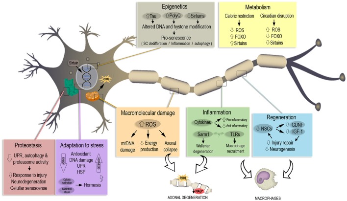
Aging constitutes the main risk factor for the development of neurodegenerative diseases. This represents a major health issue worldwide that is only expected to escalate due to the ever-increasing life expectancy of the population. Interestingly, axonal degeneration, which occurs at early stages of neurodegenerative disorders (ND) such as Alzheimer's disease, Amyotrophic lateral sclerosis, and Parkinson's disease, also takes place as a consequence of normal aging. Moreover, the alteration of several cellular processes such as proteostasis, response to cellular stress and mitochondrial homeostasis, which have been described to occur in the aging brain, can also contribute to axonal pathology. Compelling evidence indicate that the degeneration of axons precedes clinical symptoms in NDs and occurs before cell body loss, constituting an early event in the pathological process and providing a potential therapeutic target to treat neurodegeneration before neuronal cell death. Although, normal aging and the development of neurodegeneration are two processes that are closely linked, the molecular basis of the switch that triggers the transition from healthy aging to neurodegeneration remains unrevealed. In this review we discuss the potential role of axonal degeneration in this transition and provide a detailed overview of the literature and current advances in the molecular understanding of the cellular changes that occur during aging that promote axonal degeneration and then discuss this in the context of ND.
Overview
The claims and statistics of this paper haven’t been checked yet.
Run the check — free, no account needed. It reads the open-access text and checks the sources it cites.
Integrity signals
Registry
Cites
Statistics
Figures
Verification
Claims the paper makes
The claims and statistics of this paper haven’t been checked yet.
Run the check — free, no account needed. It reads the open-access text and checks the sources it cites.
Claims other papers make about this one
Each is checked against this paper’s own text: does it say what the citing paper says it does?
In the citing paper
“First, axonal degeneration, which is a precursor to many age-related neuronal disorders140,141, is characterized by rapid NAD+ depletion.”
From NAD+ metabolism and its roles in cellular processes during ageing (2021) · cites this paper as [140]
Self-check
Percentages, sums and reported differences re-computed from the paper’s own numbers.
The self-check needs the paper’s full text.
Citations
Cites
1 retracted
Poulsen, E. T., Iannuzzi, F., Rasmussen, H. F., Maier, T. J., Enghild, J. J., Jørgensen, A. L., & Matrone, C. (2017) · Frontiers in Molecular Neuroscience · doi:10.3389/fnmol.2017.00059 · Cited by 31
RetractedAging: a theory based on free radical and radiation chemistry
Harman, D. (1956) · Journal of Gerontology · doi:10.1093/geronj/11.3.298 · Cited by 8,567
Rosen, D. R., Siddique, T., Patterson, D., Figlewicz, D. A., Sapp, P., Hentati, A., Donaldson, D., Goto, J., O'Regan, J. P., Deng, H.-X., et al. (1993) · Nature · doi:10.1038/362059a0 · Cited by 7,062
The amyloid hypothesis of Alzheimer's disease at 25 years
Selkoe, D. J., & Hardy, J. (2016) · EMBO Molecular Medicine · doi:10.15252/emmm.201606210 · Cited by 6,164
2016 Alzheimer's disease facts and figures
Alzheimer's Association (2016) · Alzheimer's & Dementia · doi:10.1016/j.jalz.2016.03.001 · Cited by 5,069
Neuropathological alterations in Alzheimer disease
Serrano-Pozo, A., Frosch, M. P., Masliah, E., & Hyman, B. T. (2011) · Cold Spring Harbor Perspectives in Medicine · doi:10.1101/cshperspect.a006189 · Cited by 3,345
Protein translation and folding are coupled by an endoplasmic-reticulum-resident kinase
Harding, H. P., Zhang, Y., & Ron, D. (1999) · Nature · doi:10.1038/16729 · Cited by 3,310
Geroscience: Linking Aging to Chronic Disease
Kennedy, B. K., Berger, S. L., Brunet, A., Campisi, J., Cuervo, A. M., Epel, E. S., Franceschi, C., Lithgow, G. J., Morimoto, R. I., Pessin, J. E., et al. (2014) · Cell · doi:10.1016/j.cell.2014.10.039 · Cited by 2,764
Variant of TREM2 Associated with the Risk of Alzheimer's Disease
Jonsson, T., Stefansson, H., Steinberg, S., Jonsdottir, I., Jonsson, P. V., Snaedal, J., Bjornsson, S., Huttenlocher, J., Levey, A. I., Lah, J. J., et al. (2013) · New England Journal of Medicine · doi:10.1056/nejmoa1211103 · Cited by 2,558
Hypoxia-inducible factors and the response to hypoxic stress
Majmundar, A. J., Wong, W. J., & Simon, M. C. (2010) · Molecular Cell · doi:10.1016/j.molcel.2010.09.022 · Cited by 2,380
Cited by
Sorted by citations
Items 101–182 of 182
Can repetitive mechanical motion cause structural damage to axons?
Coppini, A., Falconieri, A., Mualem, O., Nasrin, S. R., Roudon, M., Saper, G., Hess, H., Kakugo, A., Raffa, V., & Shefi, O. (2024) · Frontiers in Molecular Neuroscience · doi:10.3389/fnmol.2024.1371738 · Cited by 12
Structural aspects of the aging invertebrate brain
Koch, S. C., Nelson, A., & Hartenstein, V. (2021) · Cell and Tissue Research · doi:10.1007/s00441-020-03314-6 · Cited by 12
Regulation of neuronal/axonal degeneration by ZNRF1 ubiquitin ligase
Araki, T., & Wakatsuki, S. (2018) · Neuroscience Research · doi:10.1016/j.neures.2018.07.008 · Cited by 12
Pieger, K., Schmitt, V., Gauer, C., Gießl, N., Prots, I., Winner, B., Winkler, J., Brandstätter, J. H., & Xiang, W. (2022) · Biomolecules · doi:10.3390/biom12081108 · Cited by 11
SARM1: The Checkpoint of Axonal Degeneration in the Nervous System Disorders
Karnik, A., & Joshi, A. R. (2025) · Molecular Neurobiology · doi:10.1007/s12035-025-04835-3 · Cited by 12
Ugbaja, S. C., Appiah‐Kubi, P., Lawal, M. M., Gumede, N. S., & Kumalo, H. M. (2021) · Journal of Biomolecular Structure and Dynamics · doi:10.1080/07391102.2020.1869099 · Cited by 11
Deep Learning to Decipher the Progression and Morphology of Axonal Degeneration
Palumbo, A., Grüning, P., Landt, S. K., Heckmann, L., Bartram, L., Pabst, A., Flory, C., Ikhsan, M., Pietsch, S., Schulz, R., et al. (2021) · Cells · doi:10.3390/cells10102539 · Cited by 11
Puvogel, S., Alsema, A. M., North, H. F., Webster, M. J., Weickert, C. S., & Eggen, B. J. L. (2024) · eNeuro · doi:10.1523/eneuro.0246-23.2024 · Cited by 12
Pathways to neurodegeneration: lessons learnt from unbiased genetic screens in Drosophila
Singhal, N., & Jaiswal, M. (2018) · Journal of Genetics · doi:10.1007/s12041-018-0954-4 · Cited by 11
Pottorf, T. S., Mann, A., Fross, S., Mansel, C. O., & Vohra, B. P. S. (2018) · Neurochemistry International · doi:10.1016/j.neuint.2018.09.010 · Cited by 11
Lee, N., Kim, S., & Lee, J.-H. (2022) · Heliyon · doi:10.1016/j.heliyon.2022.e11411 · Cited by 10
Bioenergetics of Axon Integrity and Its Regulation by Oligodendrocytes and Schwann Cells
Mishra, S. K., & Tiwari, S. P. (2024) · Molecular Neurobiology · doi:10.1007/s12035-024-03950-x · Cited by 10
Latham, A. S., Rocha, S. M., McDermott, C. P., Reigan, P., Slayden, R. A., & Tjalkens, R. B. (2024) · NeuroToxicology · doi:10.1016/j.neuro.2024.06.017 · Cited by 12
Phosphorylated SARM1 is involved in the pathological process of rotenone-induced neurodegeneration
Murata, H., Phoo, M. T. Z., Ochi, T., Tomonobu, N., Yamamoto, K., Kinoshita, R., Miyazaki, I., Nishibori, M., Asanuma, M., & Sakaguchi, M. (2023) · The Journal of Biochemistry · doi:10.1093/jb/mvad068 · Cited by 10
Longitudinal in vivo monitoring of axonal degeneration after brain injury
Chornyy, S., Borovicka, J. A., Patel, D., Shin, M., Vázquez‐Rosa, E., Miller, E., Wilson, B. M., Pieper, A. A., & Dana, H. (2023) · Cell Reports Methods · doi:10.1016/j.crmeth.2023.100481 · Cited by 11
Reijner, N., Frigerio, I., Bouwman, M. M. A., Boon, B. D. C., Guizard, N., Jubault, T., Hoozemans, J. J. M., Rozemüller, A. J. M., Bouwman, F. H., Barkhof, F., et al. (2025) · Alzheimer s Research & Therapy · doi:10.1186/s13195-025-01727-5 · Cited by 9
Effects of NAD+ in Caenorhabditis elegans Models of Neuronal Damage
Lee, Y., Jeong, H., Park, K. H., & Kim, K. W. (2020) · Biomolecules · doi:10.3390/biom10070993 · Cited by 8
Programmed axon degeneration: mechanism, inhibition and therapeutic potential
Loreto, A., & Neukomm, L. J. (2025) · Nature reviews. Neuroscience · doi:10.1038/s41583-025-00986-3 · Cited by 8
Chau, D. D., Li, W., Chan, W. W. R., Sun, J. K., Zhai, Y., Chow, H., & Lau, K. (2022) · The FASEB Journal · doi:10.1096/fj.202200757r · Cited by 8
Upregulation of Neuronal Rheb(S16H) for Hippocampal Protection in the Adult Brain
Moon, G. J., Shin, M., & Kim, S. R. (2020) · International Journal of Molecular Sciences · doi:10.3390/ijms21062023 · Cited by 8
Arun, P., Rossetti, F., Wilder, D. M., Wang, Y., Gist, I. D., & Long, J. B. (2021) · Frontiers in Neurology · doi:10.3389/fneur.2021.652190 · Cited by 7
Siddiqui, S., Khan, F., Jamali, K. S., & Musharraf, S. G. (2019) · CNS & Neurological Disorders - Drug Targets · doi:10.2174/1871527318666191111105508 · Cited by 7
Kaur, S., Sang, Y., & Aballay, A. (2022) · Journal of Biological Chemistry · doi:10.1016/j.jbc.2022.101614 · Cited by 6
Kumar, J. S. D., Molotkov, A., Kim, J., Carberry, P., Idumonyi, S., Castrillon, J., Duff, K., Shneider, N. A., & Mintz, A. (2022) · Pharmacological Reports · doi:10.1007/s43440-022-00359-y · Cited by 7
Adult Goat Retinal Neuronal Culture: Applications in Modeling Hyperglycemia
Sharma, S., Chakravarthy, H., Suresh, G., & Devanathan, V. (2019) · Frontiers in Neuroscience · doi:10.3389/fnins.2019.00983 · Cited by 6
Abdelwahab, T., Stadler, D., Knöpper, K., Arampatzi, P., Saliba, A., Kastenmüller, W., Martini, R., & Groh, J. (2023) · iScience · doi:10.1016/j.isci.2023.106698 · Cited by 6
The rise and fall of SARM1 base-exchange inhibitors
Lundbäck, T., Chandrasekar, V., Gu, C., Ju, H., McAdam, R. L., Palomero, M., Sader, K., Peter, B., Wissler, L., Nevin, P., et al. (2026) · Communications Chemistry · doi:10.1038/s42004-026-02074-8 · Cited by 7
Loss and gain of motor protein function cause microtubule bundle damage in Drosophila axons
Liew, Y.-T., Owens, M., Bailey, D. M. D., Cairns, W., Day, M., Jones, E., McCann, S., Lorenzo-Cisneros, L., Murphy, T., Parkin, J., et al. (2026) · Current Biology · doi:10.1016/j.cub.2025.12.038 · Cited by 5
Neuropathy target esterase as a biomarker and biosensor of delayed neuropathic agents
Richardson, R. J., Fink, J. K., Worden, R. M., Wijeyesakere, S. J., & Махаева, Г. Ф. (2020) · Elsevier eBooks · doi:10.1016/b978-0-12-819090-6.00058-1 · Cited by 4
Elmers, J., Colzato, L. S., Ziemssen, F., Ziemssen, T., & Beste, C. (2024) · Ageing Research Reviews · doi:10.1016/j.arr.2024.102280 · Cited by 4
Michalettos, G., Clausen, F., Rostami, E., & Marklund, N. (2024) · Neurotherapeutics · doi:10.1016/j.neurot.2024.e00472 · Cited by 3
Cdc42 Promotes Axonogenesis of Primary Hippocampal Neurons by Inhibiting Glycogen Synthase Kinase-3β
Li, Y., Chen, F.-Z., Chen, W., Zhu, H.-M., Chen, Y., Li, Z., Yan, F., Liu, Z., Dong, W.-R., Zhang, L., et al. (2022) · Journal of Integrative Neuroscience · doi:10.31083/j.jin2105133 · Cited by 3
Arnold, R. E., Saska, J., Mesquita-Ribeiro, R., Dajas‐Bailador, F., Taylor, L. J., Lewis, W., Argent, S. P., Shao, H., Houk, K. N., & Denton, R. M. (2024) · Chemical Science · doi:10.1039/d4sc03232b · Cited by 3
Understanding the underlying causes of degenerative neural diseases
Shields, B., Silverdale, M., & Prokop, A. (2024) · InnovAiT Education and inspiration for general practice · doi:10.1177/17557380241298477 · Cited by 3
Cytoplasmic mRNA Transport: Adaptors of mRNA Binding to Microtubule Motor Proteins
Vdovina, Y. A., Georgieva, S. G., & Kopytova, D. V. (2024) · Molecular Biology · doi:10.1134/s0026893324700018 · Cited by 2
Analysis of Brain Protein Stability Changes in a Mouse Model of Alzheimer’s Disease
Tang, Y., Park, H.-J., Li, S., & Fitzgerald, M. C. (2024) · Journal of Proteome Research · doi:10.1021/acs.jproteome.4c00406 · Cited by 2
Inflammaging and the sex-frailty paradox
Arosio, B., Salafia, G., Ferri, E., Mari, D., Tobaldini, E., Vitale, G., & Montano, N. (2025) · Aging Clinical and Experimental Research · doi:10.1007/s40520-025-03181-7 · Cited by 2
Zheng, M., Gao, K., Hong, Y., Le, J., Cai, J., Liang, H., & Cui, W. (2025) · Pharmaceuticals · doi:10.3390/ph18111751 · Cited by 2
Myelin sheaths persist for weeks following axon degeneration
Doty, M. E., Tuckerman, E. C., Piedra, E. T., & Hill, R. (2026) · Science Advances · doi:10.1126/sciadv.aeb2628 · Cited by 2
Do oligodendrocytes regulate axonal glucose uptake and consumption?
Fletcher, J. L., & Young, K. M. (2024) · Trends in Neurosciences · doi:10.1016/j.tins.2024.06.001 · Cited by 2
Gunaseelan, S., Ariffin, M. Z., Khanna, S., Ooi, M. H., Perera, D., Chu, J. J. H., & Chua, J. J. E. (2022) · Nature Communications · doi:10.1038/s41467-022-28533-z · Cited by 2
RetractedEpImAge: An Epigenetic-Immune Clock for Disease-Associated Biological AgingPreprint
Kalyakulina, A., Yusipov, I., Trukhanov, A., Franceschi, C., Moskalev, A., & Ivanchenko, M. (2025) · bioRxiv (Cold Spring Harbor Laboratory) · doi:10.1101/2025.03.11.642648 · Cited by 2
Anatomic and Histological Changes of the Aging Brain
Yachnis, A. T. (2019) · Cambridge University Press eBooks · doi:10.1017/9781108554350.002 · Cited by 1
Axonal transport of CHMP2b is regulated by kinesin-binding protein and disrupted by CHMP2b intron5
Kirwan, K., Puerta-Alvarado, V., & Waites, C. L. (2025) · Life Science Alliance · doi:10.26508/lsa.202402934 · Cited by 2
von Kügelgen, N., Ludwik, K. A., Mendonsa, S., Römer, C., Becher, E., Breimann, L., Strauch, M., Mari, T., Mongellaz, S., Zuckerman, B., et al. (2025) · NAR Molecular Medicine · doi:10.1093/narmme/ugaf005 · Cited by 1
The Role of Calcium-Independent Phospholipase A2 in the Molecular Mechanisms of Schizophrenia
Nakamura, S. (2025) · Cells · doi:10.3390/cells14171348 · Cited by 1
Kolinko, Y. (2026) · Contemporary clinical neuroscience · doi:10.1007/978-3-032-18296-8_11 · Cited by 1
Mishra, V. R., & Szaflarski, J. P. (2026) · Epilepsia · doi:10.1002/epi.70464 · Cited by 1
Liu, J., Ong, K., Meng, A., Stokin, G. B., Loeb, J. A., Ravits, J., & Song, F. (2026) · Acta Neuropathologica · doi:10.1007/s00401-026-03072-9 · Cited by 1
Nakamura, S. (2022) · IntechOpen eBooks · doi:10.5772/intechopen.102340 · Cited by 1
Deep learning to decipher the progression and morphology of axonal degenerationPreprint
Palumbo, A., Grüning, P., Landt, S. K., Heckmann, L., Bartram, L., Pabst, A., Flory, C., Ikhsan, M., Pietsch, S., Schulz, R., et al. (2020) · bioRxiv (Cold Spring Harbor Laboratory) · doi:10.1101/2020.08.26.269092 · Cited by 1
Alsameen, M. H., Gong, Z., Qian, W., Kiely, M., Triebswetter, C., Bergeron, C. M., Cortina, L. E., Faulkner, M. E., Laporte, J. P., & Bouhrara, M. (2023) · medRxiv · doi:10.1101/2023.03.06.23286818 · Cited by 1
The Development and Application of a Human Stem Cell-Based Model of Chemotherapy-Induced Neuropathy
Snavely, A. R. (2018) · Digital Access to Scholarship at Harvard (DASH) (Harvard University)
do Amaral, L. (2021) · Digital Library of Theses and Dissertations (Universidade de São Paulo) · doi:10.11606/t.60.2021.tde-27092021-115850
Sisti, F. (2021) · Digital Library of Theses and Dissertations (Universidade de São Paulo) · doi:10.11606/t.60.2021.tde-22032022-075600
Neuroprotective Effects of Curcumin-Loaded Emulsomes on Laser Axotomy-Induced CNS Injury ModelPreprint
Yılmaz, E. N., Bay, S., Öztürk, G., & Üçışık, M. H. (2020) · doi:10.21203/rs.3.rs-38217/v1
Axon maintenance and degeneration
Wang, F., Yang, J. H., & He, Z. (2020) · Elsevier eBooks · doi:10.1016/b978-0-12-814407-7.00010-9
Microtubule decay is a driver of neuronal ageing and a promising target for interventionPreprint
Okenve-Ramos, P., Gosling, R., Chojnowska-Monga, M., Gupta, K., Shields, S., & Sánchez‐Soriano, N. (2023) · bioRxiv (Cold Spring Harbor Laboratory) · doi:10.1101/2023.01.11.523590
Suciati, S., Suharyoto, S., Lasman, L., Nurhidayati, N., Islamy, A., & Masruroh, E. (2022) · Lumbung Inovasi Jurnal Pengabdian kepada Masyarakat · doi:10.36312/linov.v7i4.988
Computational Models of Age-Associated Cognitive SlowingPreprint
Ahmed, S., Lytton, W. W., Stewart, T. C., & Crystal, H. (2024) · bioRxiv (Cold Spring Harbor Laboratory) · doi:10.1101/2024.06.24.600545
Age-related energetic reprogramming in glial cells: possible correlations with Parkinson’s disease
Chu, B., Xiang, H., Wang, H., Lin, Y.-T., Li, R., Hu, J. R., & Qian, H. (2024) · Ageing and Neurodegenerative Diseases · doi:10.20517/and.2024.11
Fialová, L., Bartoš, A., Kalousová, M., Nosková, L., Zelenková, M., Slukova, M., & Zima, T. (2024) · Biomedical Papers · doi:10.5507/bp.2024.032
Cytoplasmic mRNA transport: adaptors of mRNA binding to microtubule motor proteins
Vdovina, Y. A., Georgieva, S. G., & Kopytova, D. V. (2024) · Molecular Biology · doi:10.31857/s0026898424030018
The Robustness of White Matter Brain Networks Decreases with Aging
Huang, C., Xie, W., & Xie, D. (2025) · Journal of Integrative Neuroscience · doi:10.31083/jin25816
Sathe, A., Chatterjee, A., Shashikumar, N., Pechman, K. R., Schilling, K. G., Landman, B. A., Bolton, C. J., Zhang, P., Bender, A. R., Hohman, T. J., et al. (2025) · medRxiv · doi:10.1101/2025.05.29.25328528
TRI-COMPARTMENT CHIP WITH MICROELECTRODE ARRAY DR1-GLASS GROOVES FOR NEURONAL CELL ALIGNMENT
Ryynänen, T., Fedele, C., Moilanen, A.-M., Vihinen, J., Sukki, L., Tornberg, K., Haikka, S., Narkilahti, S., Priimägi, A., & Kallio, P. J. (2024) · Solid-State Sensors, Actuators, and Microsystems Workshop (Hilton Head Workshop) · doi:10.31438/trf.hh2024.88
The cellular associates of late life changes in white matter microstructurePreprint
Chin, R., Zhang, X.-H., Anderson, K. M., Yendiki, A., & Holmes, A. J. (2025) · bioRxiv (Cold Spring Harbor Laboratory) · doi:10.1101/2025.09.02.673734 · Cited by 1
Targeting Poly (ADP‐Ribose) Polymerase‐1 for the Treatment of Neurodegenerative Diseases
Polk, S., Rassaeikashuk, M., Thilagavathi, R., & Selvam, C. (2025) · Chemical Biology & Drug Design · doi:10.1111/cbdd.70189
Sadikoglou, E., Domingo‐Fernándéz, D., Savytska, N., Fernandes, N., Rizzu, P., Illarionova, A., Strauß, T., Schwarz, S. C., Kodamullil, A. T., Höglinger, G. U., et al. (2025) · Cell Death and Disease · doi:10.1038/s41419-025-08147-1 · Cited by 1
Muñoz-Carvajal, F., Sanhueza, N., Sanhueza, M., & Court, F. A. (2026) · eLife · doi:10.7554/elife.103118
Self-citationMartínez, N. W., Manzano, C. M., Trujillo, A., Pizarro, I. S., Barrera, D. P., Calvo, M., & Matus, S. (2026) · Research Square · doi:10.21203/rs.3.rs-9204935/v1
Drug Discovery for Alzheimer’s Disease: Current Status
Wolfe, M. S. (2026) · Topics in medicinal chemistry · doi:10.1007/7355_2026_186
Comparative transcriptomics reveal molecular convergence and divergence in parkinsonian disorders
Geithus, S., Flønes, I. H., Alves, G. W., Tysnes, O., van de Berg, W. D. J., Pihlstrøm, L., Nido, G. S., & Tzoulis, C. (2026) · Brain · doi:10.1093/brain/awag153
Muhammad, H., Yasin, M. A. B. M., Jaafar, J., Hou, L. J., & Hitam, W. H. W. (2026) · Taiwan Journal of Ophthalmology · doi:10.4103/tjo.tjo-d-25-00165
Єна, М. С., Pyatnitsa-Gorpynchenko, N. K., Dziubenko, N., & Halushko, O. A. (2026) · Problems of ageing and longevity (Ukraine) · doi:10.71012/pro-ageing-2026-2-07
Kavoi, B. M., & Boateng, K. O. (2026) · The Journal of Basic and Applied Zoology · doi:10.1186/s41936-026-00615-0
Lossi, L., Sbriz, M., Castagna, C., & Merighi, A. (2026) · Frontiers in Cell Death · doi:10.3389/fceld.2026.1823132
Ye, C., Tang, F., & Li, X. (2026) · Frontiers in Cell and Developmental Biology · doi:10.3389/fcell.2026.1943282
Spatial patterns of Wnt/β-catenin signaling in the adult and aging Nothobranchius furzeri brain
Whisenant, E., & Lekven, A. C. (2026) · Aging Brain · doi:10.1016/j.nbas.2026.100170
Chakraborty, W., Anand, A., Kumar, S., & Roy, K. K. (2026) · Frontiers in Neuroendocrinology · doi:10.1016/j.yfrne.2026.101292
Metadata
- Authors
- Salvadores, N., Sanhueza, M., Manque, P., & Court, F. A.
- Affiliations
- Universidad Mayor (CL)
- Corresponding author
- Court, Felipe A.
- Venue
- Frontiers in Neuroscience
- Publisher
- Frontiers Media SA
- Published
- 2017-09-04
- Volume
- 11
- Pages
- 451-451
- ISSN
- 1662-453X (ISSN-L)
- PMC
- PMC5591337
- OpenAlex
- W2740768704
- Semantic Scholar
- 6954583ab5a9e498ecf5cde38c09b1e4621405f8
- Cited by
- 182
- References
- 330
- Citations by year
- 2026: 17 · 2025: 18 · 2024: 21 · 2023: 18 · 2022: 30 · 2021: 24 · 2020: 25 · 2019: 15 · 2018: 14
- FWCI
- 1.81
- Type
- journal article
- Language
- en
- Open access
- gold
- doi.org
- Licence
- cc-by
- Locations
- Frontiers in Neuroscience (publishedVersion, cc-by)
- PubMed Central (publishedVersion, cc-by)
- PubMed (publishedVersion)
- Americanae (AECID Library) (submittedVersion, cc-by-nc-nd)
- DOAJ (DOAJ: Directory of Open Access Journals) (submittedVersion)
- LA Referencia (Red Federada de Repositorios Institucionales de Publicaciones Científicas) (submittedVersion, cc-by-nc-nd)
- Funders
- International Development Research Centre
- Israel Science Foundation
- Azrieli Foundation
- Canadian Institutes of Health Research
- Fondo de Financiamiento de Centros de Investigación en Áreas Prioritarias (15150012, /FONDAP/15150012)
- Topics
- Life Sciences › Neuroscience › Cellular and Molecular Neuroscience › Alzheimer's disease research and treatments
- Life Sciences › Biochemistry, Genetics and Molecular Biology › Aging › Genetics, Aging, and Longevity in Model Organisms
- Life Sciences › Biochemistry, Genetics and Molecular Biology › Molecular Biology › Mitochondrial Function and Pathology
- Keywords
- axonal degeneration, aging, neurodegenerative diseases, Alzheimer's disease, amyotrophic lateral sclerosis, Parkinson's disease, proteostasis, cellular stress response, mitochondrial homeostasis, therapeutic target
About these checks
Registry, retraction, Cites and statistics results are computed, not judged, with one exception: a printed reference that no index lists is read once by a model before it counts as a citation error. Claims are extracted by a model and kept only when their quote appears verbatim in the text; each cited source is then checked, with a model’s help, for whether it says what the claim relies on. Those verdicts can be wrong — read the claim’s quote and the cited source before relying on one.
Statistics and arithmetic are recomputed, not judged.
Confidence bands are shown only for fields calibrated on our annotated test set. None is calibrated yet, so no band is shown.
What counts as a claim ›How we measure confidence ›How we check this ›
Are you an author, or do you see a problem with this page? Request a review
