

The DOI matches its Crossref record; 190 of the 190 references it cites were found in a registry or an index.
Microglia and Astrocyte Function and Communication: What Do We Know in Humans?
Open accessAbstract
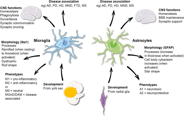
Microglia and astrocytes play essential roles in the central nervous system contributing to many functions including homeostasis, immune response, blood–brain barrier maintenance and synaptic support. Evidence has emerged from experimental models of glial communication that microglia and astrocytes influence and coordinate each other and their effects on the brain environment. However, due to the difference in glial cells between humans and rodents, it is essential to confirm the relevance of these findings in human brains. Here, we aim to review the current knowledge on microglia-astrocyte crosstalk in humans, exploring novel methodological techniques used in health and disease conditions. This will include an in-depth look at cell culture and iPSCs, post-mortem studies, imaging and fluid biomarkers, genetics and transcriptomic data. In this review, we will discuss the advantages and limitations of these methods, highlighting the understanding these methods have brought the field on these cells communicative abilities, and the knowledge gaps that remain.
Overview
The claims and statistics of this paper haven’t been checked yet.
Run the check — free, no account needed. It reads the open-access text and checks the sources it cites.
Integrity signals
Registry
Cites
Statistics
Figures
Verification
Claims the paper makes
The claims and statistics of this paper haven’t been checked yet.
Run the check — free, no account needed. It reads the open-access text and checks the sources it cites.
Claims other papers make about this one
Each is checked against this paper’s own text: does it say what the citing paper says it does?
The source backs part of the claim, not all of it — or backs it for a different population, condition or comparison.
In the citing paper
“Astrocyte and microglia iPSC models partially recapitulate the transcriptomes of their in vivo counterparts and may capture authentic neurodegenerative disease phenotypes [345,406,480,481,482,483,484].”
From Revisiting the Pathogenesis of X-Linked Adrenoleukodystrophy (2025) · cites this paper as [345]
Experimental — accuracy not yet measured
Self-check
Percentages, sums and reported differences re-computed from the paper’s own numbers.
The self-check needs the paper’s full text.
Citations
Cites
Takahashi, K., & Yamanaka, S. (2006) · Cell · doi:10.1016/j.cell.2006.07.024 · Cited by 26,673
Exploring the full spectrum of macrophage activation
Mosser, D. M., & Edwards, J. P. (2008) · Nature Reviews Immunology · doi:10.1038/nri2448 · Cited by 9,144
Polazzi, E., Mengoni, I., Caprini, M., Peña‐Altamira, E., Kurtys, E., & Monti, B. (2012) · Neurosignals · doi:10.1159/000337115 · Cited by 9,023
Neurotoxic reactive astrocytes are induced by activated microglia
Liddelow, S. A., Guttenplan, K. A., Clarke, L. E., Bennett, F. C., Bohlen, C. J., Schirmer, L., Bennett, M. L., Münch, A. E., Chung, W.-S., Peterson, T. C., et al. (2017) · Nature · doi:10.1038/nature21029 · Cited by 7,971
Resting microglial cells are highly dynamic surveillants of brain parenchyma in vivo
Nimmerjahn, A., Kirchhoff, F., & Helmchen, F. (2005) · Science · doi:10.1126/science.1110647 · Cited by 5,827
A Unique Microglia Type Associated with Restricting Development of Alzheimer’s Disease
Keren-Shaul, H., Spinrad, A., Weiner, A., Matcovitch-Natan, O., Dvir-Szternfeld, R., Ulland, T. K., David, E., Baruch, K., Lara-Astaiso, D., Toth, B., et al. (2017) · Cell · doi:10.1016/j.cell.2017.05.018 · Cited by 5,493
Astrocytes: Biology and pathology
Sofroniew, M. V., & Vinters, H. V. (2010) · Acta Neuropathologica · doi:10.1007/s00401-009-0619-8 · Cited by 5,196
Fate Mapping Analysis Reveals That Adult Microglia Derive from Primitive Macrophages
Ginhoux, F., Greter, M., Leboeuf, M., Nandi, S., See, P., Gokhan, S., Mehler, M. F., Conway, S. J., Ng, L. G., Stanley, E. R., et al. (2010) · Science · doi:10.1126/science.1194637 · Cited by 5,175
Meta-Analysis of 74,046 Individuals Identifies 11 New Susceptibility Loci for Alzheimer’s Disease
(EADI), E. A. D. I., Lambert, J.-C., (GERAD), G. A. E. R. I. A. D., Alzheimer's Disease Genetic Consortium (ADGC), (CHARGE), C. F. H. A. A. R. I. G. E., Ibrahim-Verbaas, C. A., Harold, D., Naj, A. C., Sims, R., Bellenguez, C., et al. (2013) · Nature Genetics · doi:10.1038/ng.2802 · Cited by 4,336
Microglia Sculpt Postnatal Neural Circuits in an Activity and Complement-Dependent Manner
Schafer, D. P., Lehrman, E. K., Kautzman, A. G., Koyama, R., Mardinly, A. R., Yamasaki, R., Ransohoff, R. M., Greenberg, M. E., Barres, B. A., & Stevens, B. (2012) · Neuron · doi:10.1016/j.neuron.2012.03.026 · Cited by 4,089
Cited by
Sorted by citations
Items 101–165 of 165
Gong, M., Lan, J. X., & Miao, J. (2026) · Frontiers in Aging Neuroscience · doi:10.3389/fnagi.2026.1908574 · Cited by 3
Gazerani, P. (2023) · Neuroglia · doi:10.3390/neuroglia4010002 · Cited by 3
Role of Glial Cells and Receptors in Schizophrenia Pathogenesis
Tizabi, Y., Antonelli, M. C., Tizabi, D., & Aschner, M. (2025) · Neurochemical Research · doi:10.1007/s11064-025-04336-8 · Cited by 2
Grzych, H., Fee, E. L., Kemp, M. W., Royse, E. X., Usada, H., Ikeda, H., Takahashi, Y., Takahashi, T., Jobe, A. H., & Hillman, N. H. (2025) · Pediatric Research · doi:10.1038/s41390-025-04172-0 · Cited by 2
Age-Related Changes in Neuroinflammation and Epigenetic Regulation in Mouse Ischemic Stroke Model
Kondo, M., Tamura, H., Segi‐Nishida, E., & Hasegawa, H. (2025) · Brain Sciences · doi:10.3390/brainsci15080810 · Cited by 3
Astrocitos: acción e interacción en el Sistema Nervioso
Padilla-Gil, D. N. (2024) · Revista Med · doi:10.18359/rmed.6870 · Cited by 1
Neuroglia in Tourette syndrome and obsessive-compulsive disorder
Frick, L. R. (2025) · Handbook of clinical neurology · doi:10.1016/b978-0-443-19102-2.00005-3 · Cited by 1
Solis, B. I. H., Guerrero-Padilla, F., Jiménez, E. M., López, J. M. V., Perea‐Flores, M. D. J., Rodríguez‐Cortés, O., Macías-Pérez, M. E., & Rodríguez, M. H. (2025) · Brain Sciences · doi:10.3390/brainsci15090952 · Cited by 1
Agarwal, U., Paliwal, S., Yadav, V., Pannu, A. S., Tonk, R. K., & Verma, S. K. (2025) · CNS & Neurological Disorders - Drug Targets · doi:10.2174/0118715273402657250905055635 · Cited by 2
Breivik, T. J., Gjermo, P. E., Opstad, P. K., Murison, R. C. C., von Hörsten, S., & Fristad, I. (2025) · Life · doi:10.3390/life15101572 · Cited by 2
Pushchina, E. V., Vekhova, E. E., Pimenova, E., Akhmadieva, A., & Bykova, M. E. (2025) · International Journal of Molecular Sciences · doi:10.3390/ijms262211123 · Cited by 1
Ebdah, W., Thomas, S. D., Eissa, N., Jayaprakash, P., Łażewska, D., Kieć‐Kononowicz, K., & Sadek, B. S. (2026) · Frontiers in Behavioral Neuroscience · doi:10.3389/fnbeh.2026.1722019 · Cited by 1
Weathered plastic particles negatively affect mouse primary neurons and glial cells
Shin, H. S., So, Y. H., Lee, D. H., Chang, Y., Kim, M., Joung, D., Youn, B., Lee, E.-H., & Jung, E.-M. (2026) · Archives of Toxicology · doi:10.1007/s00204-026-04334-x · Cited by 1
Kumar, S., Singh, J., Maurya, V. K., Ansari, S., Srivastava, C., Malhotra, H., Babu, S., & Saxena, S. K. (2026) · Journal of Neuropathology & Experimental Neurology · doi:10.1093/jnen/nlag025 · Cited by 1
Upadhyay, R., Kar, S., & Sevanan, M. (2026) · Frontiers in Chemical Biology · doi:10.3389/fchbi.2026.1836070 · Cited by 1
Heme oxygenase/carbon monoxide in thermoregulation and neuroenergetics
Cárnio, E. C., & Branco, L. G. S. (2026) · Neurochemistry International · doi:10.1016/j.neuint.2026.106205 · Cited by 1
Brain Aging and Cellular Senescence Trigger Neuroimmunological Dysregulation and Neurodegeneration
Vega-García, A., Apolinar, L. M., Feria-Romero, I. A., Campos‐Bedolla, P., Alberto, G.-G. C., Luna‐Munguía, H., Sofía, R.-H., & Orozco-Suárez, S. A. (2026) · Brain Sciences · doi:10.3390/brainsci16101019 · Cited by 1
Gut Microbiota and Altered Behaviour: Target on Neuroimmune Interplays
Jayaraman, S., & Paul, S. (2024) · Springer eBooks · doi:10.1007/978-981-99-8803-7_9 · Cited by 5
Pak, V., Adewale, Q., Bzdok, D., Dadar, M., Zeighami, Y., & Iturria‐Medina, Y. (2023) · bioRxiv (Cold Spring Harbor Laboratory) · doi:10.1101/2023.06.08.544227 · Cited by 1
Mhatre‐Winters, I., Eid, A., Blum, N., Han, Y., Sammoura, F. M., Wu, L., & Richardson, J. R. (2025) · bioRxiv (Cold Spring Harbor Laboratory) · doi:10.1101/2025.02.14.638293 · Cited by 1
Pradhan, A. K., Shi, Q., Tartler, K. J., & Rammes, G. (2022) · bioRxiv (Cold Spring Harbor Laboratory) · doi:10.1101/2022.05.09.491159
SARS-CoV-2 Envelope protein triggers depression and dysosmia via TLR2 mediated neuroinflammationPreprint
Su, W., Ju, J., Gu, M., Wang, X., Liu, S., Yu, J., & Mu, D.-L. (2023) · bioRxiv (Cold Spring Harbor Laboratory) · doi:10.1101/2023.01.15.524078
Rurak, G. M. (2023) · Carleton University's Institutional Repository (MacOdrum Library, Carleton University) · doi:10.22215/etd/2023-15750
Kusuma, R. B., Joewono, H. T., & Zanudin, A. (2023) · Biomolecular and Health Science Journal · doi:10.4103/bhsj.bhsj_29_23
Rat primary cortical cell tri-culture to study effects of amyloid-beta on microglia functionPreprint
Kim, H., Le, B., Goshi, N., Zhu, K., Grodzki, A. C., Lein, P. J., Zhao, M., & Şeker, E. (2024) · bioRxiv (Cold Spring Harbor Laboratory) · doi:10.1101/2024.03.15.584736
Su, W., Ju, J., Gu, M., Wang, X., Liu, S., Yu, J., & Mu, D.-L. (2023) · Figshare · doi:10.6084/m9.figshare.22790549
Use of Artificial Intelligence in Preventing and Treating Neuronal Cancer
Ward, K., Liu, K., Pathak, S., Pondugula, S. R., Alharbi, H. F., Mohandoss, K., Pole, S. S., Bhupathyraaj, M., & Dhanasekaran, M. (2024) · Nutritional neurosciences · doi:10.1007/978-981-97-2577-9_2
Modulating disease states of the central nervous system: Outcomes of neuromodulation on microglia
Bobotis, B. C., Dolhan, K., & Tremblay, M. (2024) · Elsevier eBooks · doi:10.1016/b978-0-323-95730-4.00015-9
Brain Injury and Neurodegeneration: Molecular, Functional, and Translational Approach 2.0
Ahluwalia, P. K., Gaur, P., Ahluwalia, M., & Vaibhav, K. (2024) · Biomedicines · doi:10.3390/biomedicines12112586
Johnson, S. M., Rastas, J. P., Desai, P. S., Baker, T. L., & Watters, J. J. (2025) · Respiratory Physiology & Neurobiology · doi:10.1016/j.resp.2025.104435
Characterization of active kinase signaling pathways in astrocytes and microgliaPreprint
Ryan, W. G., Eby, H., Bearss, N. R., Imami, A. S., Hamoud, A.-R. A., Pulvender, P., Bollinger, J. L., Wohleb, E. S., & McCullumsmith, R. E. (2025) · bioRxiv (Cold Spring Harbor Laboratory) · doi:10.1101/2025.04.18.649617
Kuter, K. Z., Chwastek, A. M., Paleczna, M., Kadłuczka, J., Kosmowska, B., Leonovich, I., Maciasz, W., Lenda, T., & Kula, J. (2025) · bioRxiv (Cold Spring Harbor Laboratory) · doi:10.1101/2025.06.16.659904
Anzilotti, S., Scarcella, M., Ciampa, M., Muraglia, N. D., Anastasio, C., Fiorentino, C., Rossin, F., Avallone, L., Pignataro, G., Pavone, L. M., et al. (2025) · Cell Death Discovery · doi:10.1038/s41420-025-02648-w
Sepulveda, J., Kim, J. Y., Binder, J., Vicini, S., & Rebeck, G. W. (2024) · Figshare · doi:10.6084/m9.figshare.26691931
Sepulveda, J., Kim, J. Y., Binder, J., Vicini, S., & Rebeck, G. W. (2024) · Figshare · doi:10.6084/m9.figshare.26691937
Su, W., Ju, J., Gu, M., Wang, X., Liu, S., Yu, J., & Mu, D. (2023) · Figshare · doi:10.6084/m9.figshare.22790546
Su, W., Ju, J., Gu, M., Wang, X., 刘少壮, Yu, J., & Mu, D. (2023) · Figshare · doi:10.6084/m9.figshare.22790543
Su, W., Ju, J., Gu, M., Wang, X., Liu, S., Yu, J., & Mu, D. (2023) · Figshare · doi:10.6084/m9.figshare.22790540
Sepulveda, J., Kim, J. Y., Binder, J., Vicini, S., & Rebeck, G. W. (2024) · Figshare · doi:10.6084/m9.figshare.26691934
Gautam, A. S., & Singh, R. K. (2026) · Journal of Biochemical and Molecular Toxicology · doi:10.1002/jbt.70721
How molecular mechanisms of aging drive Alzheimer's disease pathology
Chang, J., Liu, T., Cheng, X., & Wei, J. (2026) · Cellular Signalling · doi:10.1016/j.cellsig.2026.112426
Xu, Z., & Deng, Y. (2026) · Elsevier eBooks · doi:10.1016/b978-0-443-27340-7.00012-7
Chronic short sleep in early life accelerates cognitive decline via disrupted proteostasisPreprint
Komlo, R., Sengupta, K., Strus, E., & Naidoo, N. N. (2026) · bioRxiv (Cold Spring Harbor Laboratory) · doi:10.64898/2026.03.26.714554
Scenna, M. S., Bej, E., Cesare, P., Persichitti, I., Cimini, A., d’Angelo, M., & Castelli, V. (2026) · Frontiers in Pharmacology · doi:10.3389/fphar.2026.1783888 · Cited by 1
Greco, D., Das, D., Novotný, J., Olsen, M. J., & Telenský, P. (2026) · Scientific Reports · doi:10.1038/s41598-026-47881-0
Lei, S., Huang, Z. G., Wang, W., & Meng, Y. (2026) · Neuroprotection/Neuroprotection (Chichester, England. Print) · doi:10.1002/nep3.70037
Pericytes are organ-specific regulators of tissue morphogenesis
Rasouli, S. J., Kruse, K., Diéguez‐Hurtado, R., Ghanbari, P., Aravamudhan, A., Pitulescu, M. E., & Adams, R. H. (2026) · Nature Communications · doi:10.1038/s41467-026-71643-1
Meirinho, S., Ferreira, S. A., Diniz, C., Alves, G., Zhang, S., Kumar, N., Sitruk-Ware, R. L., Di, G., Navarro-Oviedo, M., Pradillo, J. M., et al. (2026) · Drug Delivery and Translational Research · doi:10.1007/s13346-026-02150-5
Perioperative Modulation of Microglia in Glioblastoma Resection
Kršek, A., Koruga, N., & Batičić, L. (2026) · Biologics · doi:10.3390/biologics6020017
Zhou, H., Xiao, X., Lu, W., & He, Y. (2026) · Veterinary Sciences · doi:10.3390/vetsci13060565
Swami, D., Sureshchandra, S., Vinnakota, J. M., Zeiser, R., Othy, S., & Acharya, M. M. (2026) · bioRxiv (Cold Spring Harbor Laboratory) · doi:10.64898/2026.06.10.730982
Neuroinflammation and Tauopathies
Zeenny, M.-N., Salame, I., Maatouk, L., & Akoury, E. (2026) · Molecular Biology Reports · doi:10.1007/s11033-026-12253-4
Grosmane, A., Saluvēra, E., Roddate, M., Krutovs, V., Blennow, K., Zetterberg, H., Zolovs, M., Kurjāne, N., & Ķēniņa, V. (2026) · PLoS ONE · doi:10.1371/journal.pone.0352017
Endogenous Neurotoxicity: A Pathophysiological Consequence of Homeostatic Dysfunction
Yanglem, S., Loushambam, B., Singh, S. M., & Vijayaraghavalu, S. (2026) · Neuroglia · doi:10.3390/neuroglia7030029
Lopes, C. R., Ferreira, S. G., Cunha, R. A., & Agostinho, P. M. (2026) · Glia · doi:10.1002/glia.70197
The Environmental Contribution to Health and Disease: Analytical Tools for Monitoring and Diagnosis
Aranda‐Merino, N., Heras, C. G. D. L., Ramos‐Payán, M., Fernández‐Torres, R., Bello‐López, M. Á., & Gómez‐Ariza, J. L. (2026) · Environments · doi:10.3390/environments13080416
Glutamate, Immune Cells and Alzheimer’s Disease: An Attempt to Link the Triad
Chinas, N. A., & Alexopoulos, H. (2026) · Cellular and Molecular Neurobiology · doi:10.1007/s10571-026-01784-w
Sreeram, S., 陈雅, Bury, L. A. D., Leskov, K., Ye, F., García‐Mesa, Y., Luttge, B. G., Eum, J., 黄军友, Kallianpur, A. R., et al. (2026) · bioRxiv (Cold Spring Harbor Laboratory) · doi:10.64898/2026.08.10.743763
Na, 李. L., Tianyi, 周. Z., Chunyan, 马. M., Yutong, 叶. Y., Zixing, 李. L., Yi, 刘. L., & Hongli, 陈. C. (2026) · Chinese Journal of Lasers · doi:10.3788/cjl251439
Saha, P. K., Rowe, R. K., Boison, D., & Shandra, O. O. (2026) · Experimental Neurology · doi:10.1016/j.expneurol.2026.115981
Organization of the forebrain cholinergic system and its glial interactions
Záborszky, L., & Gyengési, E. (2026) · Elsevier eBooks · doi:10.1016/b978-0-443-24078-2.00018-8
Murakami, K., Shimizu, T., Kido, T., & Isohama, Y. (2026) · Frontiers in Neuroscience · doi:10.3389/fnins.2026.1860290
Habib, Y. H., Mansy, B. M., & Ali, M. A. (2026) · Naunyn-Schmiedeberg s Archives of Pharmacology · doi:10.1007/s00210-026-05859-9
Swami, D., El-Khatib, S. M., Sureshchandra, S., Nguyen, E., Vinnakota, J. M., Zeiser, R., Othy, S., & Acharya, M. M. (2026) · Advanced Science · doi:10.1002/advs.77733
Metadata
- Authors
- Garland, E. F., Hartnell, I. J., & Boche, D.
- Affiliations
- University of Southampton (GB)
- Corresponding author
- Boche, Delphine
- Venue
- Frontiers in Neuroscience
- Publisher
- Frontiers Media SA
- Published
- 2022-02-16
- Volume
- 16
- Pages
- 824888-824888
- ISSN
- 1662-453X (ISSN-L)
- PMC
- PMC8888691
- OpenAlex
- W4212929528
- Cited by
- 165
- References
- 190
- Citations by year
- 2026: 39 · 2025: 40 · 2024: 50 · 2023: 28 · 2022: 8
- FWCI
- 3.27
- Type
- journal article
- Language
- en
- Open access
- gold
- doi.org
- Licence
- Locations
- Frontiers in Neuroscience (publishedVersion, cc-by)
- PubMed Central (publishedVersion, cc-by)
- PubMed (publishedVersion)
- White Rose Research Online (University of Leeds, The University of Sheffield, University of York) (acceptedVersion)
- ePrints Soton (University of Southampton) (acceptedVersion)
- DOAJ (DOAJ: Directory of Open Access Journals) (submittedVersion, cc-by-sa)
- Funders
- Alzheimer’s Research UK
- Topics
- Life Sciences › Neuroscience › Neurology › Neuroinflammation and Neurodegeneration Mechanisms
- Life Sciences › Neuroscience › Cellular and Molecular Neuroscience › Neuroscience and Neuropharmacology Research
- Life Sciences › Neuroscience › Neurology › Barrier Structure and Function Studies
- Keywords
- microglia, astrocytes, human brain, blood-brain barrier, neuroinflammation, induced pluripotent stem cells, cell culture model, fluid biomarkers, transcriptomics, neurodegenerative diseases
About these checks
Registry, retraction, Cites and statistics results are computed, not judged, with one exception: a printed reference that no index lists is read once by a model before it counts as a citation error. Claims are extracted by a model and kept only when their quote appears verbatim in the text; each cited source is then checked, with a model’s help, for whether it says what the claim relies on. Those verdicts can be wrong — read the claim’s quote and the cited source before relying on one.
Statistics and arithmetic are recomputed, not judged.
Confidence bands are shown only for fields calibrated on our annotated test set. None is calibrated yet, so no band is shown.
What counts as a claim ›How we measure confidence ›How we check this ›
Are you an author, or do you see a problem with this page? Request a review
