

The DOI matches its Crossref record; 190 of the 190 references it cites were found in a registry or an index.
Microglia and Astrocyte Function and Communication: What Do We Know in Humans?
Open accessAbstract
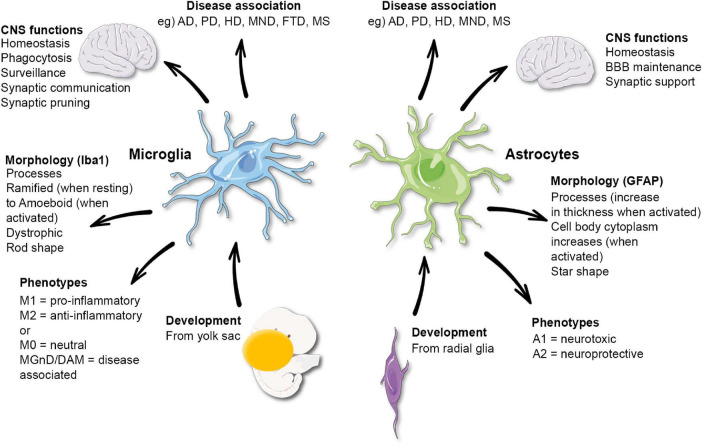
Microglia and astrocytes play essential roles in the central nervous system contributing to many functions including homeostasis, immune response, blood–brain barrier maintenance and synaptic support. Evidence has emerged from experimental models of glial communication that microglia and astrocytes influence and coordinate each other and their effects on the brain environment. However, due to the difference in glial cells between humans and rodents, it is essential to confirm the relevance of these findings in human brains. Here, we aim to review the current knowledge on microglia-astrocyte crosstalk in humans, exploring novel methodological techniques used in health and disease conditions. This will include an in-depth look at cell culture and iPSCs, post-mortem studies, imaging and fluid biomarkers, genetics and transcriptomic data. In this review, we will discuss the advantages and limitations of these methods, highlighting the understanding these methods have brought the field on these cells communicative abilities, and the knowledge gaps that remain.
Overview
The claims and statistics of this paper haven’t been checked yet.
Run the check — free, no account needed. It reads the open-access text and checks the sources it cites.
Integrity signals
Registry
Cites
Statistics
Figures
Verification
Claims the paper makes
The claims and statistics of this paper haven’t been checked yet.
Run the check — free, no account needed. It reads the open-access text and checks the sources it cites.
Claims other papers make about this one
Each is checked against this paper’s own text: does it say what the citing paper says it does?
The source backs part of the claim, not all of it — or backs it for a different population, condition or comparison.
In the citing paper
“Astrocyte and microglia iPSC models partially recapitulate the transcriptomes of their in vivo counterparts and may capture authentic neurodegenerative disease phenotypes [345,406,480,481,482,483,484].”
From Revisiting the Pathogenesis of X-Linked Adrenoleukodystrophy (2025) · cites this paper as [345]
Experimental — accuracy not yet measured
Self-check
Percentages, sums and reported differences re-computed from the paper’s own numbers.
The self-check needs the paper’s full text.
Citations
Cites
Takahashi, K., & Yamanaka, S. (2006) · Cell · doi:10.1016/j.cell.2006.07.024 · Cited by 26,673
Exploring the full spectrum of macrophage activation
Mosser, D. M., & Edwards, J. P. (2008) · Nature Reviews Immunology · doi:10.1038/nri2448 · Cited by 9,144
Polazzi, E., Mengoni, I., Caprini, M., Peña‐Altamira, E., Kurtys, E., & Monti, B. (2012) · Neurosignals · doi:10.1159/000337115 · Cited by 9,023
Neurotoxic reactive astrocytes are induced by activated microglia
Liddelow, S. A., Guttenplan, K. A., Clarke, L. E., Bennett, F. C., Bohlen, C. J., Schirmer, L., Bennett, M. L., Münch, A. E., Chung, W.-S., Peterson, T. C., et al. (2017) · Nature · doi:10.1038/nature21029 · Cited by 7,971
Resting microglial cells are highly dynamic surveillants of brain parenchyma in vivo
Nimmerjahn, A., Kirchhoff, F., & Helmchen, F. (2005) · Science · doi:10.1126/science.1110647 · Cited by 5,827
A Unique Microglia Type Associated with Restricting Development of Alzheimer’s Disease
Keren-Shaul, H., Spinrad, A., Weiner, A., Matcovitch-Natan, O., Dvir-Szternfeld, R., Ulland, T. K., David, E., Baruch, K., Lara-Astaiso, D., Toth, B., et al. (2017) · Cell · doi:10.1016/j.cell.2017.05.018 · Cited by 5,493
Astrocytes: Biology and pathology
Sofroniew, M. V., & Vinters, H. V. (2010) · Acta Neuropathologica · doi:10.1007/s00401-009-0619-8 · Cited by 5,196
Fate Mapping Analysis Reveals That Adult Microglia Derive from Primitive Macrophages
Ginhoux, F., Greter, M., Leboeuf, M., Nandi, S., See, P., Gokhan, S., Mehler, M. F., Conway, S. J., Ng, L. G., Stanley, E. R., et al. (2010) · Science · doi:10.1126/science.1194637 · Cited by 5,175
Meta-Analysis of 74,046 Individuals Identifies 11 New Susceptibility Loci for Alzheimer’s Disease
(EADI), E. A. D. I., Lambert, J.-C., (GERAD), G. A. E. R. I. A. D., Alzheimer's Disease Genetic Consortium (ADGC), (CHARGE), C. F. H. A. A. R. I. G. E., Ibrahim-Verbaas, C. A., Harold, D., Naj, A. C., Sims, R., Bellenguez, C., et al. (2013) · Nature Genetics · doi:10.1038/ng.2802 · Cited by 4,336
Microglia Sculpt Postnatal Neural Circuits in an Activity and Complement-Dependent Manner
Schafer, D. P., Lehrman, E. K., Kautzman, A. G., Koyama, R., Mardinly, A. R., Yamasaki, R., Ransohoff, R. M., Greenberg, M. E., Barres, B. A., & Stevens, B. (2012) · Neuron · doi:10.1016/j.neuron.2012.03.026 · Cited by 4,089
Cited by
Sorted by citations
The duality of amyloid-β: its role in normal and Alzheimer’s disease states
Azargoonjahromi, A. (2024) · Molecular Brain · doi:10.1186/s13041-024-01118-1 · Cited by 214
Precision drug delivery to the central nervous system using engineered nanoparticles
Gao, J., Xia, Z. J., Gunasekar, S., Jiang, C., Karp, J. M., & Joshi, N. (2024) · Nature Reviews Materials · doi:10.1038/s41578-024-00695-w · Cited by 173
Microglial activation states and their implications for Alzheimer's Disease
Valiukas, Z., Tangalakis, K., Apostolopoulos, V., & Feehan, J. (2025) · The Journal of Prevention of Alzheimer s Disease · doi:10.1016/j.tjpad.2024.100013 · Cited by 167
Microglia-Astrocyte Communication in Alzheimer’s Disease
Wu, Y., & Eisel, U. L. M. (2023) · Journal of Alzheimer s Disease · doi:10.3233/jad-230199 · Cited by 125
Bidirectional Communication Between Microglia and Astrocytes in Neuroinflammation
Bhusal, A., Afridi, R., Lee, W., & Suk, K. (2022) · Current Neuropharmacology · doi:10.2174/1570159x21666221129121715 · Cited by 113
Metadata
- Authors
- Garland, E. F., Hartnell, I. J., & Boche, D.
- Affiliations
- University of Southampton (GB)
- Corresponding author
- Boche, Delphine
- Venue
- Frontiers in Neuroscience
- Publisher
- Frontiers Media SA
- Published
- 2022-02-16
- Volume
- 16
- Pages
- 824888-824888
- ISSN
- 1662-453X (ISSN-L)
- PMC
- PMC8888691
- OpenAlex
- W4212929528
- Cited by
- 165
- References
- 190
- Citations by year
- 2026: 39 · 2025: 40 · 2024: 50 · 2023: 28 · 2022: 8
- FWCI
- 3.27
- Type
- journal article
- Language
- en
- Open access
- gold
- doi.org
- Licence
- Locations
- Frontiers in Neuroscience (publishedVersion, cc-by)
- PubMed Central (publishedVersion, cc-by)
- PubMed (publishedVersion)
- White Rose Research Online (University of Leeds, The University of Sheffield, University of York) (acceptedVersion)
- ePrints Soton (University of Southampton) (acceptedVersion)
- DOAJ (DOAJ: Directory of Open Access Journals) (submittedVersion, cc-by-sa)
- Funders
- Alzheimer’s Research UK
- Topics
- Life Sciences › Neuroscience › Neurology › Neuroinflammation and Neurodegeneration Mechanisms
- Life Sciences › Neuroscience › Cellular and Molecular Neuroscience › Neuroscience and Neuropharmacology Research
- Life Sciences › Neuroscience › Neurology › Barrier Structure and Function Studies
- Keywords
- microglia, astrocytes, human brain, blood-brain barrier, neuroinflammation, induced pluripotent stem cells, cell culture model, fluid biomarkers, transcriptomics, neurodegenerative diseases
About these checks
Registry, retraction, Cites and statistics results are computed, not judged, with one exception: a printed reference that no index lists is read once by a model before it counts as a citation error. Claims are extracted by a model and kept only when their quote appears verbatim in the text; each cited source is then checked, with a model’s help, for whether it says what the claim relies on. Those verdicts can be wrong — read the claim’s quote and the cited source before relying on one.
Statistics and arithmetic are recomputed, not judged.
Confidence bands are shown only for fields calibrated on our annotated test set. None is calibrated yet, so no band is shown.
What counts as a claim ›How we measure confidence ›How we check this ›
Are you an author, or do you see a problem with this page? Request a review
