

The DOI matches its Crossref record; 128 of the 128 references it cites were found in a registry or an index.
Mouse Models of Alzheimer’s Disease
Open accessAbstract
Alzheimer’s disease (AD) is a neurodegenerative disorder characterized by memory loss and personality changes, eventually leading to dementia. The pathological hallmarks of AD are senile plaques and neurofibrillary tangles, which comprise abnormally aggregated β-amyloid peptide (Aβ) and hyperphosphorylated tau protein. To develop preventive, diagnostic, and therapeutic strategies for AD, it is essential to establish animal models that recapitulate the pathophysiological process of AD. In this review, we will summarize the advantages and limitations of various mouse models of AD, including transgenic, knock-in, and injection models based on Aβ and tau. We will also discuss other mouse models based on neuroinflammation because recent genetic studies have suggested that microglia are crucial in the pathogenesis of AD. Although each mouse model has its advantages and disadvantages, further research on AD pathobiology will lead to the establishment of more accurate mouse models, and accelerate the development of innovative therapeutics.
Overview
The claims and statistics of this paper haven’t been checked yet.
Run the check — free, no account needed. It reads the open-access text and checks the sources it cites.
Integrity signals
Registry
Cites
Statistics
Figures
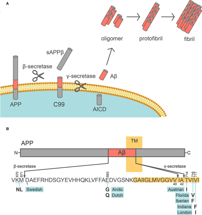
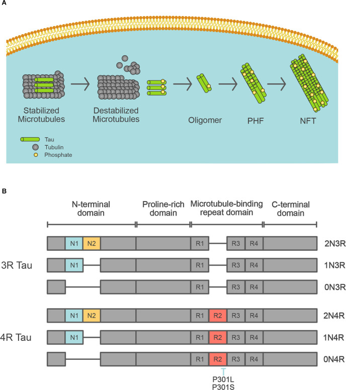
Verification
Claims the paper makes
The claims and statistics of this paper haven’t been checked yet.
Run the check — free, no account needed. It reads the open-access text and checks the sources it cites.
Claims other papers make about this one
Each is checked against this paper’s own text: does it say what the citing paper says it does?
The source backs part of the claim, not all of it — or backs it for a different population, condition or comparison.
In the citing paper
“Modeling these pathological hallmarks has largely relied on transgenic cellular and animal models harboring genetic mutations linked to ADAD (3–5).”
From Modeling late-onset Alzheimer’s disease neuropathology via direct neuronal reprogramming (2024) · cites this paper as [4]
Experimental — accuracy not yet measured
Self-check
Percentages, sums and reported differences re-computed from the paper’s own numbers.
The self-check needs the paper’s full text.
Citations
Cites
Neuropathological stageing of Alzheimer-related changes.
Braak, H., & Braak, E. (1991) · Acta Neuropathologica · doi:10.1007/bf00308809 · Cited by 16,123
The Amyloid Hypothesis of Alzheimer’s Disease: Progress and Problems on the Road to Therapeutics
Hardy, J., & Selkoe, D. J. (2002) · Science · doi:10.1126/science.1072994 · Cited by 13,864
NIA‐AA Research Framework: Toward a biological definition of Alzheimer's disease
Jack, C. R., Bennett, D. A., Blennow, K., Carrillo, M. C., Dunn, B., Haeberlein, S. B., Holtzman, D. M., Jagust, W., Jessen, F., Karlawish, J., et al. (2018) · Alzheimer's & Dementia · doi:10.1016/j.jalz.2018.02.018 · Cited by 10,537
Amyloid oligomers exacerbate tau pathology in a mouse model of tauopathy
Selenica, M.-L. B., Brownlow, M., Jimenez, J. P., Lee, D. C., Pena, G., Dickey, C. A., Gordon, M. N., & Morgan, D. (2013) · Neurodegenerative Diseases · doi:10.1159/000337230 · Cited by 10,359
The amyloid hypothesis of Alzheimer's disease at 25 years
Selkoe, D. J., & Hardy, J. (2016) · EMBO Molecular Medicine · doi:10.15252/emmm.201606210 · Cited by 6,164
A Unique Microglia Type Associated with Restricting Development of Alzheimer’s Disease
Keren-Shaul, H., Spinrad, A., Weiner, A., Matcovitch-Natan, O., Dvir-Szternfeld, R., Ulland, T. K., David, E., Baruch, K., Lara-Astaiso, D., Toth, B., et al. (2017) · Cell · doi:10.1016/j.cell.2017.05.018 · Cited by 5,493
Glenner, G. G., & Wong, C. W. (1984) · Biochemical and Biophysical Research Communications · doi:10.1016/s0006-291x(84)80190-4 · Cited by 5,404
Goate, A., Chartier-Harlin, M.-C., Mullan, M., Brown, J., Crawford, F., Fidani, L., Giuffra, L., Haynes, A., Irving, N., James, L., et al. (1991) · Nature · doi:10.1038/349704a0 · Cited by 4,659
Correlative Memory Deficits, Aβ Elevation, and Amyloid Plaques in Transgenic Mice
Hsiao, K., Chapman, P., Nilsen, S., Eckman, C., Harigaya, Y., Younkin, S., Yang, F., & Cole, G. (1996) · Science · doi:10.1126/science.274.5284.99 · Cited by 4,210
Triple-Transgenic Model of Alzheimer’s Disease with Plaques and Tangles
Oddo, S., Caccamo, A., Shepherd, J. D., Murphy, M. P., Golde, T. E., Kayed, R., Metherate, R., Mattson, M. P., Akbari, Y., & LaFerla, F. M. (2003) · Neuron · doi:10.1016/s0896-6273(03)00434-3 · Cited by 4,126
Cited by
Sorted by citations
Mitochondria in health, disease, and aging
Harrington, J. S., Ryter, S. W., Plataki, M. E., Price, D. R., & Choi, A. M. K. (2023) · Physiological Reviews · doi:10.1152/physrev.00058.2021 · Cited by 641
Magnetic iron oxide nanoparticles for brain imaging and drug delivery
Qiao, R., Fu, C., Forgham, H., Javed, I., Huang, X., Zhu, J., Whittaker, A. K., & Davis, T. P. (2023) · Advanced Drug Delivery Reviews · doi:10.1016/j.addr.2023.114822 · Cited by 257
Is Alzheimer disease a disease?
Korczyn, A. D., & Grinberg, L. T. (2024) · Nature Reviews Neurology · doi:10.1038/s41582-024-00940-4 · Cited by 198
Antidiabetic agents as a novel treatment for Alzheimer’s and Parkinson’s disease
Nowell, J., Blunt, E., Gupta, D., & Edison, P. (2023) · Ageing Research Reviews · doi:10.1016/j.arr.2023.101979 · Cited by 179
Therapeutic inhibition of ferroptosis in neurodegenerative disease
Ryan, S. K., Ugalde, C. L., Rolland, A., Skidmore, J., Devos, D., & Hammond, T. R. (2023) · Trends in Pharmacological Sciences · doi:10.1016/j.tips.2023.07.007 · Cited by 127
Neuropathogenesis-on-chips for neurodegenerative diseases
Amartumur, S., Nguyen, H. M., Huynh, T., Kim, T. S., Woo, R., Oh, E., Kim, K. K., Lee, L. P., & Heo, C. (2024) · Nature Communications · doi:10.1038/s41467-024-46554-8 · Cited by 131
The role of PI3K signaling pathway in Alzheimer’s disease
Pan, J., Yao, Q., Wang, Y., Chang, S., Li, C., Wu, Y., Shen, J., & Yang, R. (2024) · Frontiers in Aging Neuroscience · doi:10.3389/fnagi.2024.1459025 · Cited by 89
Glymphatic dysfunction in Alzheimer’s disease: A critical appraisal
Keil, S. A., Jansson, D., Braun, M., & Iliff, J. J. (2025) · Science · doi:10.1126/science.adv8269 · Cited by 100
Behaviour Hallmarks in Alzheimer’s Disease 5xFAD Mouse Model
Pádua, M. S., Guil‐Guerrero, J. L., & Lopes, P. A. (2024) · International Journal of Molecular Sciences · doi:10.3390/ijms25126766 · Cited by 87
Modeling late-onset Alzheimer’s disease neuropathology via direct neuronal reprogramming
Sun, Z., Kwon, J.-S., Ren, Y., Chen, S., Walker, C. K., Lu, X., Cates, K., Karahan, H., Sviben, S., Fitzpatrick, J. A. J., et al. (2024) · Science · doi:10.1126/science.adl2992 · Cited by 77
Immune Activation in Alzheimer Disease
Mary, A., Mancuso, R., & Heneka, M. T. (2024) · Annual Review of Immunology · doi:10.1146/annurev-immunol-101921-035222 · Cited by 82
The Importance of Complement-Mediated Immune Signaling in Alzheimer’s Disease Pathogenesis
Batista, A. F., Khan, K., Papavergi, M.-T., & Lemere, C. A. (2024) · International Journal of Molecular Sciences · doi:10.3390/ijms25020817 · Cited by 72
The Functions and Phenotypes of Microglia in Alzheimer’s Disease
Fujikawa, R., & Tsuda, M. (2023) · Cells · doi:10.3390/cells12081207 · Cited by 58
Sharma, M., Pal, P., & Gupta, S. K. (2024) · Neurochemistry International · doi:10.1016/j.neuint.2024.105761 · Cited by 49
Insights on the Use of Transgenic Mice Models in Alzheimer’s Disease Research
Pádua, M. S., Guil-Guerrero, J. L., Prates, J. A. M., & Lopes, P. A. (2024) · International Journal of Molecular Sciences · doi:10.3390/ijms25052805 · Cited by 46
Insight into the emerging and common experimental in-vivo models of Alzheimer’s disease
Dhapola, R., Kumari, S., Sharma, P., & HariKrishnaReddy, D. (2023) · Laboratory Animal Research · doi:10.1186/s42826-023-00184-1 · Cited by 53
Rodrigues, R. O., Shin, S.-R., & Bañobre-López, M. (2024) · Journal of Nanobiotechnology · doi:10.1186/s12951-024-02720-0 · Cited by 44
Polis, B., & Samson, A. O. (2024) · Journal of Alzheimer's Disease · doi:10.3233/jad-240058 · Cited by 43
Neutrophil biology in injuries and diseases of the central and peripheral nervous systems
Balog, B. M., Sonti, A., & Zigmond, R. E. (2023) · Progress in Neurobiology · doi:10.1016/j.pneurobio.2023.102488 · Cited by 46
Corraliza-Gómez, M., Bermejo, T., Lilue, J., Rodríguez-Iglesias, N., Valero, J., Cózar‐Castellano, I., Arranz, E. M., Sánchez, D., & Ganfornina, M. D. (2023) · Journal of Neuroinflammation · doi:10.1186/s12974-023-02914-7 · Cited by 39
Alzheimer’s Disease: Understanding Motor Impairments
Andrade-Guerrero, J., Martínez-Orozco, H., Villegas-Rojas, M. M., Santiago-Balmaseda, A., Delgado-Minjares, K. M., Pérez‐Segura, I., Baéz-Cortés, M. T., Toro-Colín, M. A. D., Guerra‐Crespo, M., Arias-Carrión, Ó., et al. (2024) · Brain Sciences · doi:10.3390/brainsci14111054 · Cited by 53
The reverse transcriptase inhibitor 3TC protects against age‐related cognitive dysfunction
Wahl, D., Smith, M. E., McEntee, C. M., Cavalier, A. N., Osburn, S. C., Burke, S. D., Grant, R. A., Nerguizian, D., Lark, D. S., Link, C. D., et al. (2023) · Aging Cell · doi:10.1111/acel.13798 · Cited by 40
Manfredi, J. N., Gupta, S. K., Vyavahare, S. S., Deák, F., Lu, X., Buddha, L., Wankhade, U. D., Lohakare, J., Isales, C. M., & Fulzele, S. (2024) · Physiology & Behavior · doi:10.1016/j.physbeh.2024.114778 · Cited by 42
Physical exercise regulates microglia in health and disease
Strohm, A. O., & Majewska, A. K. (2024) · Frontiers in Neuroscience · doi:10.3389/fnins.2024.1420322 · Cited by 37
In Vitro 3D Modeling of Neurodegenerative Diseases
Louit, A., Galbraith, T., & Berthod, F. (2023) · Bioengineering · doi:10.3390/bioengineering10010093 · Cited by 37
Antonioni, A., Martorana, A., Santarnecchi, E., Hampel, H. J., & Koch, G. (2025) · Alzheimer s & Dementia · doi:10.1002/alz.70337 · Cited by 31
Alzheimer’s Disease-Related Epigenetic Changes: Novel Therapeutic Targets
Paniri, A., Hosseini, M. M., & Akhavan‐Niaki, H. (2023) · Molecular Neurobiology · doi:10.1007/s12035-023-03626-y · Cited by 34
de Sousa, A. A., Dames, B. A. R., Graff, E. C., Mohamedelhassan, R., Vassilopoulos, T., & Charvet, C. J. (2023) · Communications Biology · doi:10.1038/s42003-023-05034-3 · Cited by 36
Abdelmaksoud, N. M., Sallam, A.-A. M., Abulsoud, A. I., El‐Dakroury, W. A., Mageed, S. S. A., AL‐Noshokaty, T. M., Elrebehy, M. A., Elshaer, S. S., Mahmoud, N. A., Fathi, D., et al. (2023) · Pathology - Research and Practice · doi:10.1016/j.prp.2023.155007 · Cited by 30
Guo, L., Yang, X., Zhang, Y., Xu, X., & Li, Y. (2023) · Frontiers in Aging Neuroscience · doi:10.3389/fnagi.2022.1077732 · Cited by 29
Advancements and challenges in mouse models of Alzheimer’s disease
Qian, Z., Li, Y., & Ye, K. (2024) · Trends in Molecular Medicine · doi:10.1016/j.molmed.2024.10.010 · Cited by 30
CRISPR/Cas9 and iPSC-Based Therapeutic Approaches in Alzheimer’s Disease
Raffaele, I., Cipriano, G. L., Anchesi, I., Oddo, S., & Silvestro, S. (2025) · Antioxidants · doi:10.3390/antiox14070781 · Cited by 30
Complexity of Sex Differences and Their Impact on Alzheimer’s Disease
Kadlecová, M., Freude, K., & Haukedal, H. (2023) · Biomedicines · doi:10.3390/biomedicines11051261 · Cited by 27
Differential roles of regulatory T cells in Alzheimer's disease
Jafarzadeh, A., Sheikhi, A., Jafarzadeh, Z., & Nemati, M. (2023) · Cellular Immunology · doi:10.1016/j.cellimm.2023.104778 · Cited by 26
McGeachan, R. I., Meftah, S., Taylor, L. W., Catterson, J. H., Negro, D., Bonthron, C., Holt, K., Tulloch, J., Rose, J. L., Gobbo, F., et al. (2025) · Nature Communications · doi:10.1038/s41467-025-58879-z · Cited by 23
An Alternative View of Familial Alzheimer’s Disease Genetics
Lardelli, M. T. (2023) · Journal of Alzheimer s Disease · doi:10.3233/jad-230313 · Cited by 24
Beyond Transgenic Mice: Emerging Models and Translational Strategies in Alzheimer’s Disease
Lopes, P. A., & Guil‐Guerrero, J. L. (2025) · International Journal of Molecular Sciences · doi:10.3390/ijms26125541 · Cited by 23
Switching On/Off Amyloid Plaque Formation in Transgenic Animal Models of Alzheimer’s Disease
Kozin, S. A., Kechko, O. I., Adzhubei, A. A., Makarov, A. А., & Mitkevich, V. A. (2023) · International Journal of Molecular Sciences · doi:10.3390/ijms25010072 · Cited by 21
Faraji, P., Kühn, H., & Ahmadian, S. (2024) · Journal of Molecular Neuroscience · doi:10.1007/s12031-024-02224-4 · Cited by 25
Unraveling the dual nature of brain CD8+ T cells in Alzheimer’s disease
Hu, D., & Weiner, H. L. (2024) · Molecular Neurodegeneration · doi:10.1186/s13024-024-00706-y · Cited by 19
Tauopathy promotes spinal cord-dependent production of toxic amyloid-beta in transgenic monkeys
Tu, Z., Yan, S., Han, B., Li, C., Liang, W., Lin, Y., Ding, Y., Wei, H., Wang, L., Xu, H., et al. (2023) · Signal Transduction and Targeted Therapy · doi:10.1038/s41392-023-01601-6 · Cited by 20
Optogenetic and chemogenetic approaches for modeling neurological disorders in vivo
Krut’, V. G., Kalinichenko, A. L., Maltsev, D. I., Jappy, D., Shevchenko, E. K., Podgorny, O. V., & Belousov, V. V. (2024) · Progress in Neurobiology · doi:10.1016/j.pneurobio.2024.102600 · Cited by 17
Aishwarya, R., Abdullah, C. S., Remex, N. S., Bhuiyan, M. A. N., Lu, X., Dhanesha, N., Stokes, K. Y., Orr, A. W., Kevil, C. G., & Bhuiyan, M. S. (2024) · Scientific Reports · doi:10.1038/s41598-024-67638-x · Cited by 18
Retinal dysfunction in APOE4 knock‐in mouse model of Alzheimer's disease
Abhyankar, S. D., Luo, Q., Hartman, G. D., Mahajan, N. S., Corson, T. W., Oblak, A. L., Lamb, B. T., & Bhatwadekar, A. D. (2025) · Alzheimer s & Dementia · doi:10.1002/alz.14433 · Cited by 16
Tate, M., Wijeratne, H. R. S., Kim, B., Philtjens, S., You, Y., Lee, D., Gutierrez, D. A., Sharify, D., Wells, M., Perez‐Cardelo, M., et al. (2024) · Alzheimer's & Dementia · doi:10.1002/alz.14243 · Cited by 21
Effects of Redox Homeostasis and Mitochondrial Damage on Alzheimer’s Disease
Wu, Y., & Hsieh, H. (2023) · Antioxidants · doi:10.3390/antiox12101816 · Cited by 16
Human stem cell transplantation models of Alzheimer’s disease
Ifediora, N., Canoll, P., & Hargus, G. (2024) · Frontiers in Aging Neuroscience · doi:10.3389/fnagi.2024.1354164 · Cited by 18
Liu, J.-Y., Dai, Y., He, Y.-X., & Lin, L. (2024) · Frontiers in Pharmacology · doi:10.3389/fphar.2023.1301102 · Cited by 17
The need for systems thinking to advance Alzheimer's disease research
Uleman, J. F., Quax, R., Melis, R. J. F., Hoekstra, A. G., & Rikkert, M. G. M. O. (2024) · Psychiatry Research · doi:10.1016/j.psychres.2024.115741 · Cited by 15
Numerical and Analytical Simulation of the Growth of Amyloid-β Plaques
Kuznetsov, A. V. (2024) · Journal of Biomechanical Engineering · doi:10.1115/1.4064969 · Cited by 15
Neutrophil-to-Lymphocyte Ratio in the Alzheimer’s Disease Continuum
Aprile, D., Marchi, F. D., Menegon, F., Comi, C., & Tondo, G. (2025) · International Journal of Molecular Sciences · doi:10.3390/ijms26115157 · Cited by 14
Whiteaker, P., & George, A. A. (2023) · Pharmacological Research · doi:10.1016/j.phrs.2023.106743 · Cited by 14
Sánchez-Puebla, L., López‐Cuenca, I., Salobrar‐García, E., Ramirez, A. I., Fernández‐Albarral, J. A., Matamoros, J. A., Elvira‐Hurtado, L., Salazar, J. J., Ramirez, J. M., & de Hoz, R. (2024) · Biomedicines · doi:10.3390/biomedicines12030528 · Cited by 14
The proteomic landscape of microglia in health and disease
Davis, E., & Lloyd, A. F. (2024) · Frontiers in Cellular Neuroscience · doi:10.3389/fncel.2024.1379717 · Cited by 13
Vascular Function and Ion Channels in Alzheimer's Disease
Taylor, J. L., Baudel, M. A. S. M., Nieves‐Cintrón, M., & Navedo, M. F. (2024) · Microcirculation · doi:10.1111/micc.12881 · Cited by 13
Seed structure and phosphorylation in the fuzzy coat impact tau seeding competency
Kasen, A., Lövestam, S., Breton, E., Meyerdirk, L. K., McPhail, J. A., Piché, K., Louwrier, A., Capan, C. D., Lee, H., Scheres, S. H. W., et al. (2025) · Nature Communications · doi:10.1038/s41467-025-64312-2 · Cited by 17
Zhang, S., Guo, Z., Xu, Y., Mi, J., Liu, J., Li, Z., Xie, X., & Xu, G. (2024) · Frontiers in Neuroscience · doi:10.3389/fnins.2024.1313639 · Cited by 13
Sato, K., Takayama, K., Saito, Y., & Inoue, S. (2024) · Proceedings of the National Academy of Sciences · doi:10.1073/pnas.2406854121 · Cited by 13
Zhang, X., Shi, J., Thakore, P., Gonzales, A. L., Earley, S., Chen, Q., Zhou, T., & Feng, Y. (2025) · International Journal of Molecular Sciences · doi:10.3390/ijms26073019 · Cited by 11
Metzendorf, N. G., Sehlin, D., & Hultqvist, G. (2025) · Frontiers in Neuroscience · doi:10.3389/fnins.2025.1645952 · Cited by 10
SPLICER: a highly efficient base editing toolbox that enables in vivo therapeutic exon skipping
Miskalis, A., Shirguppe, S., Winter, J., Elias, G., Swami, D., Nambiar, A., Stilger, M., Woods, W. S., Gosstola, N. C., Gapinske, M., et al. (2024) · Nature Communications · doi:10.1038/s41467-024-54529-y · Cited by 13
Microglial Responses to Alzheimer's Disease Pathology: Insights From “Omics” Studies
Reid, A. N., Jayadev, S., & Prater, K. E. (2025) · Glia · doi:10.1002/glia.24666 · Cited by 10
Chen, J., Yin, X., He, H., Lu, L. W., Wang, M., Liu, B., & Cheng, K. (2024) · Food & Function · doi:10.1039/d3fo03805j · Cited by 10
Modeling of age-related neurological disease: utility of zebrafish
Kanoh, T., Mizoguchi, T., Tonoki, A., & Itoh, M. (2024) · Frontiers in Aging Neuroscience · doi:10.3389/fnagi.2024.1399098 · Cited by 9
Singh, S., Kachhawaha, K., & Singh, S. K. (2024) · Biochemical Pharmacology · doi:10.1016/j.bcp.2024.116303 · Cited by 10
O’Hare, N. R., Millican, K., & Ebong, E. E. (2024) · Frontiers in Physiology · doi:10.3389/fphys.2024.1394725 · Cited by 10
Retinal Microstructural and Microvascular Changes in Alzheimer Disease: A Review
Ordoñez, M. A. O., Smith, R. C., Yiu, G. C., & Liu, Y. (2024) · International Ophthalmology Clinics · doi:10.1097/iio.0000000000000549 · Cited by 10
Preclinical Retinal Disease Models: Applications in Drug Development and Translational Research
Pandi, S. P. S., Winter, H., Smith, M. R., Harkin, K., & Bojdo, J. (2025) · Pharmaceuticals · doi:10.3390/ph18030293 · Cited by 9
The 3xTg-AD Mouse Model: A Comprehensive Tool for Understanding Alzheimer’s Disease
Pattanayak, A., & Firdous, S. M. (2026) · Cellular and Molecular Neurobiology · doi:10.1007/s10571-025-01655-w · Cited by 13
Papazoglou, A., Henseler, C., Weickhardt, S., Teipelke, J., Παπάζογλου, Π., Daubner, J., Schiffer, T., Krings, D., Broich, K., Hescheler, J., et al. (2024) · PLoS ONE · doi:10.1371/journal.pone.0296959 · Cited by 9
Pucci, I. M., Aguiar, A. F., Pucci, R. M., Casonatto, J., & Borghi, S. M. (2024) · Experimental Brain Research · doi:10.1007/s00221-024-06821-y · Cited by 12
McClarty, B. M., Rodríguez, G., & Dong, H. (2024) · Brain Research · doi:10.1016/j.brainres.2024.148951 · Cited by 9
Amyloid-beta precursor protein contributes to brain aging and learning declinePreprint
de Bakker, D. E. M., Mihaljević, M., Gharat, K., Richter, Y., Bagnoli, S., van Bebber, F., Adam, L., Shamim-Schulze, F., Ohlenschläger, O., Bens, M., et al. (2024) · bioRxiv (Cold Spring Harbor Laboratory) · doi:10.1101/2024.10.11.617841 · Cited by 11
Dolrahman, N., & Thong-asa, W. (2024) · EXPERIMENTAL ANIMALS · doi:10.1538/expanim.24-0021 · Cited by 8
Ren, H., Tang, L. Y., Yuan, Z., Liu, Y., Zhou, X., Xiao, X., Wu, X., Chen, W., Chen, Y., Wang, H., et al. (2024) · Frontiers in Pharmacology · doi:10.3389/fphar.2024.1492237 · Cited by 9
Geldenhuys, W. J., Wilson, G. N., Hernandez, K., Monaghan, K., Smith, K., Cicala, D. S., Poling, T. J., Walton, J. C., Han, P., & Huber, J. D. (2024) · Journal of Alzheimer s Disease · doi:10.1177/13872877241302456 · Cited by 9
Anschuetz, A., Schwab, K., Harrington, C. R., Wischik, C. M., & Riedel, G. (2025) · Journal of Alzheimer s Disease · doi:10.1177/13872877251362212 · Cited by 7
Yadav, A., Ouyang, X., Barkley, M., Watson, J. C., Madhamanchi, K., Kramer, J., Zhang, J., & Melkani, G. C. (2026) · Aging Cell · doi:10.1111/acel.70439 · Cited by 9
Limpengco, R. R., Liang, C., Sandhu, Y. K., & Mukherjee, J. (2023) · Molecules · doi:10.3390/molecules28155769 · Cited by 8
Diverse signaling mechanisms and heterogeneity of astrocyte reactivity in Alzheimer's disease
Qin, H., Zhou, L., Haque, F. T., Martín-Jiménez, C., Trang, A., Benveniste, E. N., & Wang, Q. (2023) · Journal of Neurochemistry · doi:10.1111/jnc.16002 · Cited by 10
Rodents’ visual gamma as a biomarker of pathological neural conditions
Meneghetti, N., Vannini, E., & Mazzoni, A. (2024) · The Journal of Physiology · doi:10.1113/jp283858 · Cited by 8
Grenon, M. B., Papavergi, M.-T., Bathini, P., Sadowski, M. J., & Lemere, C. A. (2024) · International Journal of Molecular Sciences · doi:10.3390/ijms25115754 · Cited by 7
Lamichhane, S., Seo, J., Jeong, J. H., Lee, S., & Lee, S. (2024) · Archives of Pharmacal Research · doi:10.1007/s12272-024-01527-9 · Cited by 8
An Overview of Transgenic Mouse Models for the Study of Alzheimer’s Disease
Lopes, P. A., Pádua, M. S., & Guil‐Guerrero, J. L. (2025) · Journal of dementia and Alzheimer's disease · doi:10.3390/jdad2010002 · Cited by 9
Calcineurin inhibition may prevent Alzheimer disease in people with Down syndrome
Dohl, J., Treadwell, Z., Norris, C. M., & Head, E. (2025) · Alzheimer s & Dementia · doi:10.1002/alz.70034 · Cited by 6
Mouse models of Anti-Aβ immunotherapies
Pikus, P., Turner, R. S., & Rebeck, G. W. (2025) · Molecular Neurodegeneration · doi:10.1186/s13024-025-00836-x · Cited by 6
Zebrafish Experimental Animal Models for AD: A Comprehensive Review
Sande, R., Godad, A. P., & Doshi, G. (2024) · Current Clinical Pharmacology · doi:10.2174/0127724328279684240104094257 · Cited by 6
Armbrust, F., Bickenbach, K., Altmeppen, H. C., Foggetti, A., Winkelmann, A., Wulff, P., Glatzel, M., Pietrzik, C. U., & Becker‐Pauly, C. (2024) · Cellular and Molecular Life Sciences · doi:10.1007/s00018-024-05139-w · Cited by 9
Illouz, T., Ascher, L. A. B., Madar, R., & Okun, E. (2024) · Scientific Reports · doi:10.1038/s41598-024-66855-8 · Cited by 7
Lau, F., Binacchi, R., Brugnara, S., Cumplido-Mayoral, A., Savino, S. D., Khan, I., Orso, A., Sartori, S., Bellosta, P., Carl, M., et al. (2025) · Neuroscience · doi:10.1016/j.neuroscience.2025.03.042 · Cited by 6
Xiao, S., Wei, X., Han, B., Shi, X., Wei, C., Liang, R., Sun, J., Zhang, Z., Han, Z., & Shen, L. (2025) · Frontiers in Aging Neuroscience · doi:10.3389/fnagi.2025.1561831 · Cited by 9
Bratke, S., Schmid, S., Ulm, B., Jungwirth, B., Blobner, M., & Borgstedt, L. (2024) · Frontiers in Medicine · doi:10.3389/fmed.2024.1342752 · Cited by 6
Matuszewska, M., Wilkaniec, A., Gąssowska-Dobrowolska, M., Cieślik, M., Olech-Kochańczyk, G., Pałasz, E., Gawinek, E., Strawski, M., & Czapski, G. A. (2025) · Frontiers in Molecular Neuroscience · doi:10.3389/fnmol.2025.1619583 · Cited by 6
Mohammed, O., & Kelemu, T. (2025) · Health Science Reports · doi:10.1002/hsr2.71065 · Cited by 5
Waqar, Z., Sethi, P., Jain, D., Singh, K., Alsaidan, O. A., Alzarea, S. I., Gupta, J. K., Saxena, S., & Sharma, M. C. (2025) · Current Genomics · doi:10.2174/0113892029372437251010114053 · Cited by 6
CX3CR1: a potential microglia-specific PET imaging target in Alzheimer’s and Parkinson’s diseases
Yang, H., Wang, Y., Xu, Y., & Wang, C. (2025) · Frontiers in Pharmacology · doi:10.3389/fphar.2025.1678159 · Cited by 6
Sampedro‐Viana, A., Fernández‐Rodicio, S., Castillo, J., Hervella, P., Alonso-Alonso, M. L., & Iglesias‐Rey, R. (2024) · International Journal of Molecular Sciences · doi:10.3390/ijms25189792 · Cited by 5
Min-Kaung-Wint-Mon, & Mitsushima, D. (2025) · Journal of dementia and Alzheimer's disease · doi:10.3390/jdad2030025 · Cited by 5
Modelling sex differences of neurological disorders in vitro
Castro‐Aldrete, L., Einsiedler, M., Cuní‐López, C., Vanhaelen, Q., Silvestri, A., Ferretti, M. T., de Gennaro, M. E., Putignano, G., Guix, M., Marino, N., et al. (2025) · Nature Reviews Bioengineering · doi:10.1038/s44222-025-00355-w · Cited by 4
Metadata
- Authors
- Yokoyama, M., Kobayashi, H., Tatsumi, L., & Tomita, T.
- Affiliations
- The University of Tokyo (JP)
- Corresponding author
- Tomita, Taisuke
- Venue
- Frontiers in Molecular Neuroscience
- Publisher
- Frontiers Media SA
- Published
- 2022-06-21
- Volume
- 15
- Pages
- 912995-912995
- ISSN
- 1662-5099 (ISSN-L)
- PMC
- PMC9254908
- OpenAlex
- W4283267746
- Cited by
- 213
- References
- 128
- Citations by year
- 2026: 40 · 2025: 63 · 2024: 77 · 2023: 31 · 2022: 1
- FWCI
- 5.91
- Type
- journal article
- Language
- en
- Open access
- gold
- doi.org
- Licence
- Locations
- Frontiers in Molecular Neuroscience (publishedVersion, cc-by)
- PubMed Central (publishedVersion, cc-by)
- PubMed (publishedVersion)
- DOAJ (DOAJ: Directory of Open Access Journals) (submittedVersion, cc-by-sa)
- Funders
- Japan Society for the Promotion of Science
- Japan Agency for Medical Research and Development
- Moonshot Research and Development Program
- Topics
- Life Sciences › Neuroscience › Cellular and Molecular Neuroscience › Alzheimer's disease research and treatments
- Life Sciences › Neuroscience › Neurology › Neuroinflammation and Neurodegeneration Mechanisms
- Health Sciences › Medicine › Psychiatry and Mental health › Dementia and Cognitive Impairment Research
- Keywords
- Alzheimer's disease, mouse model, transgenic mice, knock-in mice, amyloid-beta, tau protein, neurofibrillary tangles, amyloid plaques, neuroinflammation, microglia
About these checks
Registry, retraction, Cites and statistics results are computed, not judged, with one exception: a printed reference that no index lists is read once by a model before it counts as a citation error. Claims are extracted by a model and kept only when their quote appears verbatim in the text; each cited source is then checked, with a model’s help, for whether it says what the claim relies on. Those verdicts can be wrong — read the claim’s quote and the cited source before relying on one.
Statistics and arithmetic are recomputed, not judged.
Confidence bands are shown only for fields calibrated on our annotated test set. None is calibrated yet, so no band is shown.
What counts as a claim ›How we measure confidence ›How we check this ›
Are you an author, or do you see a problem with this page? Request a review
