

The DOI matches its Crossref record; 2 of the 2 references it cites were found in a registry or an index.
The Role of Exosomes in Pancreatic Cancer From Bench to Clinical Application: An Updated Review
Abstract
Pancreatic ductal adenocarcinoma (PDAC) remains one of the most dismal gastrointestinal malignancies with an overall 5-year survival rate of 8%–9%. The intra-tumor heterogeneity and special tumor microenvironment in PDAC make it challenging to develop effective treatment strategies. Exosomes are extracellular vesicles that originate from the endosomes and have a diameter of 40–160 nm. A growing body of evidence has shown that exosomes play vital roles in tumor initiation and development. Recently, extensive application of exosomes as biomarkers and drug carriers has rendered them attractive in the field of PDAC. This review summarizes the latest progress in the methodologies for isolation, modification, and tracking of exosomes, exosome-mediated cell-to-cell communication, clinical applications of exosome as minimally invasive liquid biopsy and drugs carriers, as well as their involvement in the angiogenic regulation in PDAC. In spite of these advancements, some obstacles are still required to be overcome to use the exosome-based technologies for early diagnosis or improvement of prognosis of patients with PDAC.
Overview
The claims and statistics of this paper haven’t been checked yet.
Run the check — free, no account needed. It reads the open-access text and checks the sources it cites.
Integrity signals
Registry
Cites
Statistics
Figures
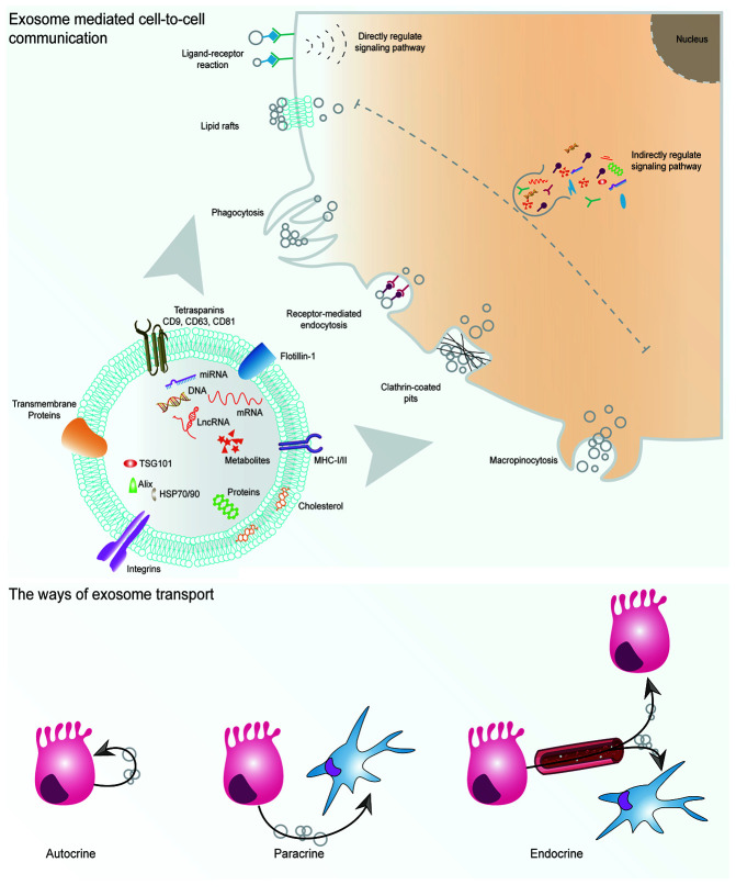
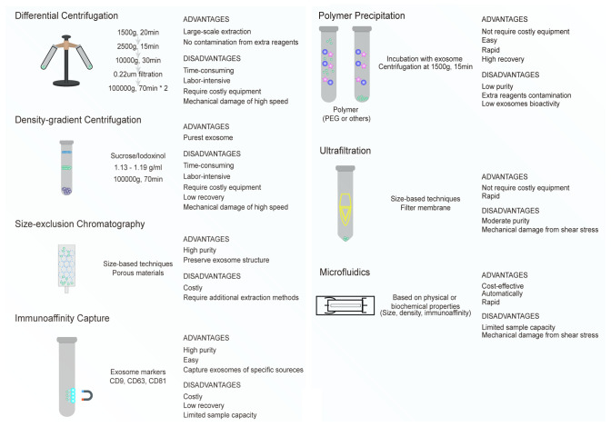
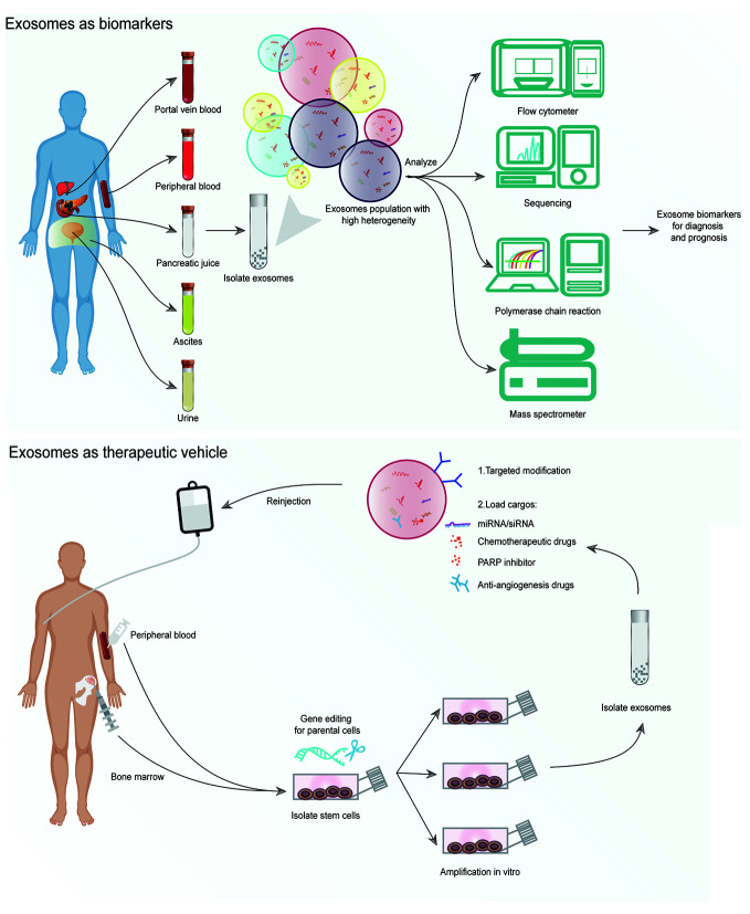
Verification
Claims the paper makes
The claims and statistics of this paper haven’t been checked yet.
Run the check — free, no account needed. It reads the open-access text and checks the sources it cites.
Claims other papers make about this one
Each is checked against this paper’s own text: does it say what the citing paper says it does?
The cited source says what the claim attributes to it.
In the citing paper
“Furthermore, this innovative strategy has progressed to a Phase I clinical trial for PDAC patients harboring the KrasG12D mutation (NCT03608631) [143].”
From Emerging role of exosomes in cancer therapy: progress and challenges (2025) · cites this paper as [143]
In the citing paper
“These engineered exosomes effectively targeted the KrasG12D mutation in PDAC cells in vivo, demonstrating a notable improvement in overall survival without causing toxicity.”
From Emerging role of exosomes in cancer therapy: progress and challenges (2025) · cites this paper as [143]
Self-check
Percentages, sums and reported differences re-computed from the paper’s own numbers.
The self-check needs the paper’s full text.
Citations
Cites
Ryan, D. P., Hong, T. S., & Bardeesy, N. (2014) · New England Journal of Medicine · doi:10.1056/nejmra1404198 · Cited by 2,348
Umezu, T., Tadokoro, H., Azuma, K., Yoshizawa, S., Ohyashiki, K., & Ohyashiki, J. H. (2014) · Blood · doi:10.1182/blood-2014-05-576116 · Cited by 600
Research works printed but not linked: 80
- Siegel, 2020, Cancer statistics, 2020, CA Cancer J Clin, 70, 7, 10.3322/caac.21590 · DOI · Paper page
- Vincent, 2011, Pancreatic cancer, Lancet, 378, 10.1016/S0140-6736(10)62307-0 · DOI
- Garrido-Laguna, 2015, Pancreatic cancer: from state-of-the-art treatments to promising novel therapies, Nat Rev Clin Oncol, 12, 10.1038/nrclinonc.2015.53 · DOI
- Yang, 2020, Current status and future prospect of surgical treatment for pancreatic cancer, Hepatobiliary Surg Nutr, 9, 89, 10.21037/hbsn.2019.12.04 · DOI
- Wunsch, 2016, Nanoscale lateral displacement arrays for the separation of exosomes and colloids down to 20 nm, Nat Nanotechnol, 11, 10.1038/nnano.2016.134 · DOI
- Li, 2018, Tumor-Secreted Exosomal miR-222 Promotes Tumor Progression via Regulating P27 Expression and Re-Localization in Pancreatic Cancer, Cell Physiol Biochem, 51, 10.1159/000495281 · DOI
- Sullivan, 2005, Role of exosomes in sperm maturation during the transit along the male reproductive tract, Blood Cells Mol Dis, 35, 1, 10.1016/j.bcmd.2005.03.005 · DOI
- Gehrmann, 2014, Harnessing the exosome-induced immune response for cancer immunotherapy, Semin Cancer Biol, 28, 58, 10.1016/j.semcancer.2014.05.003 · DOI · Paper page
- Melo, 2014, Cancer exosomes perform cell-independent microRNA biogenesis and promote tumorigenesis, Cancer Cell, 26, 10.1016/j.ccell.2014.09.005 · DOI
- Fukushima, 2020, Dysregulated Expression of the Nuclear Exosome Targeting Complex Component Rbm7 in Nonhematopoietic Cells Licenses the Development of Fibrosis, Immunity, 52, 542, 10.1016/j.immuni.2020.02.007 · DOI
- Kahlert, 2014, Identification of double-stranded genomic DNA spanning all chromosomes with mutated KRAS and p53 DNA in the serum exosomes of patients with pancreatic cancer, J Biol Chem, 289, 10.1074/jbc.C113.532267 · DOI
- Kamerkar, 2017, Exosomes facilitate therapeutic targeting of oncogenic KRAS in pancreatic cancer, Nature, 546, 498, 10.1038/nature22341 · DOI · Paper page
- Mendt, 2018, Generation and testing of clinical-grade exosomes for pancreatic cancer, JCI Insight, 3, 10.1172/jci.insight.99263 · DOI
- Li, 2018, Tumor-derived exosomal lnc-Sox2ot promotes EMT and stemness by acting as a ceRNA in pancreatic ductal adenocarcinoma, Oncogene, 37, 10.1038/s41388-018-0237-9 · DOI
- Lobb, 2015, Optimized exosome isolation protocol for cell culture supernatant and human plasma, J Extracell Vesicles, 4, 10.3402/jev.v4.27031 · DOI
- Stranska, 2018, Comparison of membrane affinity-based method with size-exclusion chromatography for isolation of exosome-like vesicles from human plasma, J Transl Med, 16, 10.1186/s12967-017-1374-6 · DOI
- Gurunathan, 2019, Review of the Isolation, Characterization, Biological Function, and Multifarious Therapeutic Approaches of Exosomes, Cells, 8, 307, 10.3390/cells8040307 · DOI
- Yang, 2020, Progress, opportunity, and perspective on exosome isolation - efforts for efficient exosome-based theranostics, Theranostics, 10, 10.7150/thno.41580 · DOI
- Zhang, 2018, Identification of distinct nanoparticles and subsets of extracellular vesicles by asymmetric flow field-flow fractionation, Nat Cell Biol, 20, 10.1038/s41556-018-0040-4 · DOI
- Niu, 2020, Integrated Immunomagnetic Bead-Based Microfluidic Chip for Exosomes Isolation, Micromachines (Basel), 11, 503, 10.3390/mi11050503 · DOI
- Lee, 2015, Acoustic purification of extracellular microvesicles, ACS Nano, 9, 10.1021/nn506538f · DOI
- Alvarez-Erviti, 2011, Delivery of siRNA to the mouse brain by systemic injection of targeted exosomes, Nat Biotechnol, 29, 10.1038/nbt.1807 · DOI · Paper page
- Ohno, 2013, Systemically injected exosomes targeted to EGFR deliver antitumor microRNA to breast cancer cells, Mol Ther, 21, 10.1038/mt.2012.180 · DOI · Paper page
- Sterzenbach, 2017, Engineered Exosomes as Vehicles for Biologically Active Proteins, Mol Ther, 25, 10.1016/j.ymthe.2017.03.030 · DOI
- Royo, 2019, Modification of the glycosylation of extracellular vesicles alters their biodistribution in mice, Nanoscale, 11, 10.1039/c8nr03900c · DOI
- Lee, 2018, pH-responsive hyaluronate-anchored extracellular vesicles to promote tumor-targeted drug delivery, Carbohydr Polym, 202, 10.1016/j.carbpol.2018.08.141 · DOI
- Hikita, 2018, Sensitive and rapid quantification of exosomes by fusing luciferase to exosome marker proteins, Sci Rep, 8, 14035, 10.1038/s41598-018-32535-7 · DOI
- Suetsugu, 2013, Imaging exosome transfer from breast cancer cells to stroma at metastatic sites in orthotopic nude-mouse models, Adv Drug Deliv Rev, 65, 10.1016/j.addr.2012.08.007 · DOI
- Takahashi, 2013, Visualization and in vivo tracking of the exosomes of murine melanoma B16-BL6 cells in mice after intravenous injection, J Biotechnol, 165, 77, 10.1016/j.jbiotec.2013.03.013 · DOI
- Lee, 2018, Facile metabolic glycan labeling strategy for exosome tracking, Biochim Biophys Acta Gen Subj, 1862, 10.1016/j.bbagen.2018.02.001 · DOI
- Busato, 2016, Magnetic resonance imaging of ultrasmall superparamagnetic iron oxide-labeled exosomes from stem cells: a new method to obtain labeled exosomes, Int J Nanomed, 11, 10.2147/IJN.S104152 · DOI
- Jiang, 2018, Gold-carbon dots for the intracellular imaging of cancer-derived exosomes, Nanotechnology, 29, 175701, 10.1088/1361-6528/aaaf14 · DOI
- Hwang, 2015, Noninvasive imaging of radiolabeled exosome-mimetic nanovesicle using (99m)Tc-HMPAO, Sci Rep, 5, 10.1038/srep15636 · DOI
- Fan, 2018, Chemoresistance Transmission via Exosome-Mediated EphA2 Transfer in Pancreatic Cancer, Theranostics, 8, 10.7150/thno.26650 · DOI
- Yang, 2020, Exosomes derived from cancer stem cells of gemcitabine-resistant pancreatic cancer cells enhance drug resistance by delivering miR-210, Cell Oncol (Dordr), 43, 10.1007/s13402-019-00476-6 · DOI
- Gao, 2019, Chronic myelogenous leukemia cells remodel the bone marrow niche via exosome-mediated transfer of miR-320, Theranostics, 9, 10.7150/thno.34813 · DOI
- Costa-Silva, 2015, Pancreatic cancer exosomes initiate pre-metastatic niche formation in the liver, Nat Cell Biol, 17, 10.1038/ncb3169 · DOI · Paper page
- Kalluri, 2016, The biology and function of fibroblasts in cancer, Nat Rev Cancer, 16, 10.1038/nrc.2016.73 · DOI · Paper page
- Mashouri, 2019, Exosomes: composition, biogenesis, and mechanisms in cancer metastasis and drug resistance, Mol Cancer, 18, 75, 10.1186/s12943-019-0991-5 · DOI · Paper page
- Valadi, 2007, Exosome-mediated transfer of mRNAs and microRNAs is a novel mechanism of genetic exchange between cells, Nat Cell Biol, 9, 654, 10.1038/ncb1596 · DOI · Paper page
- Rachagani, 2015, Clinical implications of miRNAs in the pathogenesis, diagnosis and therapy of pancreatic cancer, Adv Drug Deliv Rev, 81, 16, 10.1016/j.addr.2014.10.020 · DOI
- Wang, 2018, Hypoxic Tumor-Derived Exosomal miR-301a Mediates M2 Macrophage Polarization via PTEN/PI3Kgamma to Promote Pancreatic Cancer Metastasis, Cancer Res, 78, 10.1158/0008-5472.CAN-17-3841 · DOI
- Sun, 2019, Natural Killer Cell-Derived Exosomal miR-3607-3p Inhibits Pancreatic Cancer Progression by Targeting IL-26, Front Immunol, 10, 10.3389/fimmu.2019.02819 · DOI
- Fang, 2019, Exosomal miRNA-106b from cancer-associated fibroblast promotes gemcitabine resistance in pancreatic cancer, Exp Cell Res, 383, 10.1016/j.yexcr.2019.111543 · DOI
- Ma, 2020, Upregulation of exosomal microRNA21 in pancreatic stellate cells promotes pancreatic cancer cell migration and enhances Ras/ERK pathway activity, Int J Oncol, 56, 10.3892/ijo.2020.4986 · DOI
- Jiang, 2020, Dying tumor cell-derived exosomal miR-194-5p potentiates survival and repopulation of tumor repopulating cells upon radiotherapy in pancreatic cancer, Mol Cancer, 19, 68, 10.1186/s12943-020-01178-6 · DOI
- Kawamura, 2019, Exosome-encapsulated microRNA-4525, microRNA-451a and microRNA-21 in portal vein blood is a high-sensitive liquid biomarker for the selection of high-risk pancreatic ductal adenocarcinoma patients, J Hepatobiliary Pancreat Sci, 26, 63, 10.1002/jhbp.601 · DOI
- Goto, 2018, An elevated expression of serum exosomal microRNA-191, - 21, -451a of pancreatic neoplasm is considered to be efficient diagnostic marker, BMC Cancer, 18, 116, 10.1186/s12885-018-4006-5 · DOI
- Nakamura, 2019, Pancreatic Juice Exosomal MicroRNAs as Biomarkers for Detection of Pancreatic Ductal Adenocarcinoma, Ann Surg Oncol, 26, 10.1245/s10434-019-07269-z · DOI
- Takahasi, 2018, Usefulness of exosome-encapsulated microRNA-451a as a minimally invasive biomarker for prediction of recurrence and prognosis in pancreatic ductal adenocarcinoma, J Hepatobiliary Pancreat Sci, 25, 10.1002/jhbp.524 · DOI
- Xu, 2017, Plasma exosome miR-196a and miR-1246 are potential indicators of localized pancreatic cancer, Oncotarget, 8, 10.18632/oncotarget.20332 · DOI
- Madhavan, 2015, Combined evaluation of a panel of protein and miRNA serum-exosome biomarkers for pancreatic cancer diagnosis increases sensitivity and specificity, Int J Cancer, 136, 10.1002/ijc.29324 · DOI
- Melo, 2015, Glypican-1 identifies cancer exosomes and detects early pancreatic cancer, Nature, 523, 10.1038/nature14581 · DOI · Paper page
- Ferguson, 2016, Exosomes as therapeutics: The implications of molecular composition and exosomal heterogeneity, J Control Release, 228, 10.1016/j.jconrel.2016.02.037 · DOI
- Frampton, 2018, Glypican-1 is enriched in circulating-exosomes in pancreatic cancer and correlates with tumor burden, Oncotarget, 9, 10.18632/oncotarget.24873 · DOI
- Buscail, 2019, CD63-GPC1-Positive Exosomes Coupled with CA19-9 Offer Good Diagnostic Potential for Resectable Pancreatic Ductal Adenocarcinoma, Transl Oncol, 12, 10.1016/j.tranon.2019.07.009 · DOI
- Xiao, 2020, Combined Exosomal GPC1, CD82, and Serum CA19-9 as Multiplex Targets: A Specific, Sensitive, and Reproducible Detection Panel for the Diagnosis of Pancreatic Cancer, Mol Cancer Res, 18, 10.1158/1541-7786.MCR-19-0588 · DOI
- Ye, 2019, Systematic review: exosomal microRNAs associated with pancreatic cancer for early detection and prognosis, Eur Rev Med Pharmacol Sci, 23, 10.26355/eurrev_201911_19428 · DOI
- Jabbari, 2020, Breast cancer-derived exosomes: Tumor progression and therapeutic agents, J Cell Physiol, 235, 10.1002/jcp.29668 · DOI
- Jabbari, 2020, Tumor-derived extracellular vesicles: insights into bystander effects of exosomes after irradiation, Lasers Med Sci, 35, 10.1007/s10103-019-02880-8 · DOI
- Akbari, 2020, Free and hydrogel encapsulated exosome-based therapies in regenerative medicine, Life Sci, 249, 10.1016/j.lfs.2020.117447 · DOI
- Fitts, 2019, Exploiting Exosomes in Cancer Liquid Biopsies and Drug Delivery, Adv Healthc Mater, 8, e1801268, 10.1002/adhm.201801268 · DOI
- Zhu, 2017, Comprehensive toxicity and immunogenicity studies reveal minimal effects in mice following sustained dosing of extracellular vesicles derived from HEK293T cells, J Extracell Vesicles, 6, 10.1080/20013078.2017.1324730 · DOI
- Kordelas, 2014, MSC-derived exosomes: a novel tool to treat therapy-refractory graft-versus-host disease, Leukemia, 28, 10.1038/leu.2014.41 · DOI
- Cheng, 2014, Exosomes provide a protective and enriched source of miRNA for biomarker profiling compared to intracellular and cell-free blood, J Extracell Vesicles, 3, 10.3402/jev.v3.23743 · DOI · Paper page
- Tian, 2014, A doxorubicin delivery platform using engineered natural membrane vesicle exosomes for targeted tumor therapy, Biomaterials, 35, 10.1016/j.biomaterials.2013.11.083 · DOI
- Lopes-Bastos, 2016, Tumour-Endothelial Cell Communications: Important and Indispensable Mediators of Tumour Angiogenesis, Anticancer Res, 36
- Zhang, 2018, Role of angiogenesis in pancreatic cancer biology and therapy, BioMed Pharmacother, 108, 10.1016/j.biopha.2018.09.136 · DOI
- Provenzano, 2012, Enzymatic targeting of the stroma ablates physical barriers to treatment of pancreatic ductal adenocarcinoma, Cancer Cell, 21, 10.1016/j.ccr.2012.01.007 · DOI · Paper page
- Longo, 2016, Angiogenesis in pancreatic ductal adenocarcinoma: A controversial issue, Oncotarget, 7, 10.18632/oncotarget.10765 · DOI
- Kindler, 2010, Gemcitabine plus bevacizumab compared with gemcitabine plus placebo in patients with advanced pancreatic cancer: phase III trial of the Cancer and Leukemia Group B (CALGB 80303), J Clin Oncol, 28, 10.1200/JCO.2010.28.1386 · DOI
- Goncalves, 2012, BAYPAN study: a double-blind phase III randomized trial comparing gemcitabine plus sorafenib and gemcitabine plus placebo in patients with advanced pancreatic cancer, Ann Oncol, 23, 10.1093/annonc/mds135 · DOI
- Bergmann, 2015, A prospective randomised phase-II trial with gemcitabine versus gemcitabine plus sunitinib in advanced pancreatic cancer: a study of the CESAR Central European Society for Anticancer Drug Research-EWIV, Eur J Cancer, 51, 27, 10.1016/j.ejca.2014.10.010 · DOI
- El-Khoueiry, 2012, A randomized phase II of gemcitabine and sorafenib versus sorafenib alone in patients with metastatic pancreatic cancer, Invest N Drugs, 30, 10.1007/s10637-011-9658-9 · DOI
- Fukumura, 1998, Tumor induction of VEGF promoter activity in stromal cells, Cell, 94, 10.1016/s0092-8674(00)81731-6 · DOI · Paper page
- Masamune, 2008, Hypoxia stimulates pancreatic stellate cells to induce fibrosis and angiogenesis in pancreatic cancer, Am J Physiol Gastrointest Liver Physiol, 295, 10.1152/ajpgi.90356.2008 · DOI
- Ahmadi, 2020, Tumor cells derived-exosomes as angiogenenic agents: possible therapeutic implications, J Transl Med, 18, 249, 10.1186/s12967-020-02426-5 · DOI
- Hsu, 2017, Hypoxic lung cancer-secreted exosomal miR-23a increased angiogenesis and vascular permeability by targeting prolyl hydroxylase and tight junction protein ZO-1, Oncogene, 36, 10.1038/onc.2017.105 · DOI
- Zhao, 2018, Hypoxic glioblastoma release exosomal VEGF-A induce the permeability of blood-brain barrier, Biochem Biophys Res Commun, 502, 10.1016/j.bbrc.2018.05.140 · DOI
- Skog, 2008, Glioblastoma microvesicles transport RNA and proteins that promote tumour growth and provide diagnostic biomarkers, Nat Cell Biol, 10, 10.1038/ncb1800 · DOI · Paper page
Cited by
Sorted by citations
Emerging role of exosomes in cancer therapy: progress and challenges
Li, J., Wang, J., & Chen, Z. (2025) · Molecular Cancer · doi:10.1186/s12943-024-02215-4 · Cited by 297
Metadata
- Authors
- Chen, K., Wang, Q., Kornmann, M., Tian, X., & Yang, Y.
- Venue
- Frontiers in Oncology
- Publisher
- Frontiers Media SA
- Published
- 2021-02-26
- Volume
- 11
- ISSN
- 2234-943X
- PMC
- PMC7952979
- Cited by
- 24
- References
- 2
- Funders
- National Natural Science Foundation of China (81672353, 81871954)
About these checks
Registry, retraction, Cites and statistics results are computed, not judged, with one exception: a printed reference that no index lists is read once by a model before it counts as a citation error. Claims are extracted by a model and kept only when their quote appears verbatim in the text; each cited source is then checked, with a model’s help, for whether it says what the claim relies on. Those verdicts can be wrong — read the claim’s quote and the cited source before relying on one.
Statistics and arithmetic are recomputed, not judged.
Confidence bands are shown only for fields calibrated on our annotated test set. None is calibrated yet, so no band is shown.
What counts as a claim ›How we measure confidence ›How we check this ›
Are you an author, or do you see a problem with this page? Request a review
