

The DOI matches its Crossref record; 1 of the 1 references it cites were found in a registry or an index.
Madecassoside Protects Against LPS-Induced Acute Lung Injury via Inhibiting TLR4/NF-κB Activation and Blood-Air Barrier Permeability
Open accessAbstract
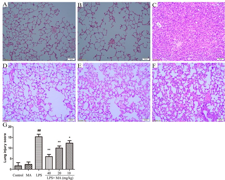
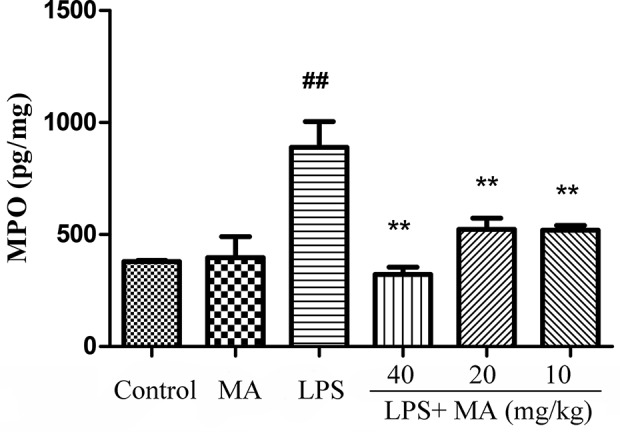
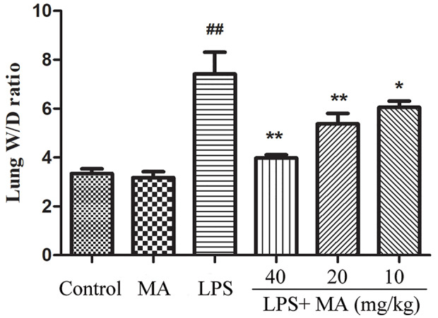
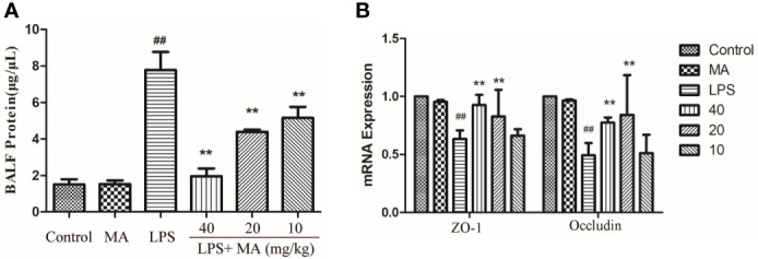
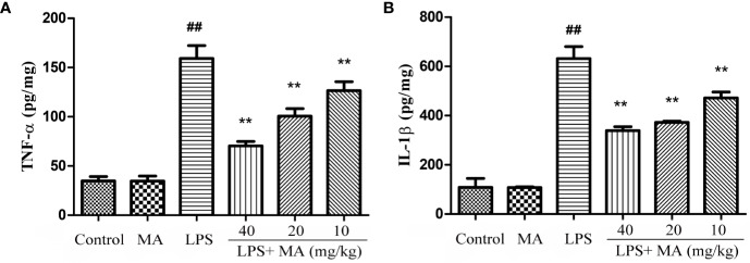
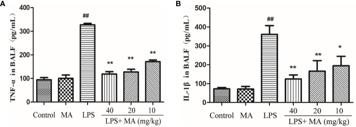
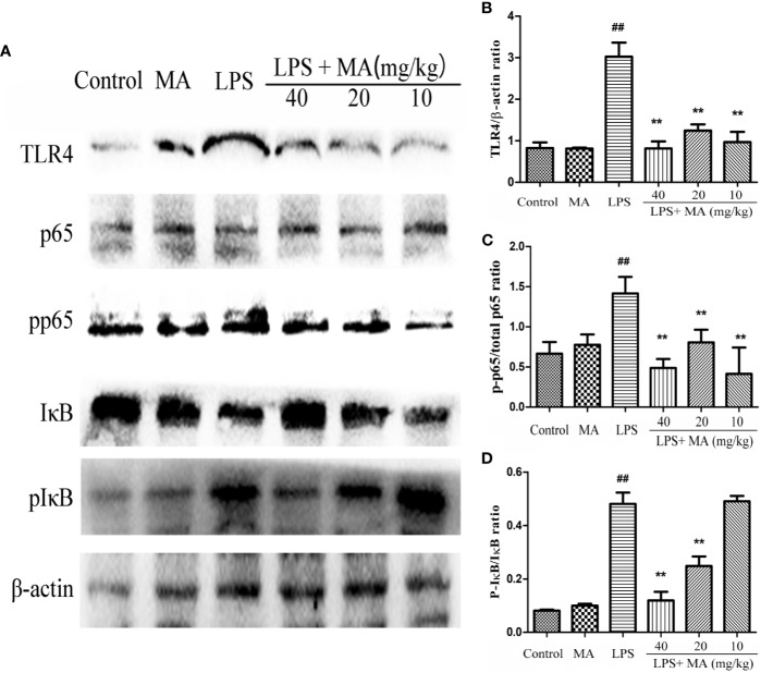
Madecassoside (MA), a crucial ingredient of Centella asiatica, has been reported to exhibit a variety of bioactivities, including anti-pulmonary fibrosis, and anti-inflammatory effects. Here we aimed to elucidate the protective activity and underlying mechanisms of MA with a focus on acute lung injury (ALI). The mice were received intratracheal of LPS to establish the ALI model and treated with MA. Then we measured the pathological changes by haematoxylin and eosin staining, the levels of pro-inflammatory cytokines and myeloperoxidase (MPO) by ELISA, the transcriptional level of tight junction proteins by qRT-PCR, as well as the expression of Toll-likereceptor4/Nuclear factor kappa-B (TLR4/NF-κB) pathway by western blot. The results indicated that MA significantly inhibited LPS-induced pathological damages, lung edema, MPO activity, and pro-inflammatory cytokines production. Furthermore, MA obviously reduced alveolar epithelium permeability which showed by reduced secretion of total proteins in the BALF and enhanced mRNA expression of tight junction as Occludin and zonula occludens-1 (ZO-1) comparing to LPS. Further research indicated that the LPS stimulation up-regulated the TLR4/NF-κB signaling pathway and the up-regulation were reversed by MA. In conclusion, these data supported that MA had protective effects on LPS-induced ALI. The therapeutic possibility may be associated with reducing the alveolar epithelium permeability and inflammatory response via repressing the activation of TLR4/NF-κB pathway.
Overview
The claims and statistics of this paper haven’t been checked yet.
Run the check — free, no account needed. It reads the open-access text and checks the sources it cites.
Integrity signals
Registry
Cites
Statistics
Figures
Verification
Claims the paper makes
The claims and statistics of this paper haven’t been checked yet.
Run the check — free, no account needed. It reads the open-access text and checks the sources it cites.
Claims other papers make about this one
Each is checked against this paper’s own text: does it say what the citing paper says it does?
The cited source says what the claim attributes to it.
In the citing paper
“madecassoside exerts anti‐inflammatory action by downregulating the TLR4/NF‐κB signalling pathway in LPS‐induced acute lung injury”
From Therapeutic Properties and Pharmacological Activities of Asiaticoside and Madecassoside: A Review (2023) · cites this paper as [114]
The cited source says what the claim attributes to it.
In the citing paper
“and also protects the lungs by repairing LPS‐induced pathological damages, lung oedema and alveolar epithelium integrity.”
From Therapeutic Properties and Pharmacological Activities of Asiaticoside and Madecassoside: A Review (2023) · cites this paper as [114]
Self-check
Percentages, sums and reported differences re-computed from the paper’s own numbers.
The self-check needs the paper’s full text.
Citations
Cites
Protective effects of madecassoside against Doxorubicin induced nephrotoxicity in vivo and in vitro
Su, Z., Ye, J., Qin, Z., & Ding, X. (2015) · Scientific Reports · doi:10.1038/srep18314 · Cited by 83
Research works printed but not linked: 26
- Akira, 2001, Toll-like receptors: critical proteins linking innate and acquired immunity, Nat. Immunol., 2, 675, 10.1038/90609 · DOI · Paper page
- Cao, 2010, Madecassoside suppresses LPS-induced TNF-alpha. production in cardiomyocytes through inhibition of ERK, p38, and NF-kappa B activity, Int. Immunopharmacol., 10, 723, 10.1016/j.intimp.2010.03.015 · DOI
- Chen, 2010, The value of the lipopolysaccharide-induced acute lung injury model in respiratory medicine, Expert Rev. Respir. Med., 4, 773, 10.1586/ers.10.71 · DOI
- Chen, 2019, Inhibition of myeloid differentiation factor 2 by baicalein protects against acute lung injury, Phytomedicine, 63, 152997, 10.1016/j.phymed.2019.152997 · DOI
- Fu, 2019, Protective effect of Cordyceps sinensis extract on lipopolysaccharide-induced acute lung injury in mice, Biosci. Rep., 39, 10.1042/BSR20190789 · DOI
- Herrero, 2019, Fas activation alters tight junction proteins in acute lung injury, Thorax, 74, 69, 10.1136/thoraxjnl-2018-211535 · DOI
- Hu, 2019, Aspirin-triggered resolvin D1 alleviates paraquat-induced acute lung injury in mice, Life Sci., 218, 38, 10.1016/j.lfs.2018.12.028 · DOI
- Jin, 2018, Osthole Protects against Acute Lung Injury by Suppressing NF-kappaB-Dependent Inflammation, Mediators Inflammation, 2018, 4934592, 10.1155/2018/4934592 · DOI
- Kristof, 1998, Role of inducible nitric oxide synthase in endotoxin-induced acute lung injury, Am. J. Respir. Crit. Care Med., 158, 1883, 10.1164/ajrccm.158.6.9802100 · DOI
- Li, 2018, Nobiletin-Ameliorated Lipopolysaccharide-Induced Inflammation in Acute Lung Injury by Suppression of NF-kappaB Pathway In Vivo and Vitro, Inflammation, 41, 996, 10.1007/s10753-018-0753-3 · DOI
- Lu, 2014, Madecassoside ameliorates bleomycin-induced pulmonary fibrosis in mice by downregulating collagen deposition, Phytother. Res., 28, 1224, 10.1002/ptr.5120 · DOI
- Maia, 2019, Effects of Obesity on Pulmonary Inflammation and Remodeling in Experimental Moderate Acute Lung Injury, Front. Immunol., 10, 10.3389/fimmu.2019.01215 · DOI
- Markopoulos, 2018, Roles of NF-kappa B Signaling in the Regulation of miRNAs Impacting on Inflammation in Cancer, Biomedicines, 6, 10.3390/biomedicines6020040 · DOI
- Meng, 2018, The protective effect of dexmedetomidine on LPS-induced acute lung injury through the HMGB1-mediated TLR4/NF-kappa B and PI3K/Akt/mTOR pathways, Mol. Immunol., 94, 7, 10.1016/j.molimm.2017.12.008 · DOI
- Miao, 2007, Evaluation of the changes of immune cells during lipopolysaccharide-induced mastitis in rats, Cytokine, 40, 135, 10.1016/j.cyto.2007.08.012 · DOI
- Qin, 2019, LL-37 and its analog FF/CAP18 attenuate neutrophil migration in sepsis-induced acute lung injury, J. Cell Biochem., 120, 4863, 10.1002/jcb.27641 · DOI
- Rittirsch, 2008, Acute lung injury induced by lipopolysaccharide is independent of complement activation, J. Immunol., 180, 7664, 10.4049/jimmunol.180.11.7664 · DOI
- Sasmita, 2018, Madecassoside activates anti-neuroinflammatory mechanisms by inhibiting lipopolysaccharide-induced microglial inflammation, Int. J. Mol. Med., 41, 3033, 10.3892/ijmm.2018.3479 · DOI
- Tan, 2018, Protective Effect of 2-Hydroxymethyl Anthraquinone from Hedyotis diffusa Willd in Lipopolysaccharide-Induced Acute Lung Injury Mediated by TLR4-NF-kappaB Pathway, Inflammation, 41, 2136, 10.1007/s10753-018-0857-9 · DOI
- Tian, 2019, The protective effect of the flavonoid fraction of Abutilon theophrasti Medic. leaves on LPS-induced acute lung injury in mice via the NF-kappaB and MAPK signalling pathways, BioMed. Pharmacother., 109, 1024, 10.1016/j.biopha.2018.10.197 · DOI
- Togbe, 2007, Toll-like receptor and tumour necrosis factor dependent endotoxin-induced acute lung injury, Int. J. Exp. Pathol., 88, 387, 10.1111/j.1365-2613.2007.00566.x · DOI
- Wang, 2018, Madecassoside prevents acute liver failure in LPS/D-GalN-induced mice by inhibiting p38/NF-kappa B and activating Nrf2/HO-1 signaling, Biomed. Pharmacother., 103, 1137, 10.1016/j.biopha.2018.04.162 · DOI · Paper page
- Xia, 2009, Phosphorylation of p53 by IkappaB kinase 2 promotes its degradation by beta-TrCP, Proc. Natl. Acad. Sci. U. S. A, 106, 2629, 10.1073/pnas.0812256106 · DOI
- Xia, 2016, Madecassoside ameliorates bleomycin-induced pulmonary fibrosis in mice through promoting the generation of hepatocyte growth factor via PPAR-gamma in colon, Br. J. Pharmacol., 173, 1219, 10.1111/bph.13421 · DOI
- Yang, 2019, Reverse-D-4F improves endothelial progenitor cell function and attenuates LPS-induced acute lung injury, Respir. Res., 20, 131, 10.1186/s12931-019-1099-6 · DOI
- Ye, 2019, ACE2 exhibits protective effects against LPS-induced acute lung injury in mice by inhibiting the LPS-TLR4 pathway, Exp. Mol. Pathol., 113, 104350, 10.1016/j.yexmp.2019.104350 · DOI
Cited by
Therapeutic Properties and Pharmacological Activities of Asiaticoside and Madecassoside: A Review
Bandopadhyay, S., Mandal, S., Ghorai, M., Jha, N. K., Kumar, M., Radha, Ghosh, A., Proćków, J., Pérez de la Lastra, J. M., & Dey, A. (2023) · Journal of Cellular and Molecular Medicine · doi:10.1111/jcmm.17635 · Cited by 220
Metadata
- Authors
- Peng, L.-Y., Shi, H.-T., Yuan, M., Li, J.-H., Song, K., Huang, J.-N., Yi, P.-F., Shen, H.-Q., & Fu, B.-D.
- Affiliations
- Jilin University (CN)
- Corresponding author
- Fu, Ben-Dong
- Venue
- Frontiers in Pharmacology
- Publisher
- Frontiers Media SA
- Published
- 2020-06-05
- Volume
- 11
- Pages
- 807-807
- ISSN
- 1663-9812 (ISSN-L)
- PMC
- PMC7289980
- OpenAlex
- W3033663687
- Cited by
- 26
- References
- 1
- Citations by year
- 2026: 3 · 2025: 5 · 2024: 3 · 2023: 5 · 2022: 5 · 2021: 5
- FWCI
- 2.11
- Type
- journal article
- Language
- en
- Open access
- gold
- doi.org
- Licence
- Locations
- Frontiers in Pharmacology (publishedVersion, cc-by)
- PubMed Central (publishedVersion, cc-by)
- PubMed (publishedVersion)
- DOAJ (DOAJ: Directory of Open Access Journals) (submittedVersion, cc-by-sa)
- Funders
- National Natural Science Foundation of China (201403051, 31972724, 31372470)
- Special Fund for Agro-scientific Research in the Public Interest (201403051)
- Topics
- Health Sciences › Medicine › Pulmonary and Respiratory Medicine › Respiratory Support and Mechanisms
- Life Sciences › Immunology and Microbiology › Immunology › Immune Response and Inflammation
- Health Sciences › Medicine › Complementary and alternative medicine › Natural Compounds in Disease Treatment
- Keywords
- madecassoside, Centella asiatica, acute lung injury, lipopolysaccharide, TLR4/NF-κB signaling, tight junction proteins, occludin, ZO-1, alveolar epithelium, inflammatory cytokines, myeloperoxidase, pulmonary edema, anti-inflammatory activity
About these checks
Registry, retraction, Cites and statistics results are computed, not judged, with one exception: a printed reference that no index lists is read once by a model before it counts as a citation error. Claims are extracted by a model and kept only when their quote appears verbatim in the text; each cited source is then checked, with a model’s help, for whether it says what the claim relies on. Those verdicts can be wrong — read the claim’s quote and the cited source before relying on one.
Statistics and arithmetic are recomputed, not judged.
Confidence bands are shown only for fields calibrated on our annotated test set. None is calibrated yet, so no band is shown.
What counts as a claim ›How we measure confidence ›How we check this ›
Are you an author, or do you see a problem with this page? Request a review
How to cite
Cite CoChat for the checks on this page, and the paper for its own findings.
- This page
- CoChat. (2026). Citation check: Madecassoside Protects Against LPS-Induced Acute Lung Injury via Inhibiting TLR4/NF-κB Activation and Blood-Air Barrier Permeability. https://app.cochat.ai/papers/10.3389/fphar.2020.00807
- The paper
- Peng, L.-Y., Shi, H.-T., Yuan, M., Li, J.-H., Song, K., Huang, J.-N., Yi, P.-F., Shen, H.-Q., & Fu, B.-D. (2020). Madecassoside Protects Against LPS-Induced Acute Lung Injury via Inhibiting TLR4/NF-κB Activation and Blood-Air Barrier Permeability. Frontiers in Pharmacology. https://doi.org/10.3389/fphar.2020.00807
Data sources
Bibliographic data: Semantic Scholar, OpenAlex, Crossref.
Contains information from the Semantic Scholar Academic Graph Datasets, made available under the ODC Attribution License (ODC-BY 1.0).
Semantic Scholar’s data is described in: Kinney, R., et al. (2023). The Semantic Scholar Open Data Platform. arXiv:2301.10140.
