

The DOI matches its Crossref record; 3 of the 3 references it cites were found in a registry or an index.
Di-Tyrosine Crosslinking and NOX4 Expression as Oxidative Pathological Markers in the Lungs of Patients with Idiopathic Pulmonary Fibrosis
Abstract
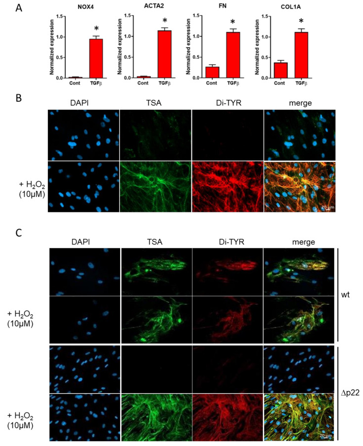
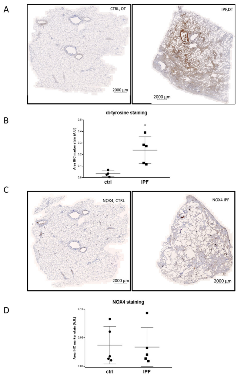
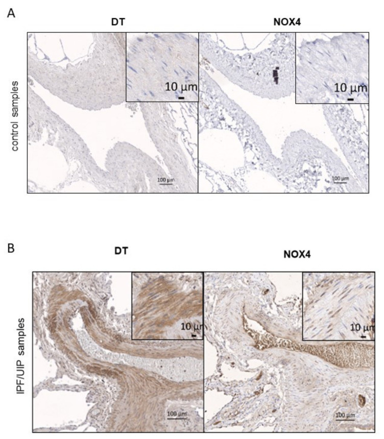
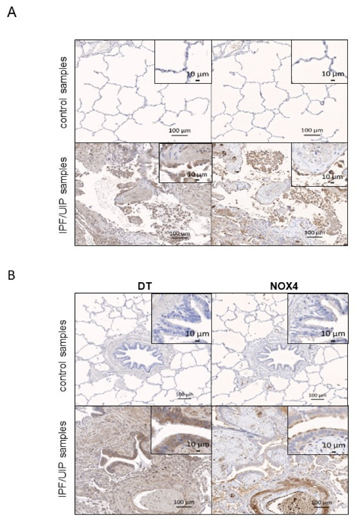
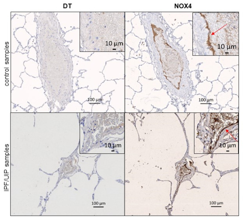
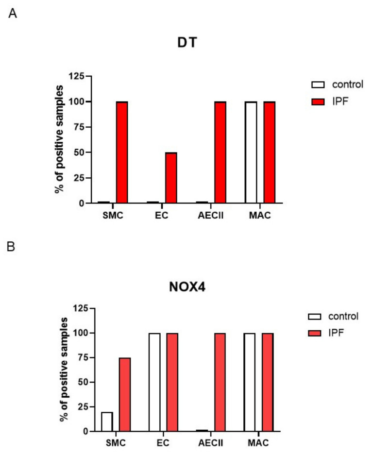
Idiopathic pulmonary fibrosis (IPF) is a noninflammatory progressive lung disease. Oxidative damage is a hallmark of IPF, but the sources and consequences of oxidant generation in the lungs are unclear. In this study, we addressed the link between the H2O2-generating enzyme NADPH oxidase 4 (NOX4) and di-tyrosine (DT), an oxidative post-translational modification in IPF lungs. We performed immunohistochemical staining for DT and NOX4 in pulmonary tissue from patients with IPF and controls using validated antibodies. In the healthy lung, DT showed little or no staining and NOX4 was mostly present in normal vascular endothelium. On the other hand, both markers were detected in several cell types in the IPF patients, including vascular smooth muscle cells and epithelium (bronchial cells and epithelial cells type II). The link between NOX4 and DT was addressed in human fibroblasts deficient for NOX4 activity (mutation in the CYBA gene). Induction of NOX4 by Transforming growth factor beta 1 (TGFβ1) in fibroblasts led to moderate DT staining after the addition of a heme-containing peroxidase in control cells but not in the fibroblasts deficient for NOX4 activity. Our data indicate that DT is a histological marker of IPF and that NOX4 can generate a sufficient amount of H2O2 for DT formation in vitro.
Overview
The claims and statistics of this paper haven’t been checked yet.
Run the check — free, no account needed. It reads the open-access text and checks the sources it cites.
Integrity signals
Registry
Cites
Statistics
Figures
Verification
Claims the paper makes
The claims and statistics of this paper haven’t been checked yet.
Run the check — free, no account needed. It reads the open-access text and checks the sources it cites.
Claims other papers make about this one
Each is checked against this paper’s own text: does it say what the citing paper says it does?
In the citing paper
“Elevated levels of dityrosine have been reported to be linked to inflammatory respiratory disease (Blaskovic et al., 2021;Pennathur et al., 2016).”
From Airborne microplastic exposure and urinary oxidative damage biomarkers in populations living in traffic-congested areas (2026) · cites this paper as Blaskovic et al., 2021
Self-check
Percentages, sums and reported differences re-computed from the paper’s own numbers.
The self-check needs the paper’s full text.
Citations
Cites
NADPH oxidase-4 mediates myofibroblast activation and fibrogenic responses to lung injury
Hecker, L., Vittal, R., Jones, T., Jagirdar, R., Luckhardt, T. R., Horowitz, J. C., Pennathur, S., Martinez, F. J., & Thannickal, V. J. (2009) · Nature Medicine · doi:10.1038/nm.2005 · Cited by 852
Wells, R. G. (2013) · Biochimica et Biophysica Acta (BBA) - Molecular Basis of Disease · doi:10.1016/j.bbadis.2013.02.007 · Cited by 384
Pennathur, S., Vivekanandan-Giri, A., Locy, M. L., Kulkarni, T., Zhi, D., Zeng, L., Byun, J., de Andrade, J. A., & Thannickal, V. J. (2016) · American Journal of Respiratory and Critical Care Medicine · doi:10.1164/rccm.201505-0992oc · Cited by 36
Research works printed but not linked: 31
- Wolters, 2014, Pathogenesis of Idiopathic Pulmonary Fibrosis, Annu. Rev. Pathol. Mech. Dis., 9, 157, 10.1146/annurev-pathol-012513-104706 · DOI
- Shumar, J., Chandel, A., and King, C. (2021). Antifibrotic Therapies and Progressive Fibrosing Interstitial Lung Disease (PF-ILD): Building on INBUILD. J. Clin. Med., 10. · DOI
- Betensley, A., Sharif, R., and Karamichos, D. (2016). A Systematic Review of the Role of Dysfunctional Wound Healing in the Patho-genesis and Treatment of Idiopathic Pulmonary Fibrosis. J. Clin. Med., 6. · DOI
- Veith, 2019, Redox Imbalance in Idiopathic Pulmonary Fibrosis: A Role for Oxidant Cross-Talk Between NADPH Oxidase Enzymes and Mitochondria, Antioxid. Redox Signal., 31, 1092, 10.1089/ars.2019.7742 · DOI
- Carnesecchi, 2011, A key role for NOX4 in epithelial cell death during development of lung fibrosis, Antioxid. Redox Signal., 15, 607, 10.1089/ars.2010.3829 · DOI · Paper page
- Bedard, 2007, The NOX Family of ROS-Generating NADPH Oxidases: Physiology and Pathophysiology, Physiol. Rev., 87, 245, 10.1152/physrev.00044.2005 · DOI · Paper page
- Sies, 2020, Reactive oxygen species (ROS) as pleiotropic physiological signalling agents, Nat. Rev. Mol. Cell Biol., 21, 363, 10.1038/s41580-020-0230-3 · DOI · Paper page
- Geiszt, 2019, The Relationship of NADPH Oxidases and Heme Peroxidases: Fallin’ in and Out, Front. Immunol., 10, 394, 10.3389/fimmu.2019.00394 · DOI
- Aeschbach, 1976, Formation of dityrosine cross-links in proteins by oxidation of tyrosine residues, Biochim. Biophys. Acta Protein Struct., 439, 292, 10.1016/0005-2795(76)90064-7 · DOI
- Wong, 2004, The Oxidative Burst at Fertilization Is Dependent upon Activation of the Dual Oxidase Udx1, Dev. Cell, 7, 801, 10.1016/j.devcel.2004.10.014 · DOI
- Hensley, 1998, Electrochemical analysis of protein nitrotyrosine and dityrosine in the Alzheimer brain indicates region-specific accumulation, J. Neurosci., 18, 8126, 10.1523/JNEUROSCI.18-20-08126.1998 · DOI
- Biasetti, 2016, The involvement of dityrosine crosslinking in alpha-synuclein assembly and deposition in Lewy Bodies in Parkinson’s disease, Sci. Rep., 6, 39171, 10.1038/srep39171 · DOI
- Leeuwenburgh, 1997, Mass Spectrometric Quantification of Markers for Protein Oxidation by Tyrosyl Radical, Copper, and Hydroxyl Radical in Low Density Lipoprotein Isolated from Human Atherosclerotic Plaques, J. Biol. Chem., 272, 3520, 10.1074/jbc.272.6.3520 · DOI
- Locy, 2020, Oxidative cross-linking of fibronectin confers protease resistance and inhibits cellular migration, Sci. Signal., 13, eaau2803, 10.1126/scisignal.aau2803 · DOI
- Brault, 2014, Optimized Generation of Functional Neutrophils and Macrophages from Patient-Specific Induced Pluripotent Stem Cells:Ex VivoModels of X0-Linked, AR220- and AR470- Chronic Granulomatous Diseases, BioRes. Open Access, 3, 311, 10.1089/biores.2014.0045 · DOI
- Prior, 2016, CRISPR/Cas9-mediated knockout of p22phox leads to loss of Nox1 and Nox4, but not Nox5 activity, Redox Biol., 9, 287, 10.1016/j.redox.2016.08.013 · DOI
- Vandesompele, 2002, Accurate normalization of real-time quantitative RT-PCR data by geometric averaging of multiple in-ternal control genes, Genome Biol., 3, RESEARCH0034, 10.1186/gb-2002-3-7-research0034 · DOI · Paper page
- Weinberg, R.A., and Comb, M.J. (2015). Chapter 12: Immunohistochemistry (IHC) in CST Guide: Pathways & Protocols, Cell Signaling Technology, Inc.. [1st ed.].
- Diebold, 2019, Guidelines for the Detection of NADPH Oxidases by Immunoblot and RT-qPCR, Methods Mol. Biol., 1982, 191, 10.1007/978-1-4939-9424-3_12 · DOI
- Augsburger, 2019, Pharmacological characterization of the seven human NOX isoforms and their inhibitors, Redox Biol., 26, 101272, 10.1016/j.redox.2019.101272 · DOI
- Rajaram, 2019, Tubular NOX4 expression decreases in chronic kidney disease but does not modify fibrosis evolution, Redox Biol., 26, 101234, 10.1016/j.redox.2019.101234 · DOI
- Kato, 2000, Immunochemical Detection of Protein Dityrosine in Atherosclerotic Lesion of Apo-E-Deficient Mice Using a Novel Monoclonal Antibody, Biochem. Biophys. Res. Commun., 275, 11, 10.1006/bbrc.2000.3265 · DOI
- Larios, 2001, Oxidative protein cross-linking reactions involving L-tyrosine in transforming growth factor-beta1-stimulated fibroblasts, J. Biol. Chem., 276, 17437, 10.1074/jbc.M100426200 · DOI
- Woods, 2003, Detection of HOCl-mediated protein oxidation products in the extracellular matrix of human atherosclerotic plaques, Biochem. J., 370, 729, 10.1042/bj20021710 · DOI
- Maher, 2019, Antifibrotic therapy for idiopathic pulmonary fibrosis: Time to treat, Respir. Res., 20, 1, 10.1186/s12931-019-1161-4 · DOI
- Mora, 2017, Emerging therapies for idiopathic pulmonary fibrosis, a progressive age-related disease, Nat. Rev. Drug Discov., 16, 755, 10.1038/nrd.2017.170 · DOI
- Modarressi, 2016, NADPH oxidase 4 deficiency leads to impaired wound repair and reduced dityrosine-crosslinking, but does not affect myofibroblast formation, Free. Radic. Biol. Med., 96, 374, 10.1016/j.freeradbiomed.2016.04.194 · DOI
- Luxen, 2009, Heterodimerization controls localization of Duox-DuoxA NADPH oxidases in airway cells, J. Cell Sci., 122, 1238, 10.1242/jcs.044123 · DOI
- Forteza, 2005, Regulated hydrogen peroxide production by Duox in human airway epithelial cells, Am. J. Respir. Cell Mol. Biol., 32, 462, 10.1165/rcmb.2004-0302OC · DOI
- Sarr, 2018, Antimicrobial actions of dual oxidases and lactoperoxidase, J. Microbiol., 56, 373, 10.1007/s12275-018-7545-1 · DOI · Paper page
- Louzada, 2021, NADPH oxidase DUOX1 sustains TGF-beta1 signalling and promotes lung fibrosis, Eur. Respir. J., 57, 1901949, 10.1183/13993003.01949-2019 · DOI
Cited by
Sorted by citations
Navasumrit, P., Chompoobut, C., Rattanayut, P., Kuntinuguntanon, A., Waeya, S., Kanitwithayanun, J., & Ruchirawat, M. (2026) · Journal of Hazardous Materials Advances · doi:10.1016/j.hazadv.2026.101571
Metadata
- Authors
- Blaskovic, S., Donati, Y., Ruchonnet-Metrailler, I., Seredenina, T., Krause, K.-H., Pache, J.-C., Adler, D., Barazzone-Argiroffo, C., & Jaquet, V.
- Affiliations
- Department of Pediatrics, Gynecology and Obstetrics, Children’s Hospital, 1211 Geneva, Switzerland
- Department of Pathology and Immunology, Medical School, University of Geneva, 1211 Geneva, Switzerland
- Service of Clinical Pathology, Department of Pathology and Immunology, Medical School, University of Geneva, 1211 Geneva, Switzerland
- Division of Pulmonary Diseases, Department of Medicine, Geneva University Hospitals, 1211 Geneva, Switzerland
- Venue
- Antioxidants
- Publisher
- MDPI AG
- Published
- 2021-11-18
- Volume
- 10
- Issue
- 11
- Pages
- 1833
- ISSN
- 2076-3921
- PMC
- PMC8615037
- Cited by
- 11
- References
- 3
- Language
- en
- Funders
- Swiss national science foundation (310030-159500/1)
About these checks
Registry, retraction, Cites and statistics results are computed, not judged, with one exception: a printed reference that no index lists is read once by a model before it counts as a citation error. Claims are extracted by a model and kept only when their quote appears verbatim in the text; each cited source is then checked, with a model’s help, for whether it says what the claim relies on. Those verdicts can be wrong — read the claim’s quote and the cited source before relying on one.
Statistics and arithmetic are recomputed, not judged.
Confidence bands are shown only for fields calibrated on our annotated test set. None is calibrated yet, so no band is shown.
What counts as a claim ›How we measure confidence ›How we check this ›
Are you an author, or do you see a problem with this page? Request a review
