

The DOI matches its Crossref record; 229 of the 232 references it cites were found in a registry or an index.
Neuroimaging of Mouse Models of Alzheimer’s Disease
Open accessAbstract
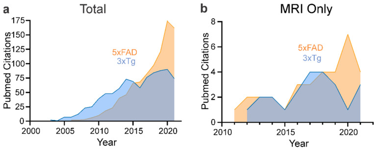
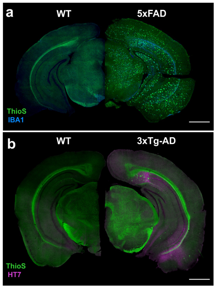
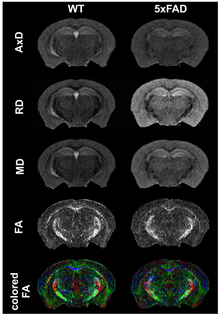
Magnetic resonance imaging (MRI) and positron emission tomography (PET) have made great strides in the diagnosis and our understanding of Alzheimer’s Disease (AD). Despite the knowledge gained from human studies, mouse models have and continue to play an important role in deciphering the cellular and molecular evolution of AD. MRI and PET are now being increasingly used to investigate neuroimaging features in mouse models and provide the basis for rapid translation to the clinical setting. Here, we provide an overview of the human MRI and PET imaging landscape as a prelude to an in-depth review of preclinical imaging in mice. A broad range of mouse models recapitulate certain aspects of the human AD, but no single model simulates the human disease spectrum. We focused on the two of the most popular mouse models, the 3xTg-AD and the 5xFAD models, and we summarized all known published MRI and PET imaging data, including contrasting findings. The goal of this review is to provide the reader with broad framework to guide future studies in existing and future mouse models of AD. We also highlight aspects of MRI and PET imaging that could be improved to increase rigor and reproducibility in future imaging studies.
Overview
The claims and statistics of this paper haven’t been checked yet.
Run the check — free, no account needed. It reads the open-access text and checks the sources it cites.
Integrity signals
Registry
Cites
Statistics
Figures
Verification
Claims the paper makes
The claims and statistics of this paper haven’t been checked yet.
Run the check — free, no account needed. It reads the open-access text and checks the sources it cites.
Claims other papers make about this one
Each is checked against this paper’s own text: does it say what the citing paper says it does?
The source backs part of the claim, not all of it — or backs it for a different population, condition or comparison.
In the citing paper
“Alongside the progression of AD pathology, age-related brain atrophy and myelin loss are observed on advanced high-field MRI (Ni, 2021; Jullienne et al., 2022; Bergamino et al., 2023).”
From Multimodal MRI combined with RNA sequencing reveals pathological signatures in the 9-month-old 3×Tg-AD mouse brain (2026) · cites this paper as Jullienne et al., 2022
Experimental — accuracy not yet measured
Self-check
Percentages, sums and reported differences re-computed from the paper’s own numbers.
The self-check needs the paper’s full text.
Citations
Cites
NIA‐AA Research Framework: Toward a biological definition of Alzheimer's disease
Jack, C. R., Bennett, D. A., Blennow, K., Carrillo, M. C., Dunn, B., Haeberlein, S. B., Holtzman, D. M., Jagust, W., Jessen, F., Karlawish, J., et al. (2018) · Alzheimer's & Dementia · doi:10.1016/j.jalz.2018.02.018 · Cited by 10,537
Scheltens, P., De Strooper, B., Kivipelto, M., Holstege, H., Chételat, G., Teunissen, C. E., Cummings, J., & van der Flier, W. M. (2021) · The Lancet · doi:10.1016/s0140-6736(20)32205-4 · Cited by 5,465
Imaging brain amyloid in Alzheimer's disease with Pittsburgh Compound‐B
Klunk, W. E., Engler, H., Nordberg, A., Wang, Y., Blomqvist, G., Holt, D. P., Bergström, M., Savitcheva, I., Huang, G., Estrada, S., et al. (2004) · Annals of Neurology · doi:10.1002/ana.20009 · Cited by 4,496
Triple-Transgenic Model of Alzheimer’s Disease with Plaques and Tangles
Oddo, S., Caccamo, A., Shepherd, J. D., Murphy, M. P., Golde, T. E., Kayed, R., Metherate, R., Mattson, M. P., Akbari, Y., & LaFerla, F. M. (2003) · Neuron · doi:10.1016/s0896-6273(03)00434-3 · Cited by 4,126
Oakley, H., Cole, S. L., Logan, S., Maus, E., Shao, P., Craft, J., Guillozet-Bongaarts, A., Ohno, M., Disterhoft, J., Van Eldik, L., et al. (2006) · The Journal of Neuroscience · doi:10.1523/jneurosci.1202-06.2006 · Cited by 3,530 · cited 3×
“Frank amyloid deposition at 9 months of age results in neurodegeneration accompanied by neuronal loss [60].”
BackgroundMethodInfluentialNeuropathological alterations in Alzheimer disease
Serrano-Pozo, A., Frosch, M. P., Masliah, E., & Hyman, B. T. (2011) · Cold Spring Harbor Perspectives in Medicine · doi:10.1101/cshperspect.a006189 · Cited by 3,345
Bassett, D. S., & Bullmore, E. (2006) · The Neuroscientist · doi:10.1177/1073858406293182 · Cited by 2,600
2021 Alzheimer's disease facts and figures
Anonymous (2021) · Alzheimer s & Dementia · doi:10.1002/alz.12328 · Cited by 2,339
Deaths: Leading Causes for 2017.
Heron, M. P. (2019) · PubMed · Cited by 2,108
Jankowsky, J. L., Fadale, D. J., Anderson, J., Xu, G. M., Gonzales, V., Jenkins, N. A., Copeland, N. G., Lee, M. K., Younkin, L. H., Wagner, S. L., et al. (2004) · Human Molecular Genetics · doi:10.1093/hmg/ddh019 · Cited by 1,575
Cited by
Sorted by citations
Alcohol Use Disorder and Dementia: A Review
Zahr, N. M. (2024) · Alcohol research · doi:10.35946/arcr.v44.1.03 · Cited by 42
Cortical cerebrovascular and metabolic perturbations in the 5xFAD mouse model of Alzheimer’s disease
Jullienne, A., Szu, J. I., Quan, R., Trinh, M. V., Norouzi, T., Noarbe, B. P., Bedwell, A. A., Eldridge, K., Persohn, S. A., Territo, P. R., et al. (2023) · Frontiers in Aging Neuroscience · doi:10.3389/fnagi.2023.1220036 · Cited by 30
Self-citationZeng, X., Cheung, S. K. K., Shi, M., Or, P. M. Y., Li, Z., Liu, J. Y. H., Ho, W. L.-H., Liu, T., Lu, K., Rudd, J. A., et al. (2023) · Journal of Neuroinflammation · doi:10.1186/s12974-023-02970-z · Cited by 28
Andrade-Guerrero, J., Rodríguez-Arellano, P., Barron-Leon, N., Orta‐Salazar, E., Ledesma-Alonso, C., Díaz-Cintra, S. Y., & Soto-Rojas, L. O. (2023) · Cells · doi:10.3390/cells12212531 · Cited by 28
Progressive Vascular Abnormalities in the Aging 3xTg-AD Mouse Model of Alzheimer’s Disease
Jullienne, A., Quan, R., Szu, J. I., Trinh, M. V., Behringer, E. J., & Obenaus, A. (2022) · Biomedicines · doi:10.3390/biomedicines10081967 · Cited by 23
Self-citationYoo, C.-H., Kim, J., Baek, H.-M., Chang, K.-A., & Choe, B.-Y. (2023) · International Journal of Molecular Sciences · doi:10.3390/ijms24065073 · Cited by 13
Age-dependent microstructure alterations in 5xFAD mice by high-resolution diffusion tensor imaging
Maharjan, S., Tsai, A. P., Lin, P. B., Ingraham, C., Jewett, M. R., Landreth, G. E., Oblak, A. L., & Wang, N. (2022) · Frontiers in Neuroscience · doi:10.3389/fnins.2022.964654 · Cited by 15
“Different MRI techniques have been used to study the brain structure and function of 5xFAD mice (Mlynarik et al., 2012; Igarashi et al., 2020; Jullienne et al., 2022).”
MethodLiang, C., Nguyen, G. A. H., Danh, T. B., Sandhu, A. K., Melkonyan, L. L., Syed, A. U., & Mukherjee, J. (2023) · Synapse · doi:10.1002/syn.22265 · Cited by 11
Characterization of cerebrovascular changes in Alzheimer’s disease mice by photoacoustic imaging
Li, X., Zhang, Z., Shi, H., & Zhou, F. (2024) · Journal of Innovative Optical Health Sciences · doi:10.1142/s179354582450007x · Cited by 9
Stapleton, M. C., Koch, S., Cortes, D. R. E., Wyman, S. K., Schwab, K. E., Mueller, S. G., McKennan, C. G., Boehm‐Sturm, P., & Wu, Y. L. (2023) · Frontiers in Neuroscience · doi:10.3389/fnins.2023.1183312 · Cited by 6
Chicote‐González, A., Solana‐Balaguer, J., Garcia‐Segura, P., Campoy‐Campos, G., Pérez, I. P., Pérez‐Navarro, E., Rodríguez, M. J., Alberch, J., Giralt, A., Sòria, G., et al. (2025) · Alzheimer s & Dementia · doi:10.1002/alz.70051 · Cited by 7
Wang, R., Li, J., Li, X., Guo, Y., Chen, P., & Tian, P. (2025) · Reviews in the Neurosciences · doi:10.1515/revneuro-2025-0013 · Cited by 7
WhatNIs N-ough for MRI-Based Animal Neuroimaging?
Grandjean, J., Lake, E. M. R., Pagani, M., & Mandino, F. (2024) · eNeuro · doi:10.1523/eneuro.0531-23.2024 · Cited by 4
Das, R. K., Sahoo, N., Roy, D., Nguyen, L., Rodrigo, H., Duttaroy, A. K., Fields, J. A., & Roy, U. (2026) · Journal of Neuroinflammation · doi:10.1186/s12974-026-03713-6 · Cited by 4
van den Broek, S. L., Sehlin, D., Andersen, J. V., Aldana, B. I., Beschörner, N., Nedergaard, M., Knudsen, G. M., Syvänen, S., & Herth, M. M. (2022) · Frontiers in Nuclear Medicine · doi:10.3389/fnume.2022.1001722 · Cited by 5
Sheppard, P. A. S., Oomen, C. A., Bussey, T. J., & Saksida, L. M. (2024) · eNeuro · doi:10.1523/eneuro.0120-24.2024 · Cited by 3
Afxenti, S., Bourdakou, M. M., Loizidou, E. M., Zachariou, M., Lambrianides, A., Pantzaris, M., & Spyrou, G. M. (2025) · Expert Review of Proteomics · doi:10.1080/14789450.2025.2584129 · Cited by 3
Copper Peptides in Regenerative Aesthetic Dermatology
Sarbaziha, R., & Goldberg, D. J. (2026) · Dermatological Reviews · doi:10.1002/der2.70067 · Cited by 4
Riffo-Lepe, N., González-Sanmiguel, J., Armijo‐Weingart, L., Saavedra, P., Hernandez, D., Ramos, G., Martín, L. S., & Aguayo, L. G. (2025) · Neural Regeneration Research · doi:10.4103/nrr.nrr-d-24-01052 · Cited by 2
Raikes, A. C., Bhattrai, A., Wang, T., Wiegand, J. L., & Brinton, R. D. (2026) · Frontiers in Neuroscience · doi:10.3389/fnins.2026.1843319 · Cited by 3
Ott, F. W., Sichler, M. E., Bouter, C., Enayati, M., Wiltfang, J., Bayer, T. A., Beindorff, N., Löw, M. J., & Bouter, Y. (2025) · Journal of Alzheimer s Disease Reports · doi:10.1177/25424823241306770 · Cited by 2
Sehlin, D., Aguilar, X., Cortés‐Canteli, M., Syvänen, S., & van den Broek, S. L. (2025) · EJNMMI Radiopharmacy and Chemistry · doi:10.1186/s41181-025-00383-2 · Cited by 1
Sha, L., Wu, M., Sun, S., Xie, Y., Liu, Y., Li, Z., Liu, R., Wanhua, G., Yan, S., Xu, B., et al. (2026) · Journal of Alzheimer s Disease · doi:10.1177/13872877261418164 · Cited by 1
Special Issue “Alzheimer’s Disease—115 Years after Its Discovery”
Cardoso, S., Carvalho, C., & Correia, S. C. (2024) · Biomedicines · doi:10.3390/biomedicines12030478 · Cited by 1
Celdran‐Bonafonte, D., O’Connell, K. A., Dieckhaus, L., Mocarski, E., Adras, N. M., Hutchinson, E. B., & Pacharinsak, C. (2025) · Journal of the American Association for Laboratory Animal Science · doi:10.30802/aalas-jaalas-24-086 · Cited by 1
A relaxation-diffusion MRI dataset of aging mouse brains at 9.4 Tesla
Ren, B., Wu, Y., Liu, Y., Bai, Y., Tao, Q., Feng, Y., & Zhang, X. (2025) · Scientific Data · doi:10.1038/s41597-025-06088-0 · Cited by 1
Li, Y., Tang, Z., Yao, M., Ran, Y., & Qiu, Z. (2026) · Neural Regeneration Research · doi:10.4103/nrr.nrr-d-25-00006
Exploring the Relation Between Aerobic Exercise, BDNF and Alzheimer’s Disease: A Research Protocol
Mahmood, R. (2022) · Undergraduate Research in Natural and Clinical Science and Technology (URNCST) Journal · doi:10.26685/urncst.427
Zeng, X., Cheung, S. K. K., Shi, M., Or, P. M. Y., Li, Z., Liu, J. Y. H., Ho, W. L.-H., Liu, T., Lu, K., Rudd, J. A., et al. (2024) · Figshare · doi:10.6084/m9.figshare.26648808
García, C. L., Anastassiadis, C., Urosevic, M., Park, M., Gallino, D., Devenyi, G. A., Tullo, S., Yee, Y., & Chakravarty, M. M. (2026) · eLife · doi:10.7554/elife.111433.1
García, C. L., Anastassiadis, C., Urosevic, M., Park, M., Gallino, D., Devenyi, G. A., Tullo, S., Yee, Y., & Chakravarty, M. M. (2026) · eLife · doi:10.7554/elife.111433
Gezginer, I., Karakatsani, M. E., Nanda, P., Chalasani, P., Kindler, D., Storz, R., Belau, M., Ni, R., Schratt, G. M., Deán‐Ben, X. L., et al. (2026) · Alzheimer s Research & Therapy · doi:10.1186/s13195-026-02109-1
Or, P., Yon, M., Narvaez, O., Sitnikova, V., Malm, T., Bouhrara, M., Sierra, A., Topgaard, D., & Benjamini, D. (2026) · bioRxiv (Cold Spring Harbor Laboratory) · doi:10.64898/2026.08.26.747377
Metadata
- Authors
- Jullienne, A., Trinh, M. V., & Obenaus, A.
- Affiliations
- University of California, Irvine (US)
- Department of Pediatrics, University of California, Irvine, CA 92697, USA
- Venue
- Biomedicines
- Publisher
- MDPI AG
- Published
- 2022-01-28
- Volume
- 10
- Issue
- 2
- Pages
- 305
- ISSN
- 2227-9059 (ISSN-L)
- PMC
- PMC8869427
- OpenAlex
- W4210462365
- Cited by
- 32
- References
- 232
- Citations by year
- 2026: 8 · 2025: 8 · 2024: 6 · 2023: 6 · 2022: 4
- FWCI
- 0.83
- Type
- journal article
- Language
- en
- Open access
- gold
- doi.org
- Licence
- Locations
- Biomedicines (publishedVersion, cc-by)
- PubMed Central (publishedVersion, cc-by)
- PubMed (publishedVersion)
- eScholarship (California Digital Library) (submittedVersion, cc-by)
- DOAJ (DOAJ: Directory of Open Access Journals) (submittedVersion, cc-by-sa)
- Funders
- National Institute on Aging (AG054345-01, AG054349-01)
- Topics
- Life Sciences › Neuroscience › Cellular and Molecular Neuroscience › Alzheimer's disease research and treatments
- Health Sciences › Medicine › Psychiatry and Mental health › Dementia and Cognitive Impairment Research
- Life Sciences › Neuroscience › Neurology › Neuroinflammation and Neurodegeneration Mechanisms
- Keywords
- Alzheimer's disease, mouse models of Alzheimer's disease, 3xTg-AD mice, 5xFAD mice, magnetic resonance imaging, positron emission tomography, imaging biomarkers
About these checks
Registry, retraction, Cites and statistics results are computed, not judged, with one exception: a printed reference that no index lists is read once by a model before it counts as a citation error. Claims are extracted by a model and kept only when their quote appears verbatim in the text; each cited source is then checked, with a model’s help, for whether it says what the claim relies on. Those verdicts can be wrong — read the claim’s quote and the cited source before relying on one.
Statistics and arithmetic are recomputed, not judged.
Confidence bands are shown only for fields calibrated on our annotated test set. None is calibrated yet, so no band is shown.
What counts as a claim ›How we measure confidence ›How we check this ›
Are you an author, or do you see a problem with this page? Request a review
