

The DOI matches its Crossref record; 229 of the 232 references it cites were found in a registry or an index.
Neuroimaging of Mouse Models of Alzheimer’s Disease
Open accessAbstract
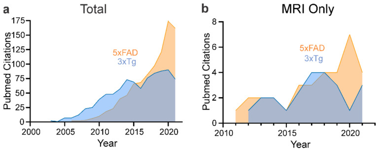
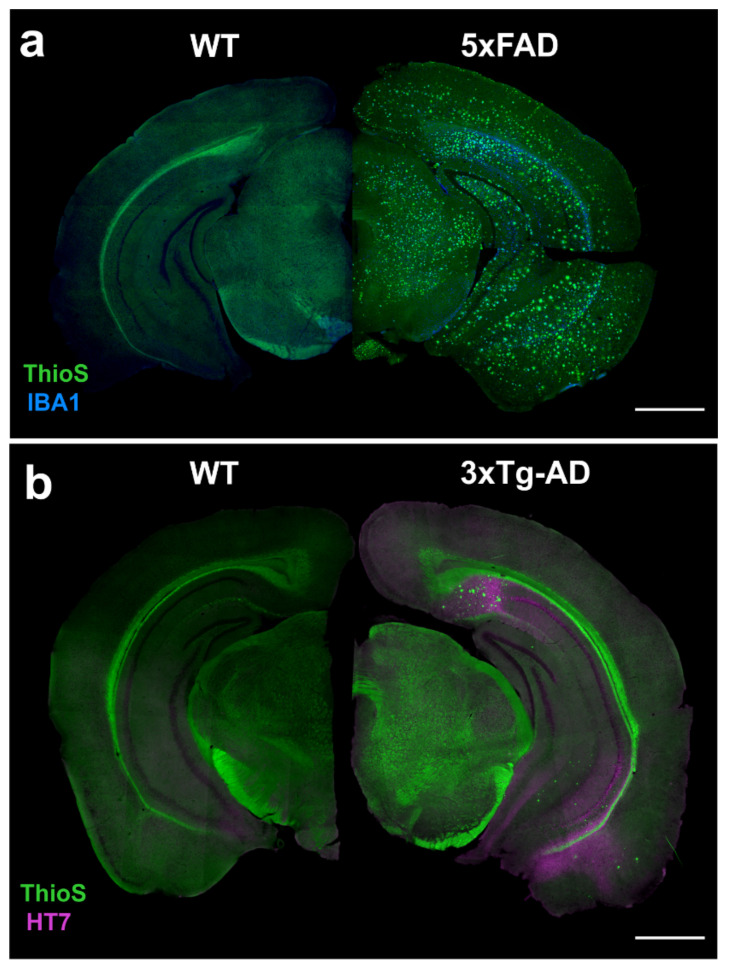
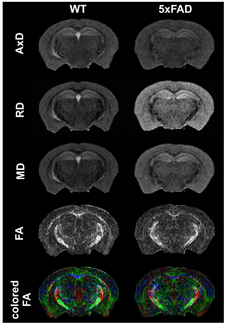
Magnetic resonance imaging (MRI) and positron emission tomography (PET) have made great strides in the diagnosis and our understanding of Alzheimer’s Disease (AD). Despite the knowledge gained from human studies, mouse models have and continue to play an important role in deciphering the cellular and molecular evolution of AD. MRI and PET are now being increasingly used to investigate neuroimaging features in mouse models and provide the basis for rapid translation to the clinical setting. Here, we provide an overview of the human MRI and PET imaging landscape as a prelude to an in-depth review of preclinical imaging in mice. A broad range of mouse models recapitulate certain aspects of the human AD, but no single model simulates the human disease spectrum. We focused on the two of the most popular mouse models, the 3xTg-AD and the 5xFAD models, and we summarized all known published MRI and PET imaging data, including contrasting findings. The goal of this review is to provide the reader with broad framework to guide future studies in existing and future mouse models of AD. We also highlight aspects of MRI and PET imaging that could be improved to increase rigor and reproducibility in future imaging studies.
Overview
The claims and statistics of this paper haven’t been checked yet.
Run the check — free, no account needed. It reads the open-access text and checks the sources it cites.
Integrity signals
Registry
Cites
Statistics
Figures
Verification
Claims the paper makes
The claims and statistics of this paper haven’t been checked yet.
Run the check — free, no account needed. It reads the open-access text and checks the sources it cites.
Claims other papers make about this one
Each is checked against this paper’s own text: does it say what the citing paper says it does?
The source backs part of the claim, not all of it — or backs it for a different population, condition or comparison.
In the citing paper
“Alongside the progression of AD pathology, age-related brain atrophy and myelin loss are observed on advanced high-field MRI (Ni, 2021; Jullienne et al., 2022; Bergamino et al., 2023).”
From Multimodal MRI combined with RNA sequencing reveals pathological signatures in the 9-month-old 3×Tg-AD mouse brain (2026) · cites this paper as Jullienne et al., 2022
Experimental — accuracy not yet measured
Self-check
Percentages, sums and reported differences re-computed from the paper’s own numbers.
The self-check needs the paper’s full text.
Citations
Cites
NIA‐AA Research Framework: Toward a biological definition of Alzheimer's disease
Jack, C. R., Bennett, D. A., Blennow, K., Carrillo, M. C., Dunn, B., Haeberlein, S. B., Holtzman, D. M., Jagust, W., Jessen, F., Karlawish, J., et al. (2018) · Alzheimer's & Dementia · doi:10.1016/j.jalz.2018.02.018 · Cited by 10,537
Scheltens, P., De Strooper, B., Kivipelto, M., Holstege, H., Chételat, G., Teunissen, C. E., Cummings, J., & van der Flier, W. M. (2021) · The Lancet · doi:10.1016/s0140-6736(20)32205-4 · Cited by 5,465
Imaging brain amyloid in Alzheimer's disease with Pittsburgh Compound‐B
Klunk, W. E., Engler, H., Nordberg, A., Wang, Y., Blomqvist, G., Holt, D. P., Bergström, M., Savitcheva, I., Huang, G., Estrada, S., et al. (2004) · Annals of Neurology · doi:10.1002/ana.20009 · Cited by 4,496
Triple-Transgenic Model of Alzheimer’s Disease with Plaques and Tangles
Oddo, S., Caccamo, A., Shepherd, J. D., Murphy, M. P., Golde, T. E., Kayed, R., Metherate, R., Mattson, M. P., Akbari, Y., & LaFerla, F. M. (2003) · Neuron · doi:10.1016/s0896-6273(03)00434-3 · Cited by 4,126
Oakley, H., Cole, S. L., Logan, S., Maus, E., Shao, P., Craft, J., Guillozet-Bongaarts, A., Ohno, M., Disterhoft, J., Van Eldik, L., et al. (2006) · The Journal of Neuroscience · doi:10.1523/jneurosci.1202-06.2006 · Cited by 3,530 · cited 3×
“Frank amyloid deposition at 9 months of age results in neurodegeneration accompanied by neuronal loss [60].”
BackgroundMethodInfluentialNeuropathological alterations in Alzheimer disease
Serrano-Pozo, A., Frosch, M. P., Masliah, E., & Hyman, B. T. (2011) · Cold Spring Harbor Perspectives in Medicine · doi:10.1101/cshperspect.a006189 · Cited by 3,345
Bassett, D. S., & Bullmore, E. (2006) · The Neuroscientist · doi:10.1177/1073858406293182 · Cited by 2,600
2021 Alzheimer's disease facts and figures
Anonymous (2021) · Alzheimer s & Dementia · doi:10.1002/alz.12328 · Cited by 2,339
Deaths: Leading Causes for 2017.
Heron, M. P. (2019) · PubMed · Cited by 2,108
Jankowsky, J. L., Fadale, D. J., Anderson, J., Xu, G. M., Gonzales, V., Jenkins, N. A., Copeland, N. G., Lee, M. K., Younkin, L. H., Wagner, S. L., et al. (2004) · Human Molecular Genetics · doi:10.1093/hmg/ddh019 · Cited by 1,575
Matthews, K. A., Xu, W., Gaglioti, A. H., Holt, J. B., Croft, J. B., Mack, D., & McGuire, L. C. (2019) · Alzheimer's & Dementia · doi:10.1016/j.jalz.2018.06.3063 · Cited by 1,124
Sex differences in Alzheimer disease — the gateway to precision medicine
for the Women’s Brain Project and the Alzheimer Precision Medicine Initiative, Ferretti, M. T., Iulita, M. F., Cavedo, E., Chiesa, P. A., Schumacher Dimech, A., Santuccione Chadha, A., Baracchi, F., Girouard, H., Misoch, S., et al. (2018) · Nature Reviews Neurology · doi:10.1038/s41582-018-0032-9 · Cited by 1,009
Tau positron emission tomographic imaging in aging and early Alzheimer disease
Johnson, K. A., Schultz, A., Betensky, R. A., Becker, J. A., Sepulcre, J., Rentz, D., Mormino, E., Chhatwal, J., Amariglio, R., Papp, K., et al. (2016) · Annals of Neurology · doi:10.1002/ana.24546 · Cited by 986
Positron Emission Tomography in Evaluation of Dementia
Silverman, D. H. S., Small, G. W., Chang, C. Y., Lu, C. S., de Aburto, M. A. K., Chen, W., Czernin, J. G., Rapoport, S. I., Pietrini, P., Alexander, G. E., et al. (2001) · JAMA · doi:10.1001/jama.286.17.2120 · Cited by 960
APP mouse models for Alzheimer’s disease preclinical studies.
Sasaguri, H., Nilsson, P., Hashimoto, S., Nagata, K., Saito, T., De Strooper, B., Hardy, J., Vassar, R., Winblad, B., & Saido, T. C. (2017) · The EMBO Journal · doi:10.15252/embj.201797397 · Cited by 793
Mesulam, M. M., Guillozet-Bongaarts, A. L., Shaw, P. L., Levey, A. I., Duysen, E. G., & Lockridge, O. (2002) · Neuroscience · doi:10.1016/s0306-4522(01)00613-3 · Cited by 662
CHANGES IN BRAIN CHOLINESTERASES IN SENILE DEMENTIA OF ALZHEIMER TYPE
Perry, E. K., Perry, R. H., Blessed, G., & Tomlinson, B. E. (1978) · Neuropathology and Applied Neurobiology · doi:10.1111/j.1365-2990.1978.tb00545.x · Cited by 560
Spread of pathological tau proteins through communicating neurons in human Alzheimer’s disease
Vogel, J. W., Iturria‐Medina, Y., Strandberg, O. T., Smith, R. A., Levitis, E., Evans, A. C., Hansson, O., Initiative, A. D. N., Weiner, M. W., Aisen, P., et al. (2020) · Nature Communications · doi:10.1038/s41467-020-15701-2 · Cited by 537
de Paula, V. D. J. R., Radanovic, M., Diniz, B. S., & Forlenza, O. V. (2012) · Sub-cellular biochemistry/Subcellular biochemistry · doi:10.1007/978-94-007-5416-4_14 · Cited by 466
Impact of animal handling on the results of 18F-FDG PET studies in mice.
Fueger, B. J., Czernin, J. G., Hildebrandt, I. J., Tran, C., Halpern, B. S., Stout, D., Phelps, M. E., & Weber, W. (2006) · PubMed · Cited by 455
Chen, M.-K., Mecca, A. P., Naganawa, M., Finnema, S. J., Toyonaga, T., Lin, S.-F., Najafzadeh, S., Ropchan, J., Lu, Y., McDonald, J. W., et al. (2018) · JAMA Neurology · doi:10.1001/jamaneurol.2018.1836 · Cited by 431
Mattsson-Carlgren, N., Andersson, E., Janelidze, S., Ossenkoppele, R., Insel, P., Strandberg, O., Zetterberg, H., Rosen, H. J., Rabinovici, G., Chai, X., et al. (2020) · Science Advances · doi:10.1126/sciadv.aaz2387 · Cited by 359
Holmes, C., Cairns, N. J., Lantos, P. L., & MANN, A. H. (1999) · The British Journal of Psychiatry · doi:10.1192/bjp.174.1.45 · Cited by 353
Schroeter, M. L., Stein, T., Maslowski, N. I., & Neumann, J. (2009) · NeuroImage · doi:10.1016/j.neuroimage.2009.05.037 · Cited by 348
Effectiveness of regional DTI measures in distinguishing Alzheimer’s disease, MCI, and normal aging
Nir, T. M., Jahanshad, N., Villalon-Reina, J. E., Toga, A. W., Jack, C. R., Weiner, M. W., & Thompson, P. M. (2013) · NeuroImage: Clinical · doi:10.1016/j.nicl.2013.07.006 · Cited by 348
The complexity of Alzheimer’s disease: an evolving puzzle
Ferrari, C., & Sorbi, S. (2021) · Physiological Reviews · doi:10.1152/physrev.00015.2020 · Cited by 343
The role of the medial prefrontal cortex in cognition, ageing and dementia
Jobson, D. D., Hase, Y., Clarkson, A. N., & Kalaria, R. N. (2021) · Brain Communications · doi:10.1093/braincomms/fcab125 · Cited by 338
Oblak, A. L., Lin, P. B., Kotredes, K. P., Pandey, R. S., Garceau, D., Williams, H. M., Uyar, A., O’Rourke, R., O’Rourke, S., Ingraham, C., et al. (2021) · Frontiers in Aging Neuroscience · doi:10.3389/fnagi.2021.713726 · Cited by 325
Sadleir, K. R., Kandalepas, P. C., Buggia-Prévot, V., Nicholson, D. A., Thinakaran, G., & Vassar, R. (2016) · Acta Neuropathologica · doi:10.1007/s00401-016-1558-9 · Cited by 294
Ibrahim, B., Suppiah, S., Ibrahim, N., Mohamad, M., Hassan, H. A., Nasser, N. S., & Saripan, M. I. (2021) · Human Brain Mapping · doi:10.1002/hbm.25369 · Cited by 272
Janelsins, M. C., Mastrangelo, M. A., Oddo, S., LaFerla, F. M., Federoff, H. J., & Bowers, W. J. (2005) · Journal of Neuroinflammation · doi:10.1186/1742-2094-2-23 · Cited by 260
Susceptibility-weighted Imaging: Technical Essentials and Clinical Neurologic Applications
Haller, S., Haacke, E. M., Thurnher, M. M., & Barkhof, F. (2021) · Radiology · doi:10.1148/radiol.2021203071 · Cited by 256
Early-Onset Familial Alzheimer's Disease (EOFAD)
Wu, L., Rosa‐Neto, P., Hsiung, G. R., Sadovnick, A. D., Masellis, M., Black, S. E., Jia, J., & Gauthier, S. G. (2012) · Canadian Journal of Neurological Sciences / Journal Canadien des Sciences Neurologiques · doi:10.1017/s0317167100013949 · Cited by 240
Colgan, N., Siow, B., O'Callaghan, J. M., Harrison, I. F., Wells, J. A., Holmes, H. E., Ismail, O., Richardson, S., Alexander, D. C., Collins, E. C., et al. (2016) · NeuroImage · doi:10.1016/j.neuroimage.2015.10.043 · Cited by 218
Redwine, J. M., Kosofsky, B. E., Jacobs, R. E., Games, D., Reilly, J. F., Morrison, J. H., Young, W. G., & Bloom, F. E. (2003) · Proceedings of the National Academy of Sciences · doi:10.1073/pnas.242746599 · Cited by 204
Marjańska, M., Curran, G. L., Wengenack, T. M., Henry, P., Bliss, R., Poduslo, J. F., Jack, C. R., Uǧurbil, K., & Garwood, M. (2005) · Proceedings of the National Academy of Sciences · doi:10.1073/pnas.0505513102 · Cited by 201
Recent developments on PET radiotracers for TSPO and their applications in neuroimaging
Zhang, L., Hu, K., Shao, T., Hou, L., Zhang, S., Ye, W., Josephson, L., Meyer, J. H., Zhang, M., Vasdev, N., et al. (2020) · Acta Pharmaceutica Sinica B · doi:10.1016/j.apsb.2020.08.006 · Cited by 195
McIntosh, A. L., Mela, V., Harty, C., Minogue, A. M., Costello, D. A., Kerskens, C., & Lynch, M. A. (2019) · Brain Pathology · doi:10.1111/bpa.12704 · Cited by 193
Functional imaging studies of episodic memory in Alzheimer's disease: a quantitative meta-analysis
Schwindt, G., & Black, S. E. (2008) · NeuroImage · doi:10.1016/j.neuroimage.2008.11.024 · Cited by 179
Yan, T., Wang, W., Liu, Y., Chen, K., Chen, R., & Han, Y. (2018) · Theranostics · doi:10.7150/thno.23772 · Cited by 178
Neuroprotective effects of CD4+CD25+Foxp3+ regulatory T cells in a 3xTg-AD Alzheimer's disease model
Baek, H., Ye, M., Kang, G., Lee, C., Lee, G., Choi, D. B., Jung, J., Kim, H., Lee, S., Kim, J. S., et al. (2016) · Oncotarget · doi:10.18632/oncotarget.12469 · Cited by 174
A multiomics approach to heterogeneity in Alzheimer’s disease: focused review and roadmap
Badhwar, A., McFall, G. P., Sapkota, S., Black, S. E., Chertkow, H. M., Duchesne, S., Masellis, M., Li, L., Dixon, R. A., & Bellec, P. (2019) · Brain · doi:10.1093/brain/awz384 · Cited by 164
Artificial Intelligence for Alzheimer’s Disease: Promise or Challenge?
Fabrizio, C., Termine, A., Caltagirone, C., & Sancesario, G. M. (2021) · Diagnostics · doi:10.3390/diagnostics11081473 · Cited by 163
Gong, N.-J., Dibb, R., Bulk, M., van der Weerd, L., & Liu, C. (2019) · NeuroImage · doi:10.1016/j.neuroimage.2019.02.019 · Cited by 162
Dedeoglu, A., Choi, J., Cormier, K., Kowall, N. W., & Jenkins, B. G. (2004) · Brain Research · doi:10.1016/j.brainres.2004.02.079 · Cited by 161
In vivo axonal transport rates decrease in a mouse model of Alzheimer's disease
Smith, K. D. B., Kallhoff, V., Zheng, H., & Pautler, R. G. (2007) · NeuroImage · doi:10.1016/j.neuroimage.2007.01.046 · Cited by 153
Lau, J. C., Lerch, J. P., Sled, J. G., Henkelman, R. M., Evans, A. C., & Bedell, B. J. (2008) · NeuroImage · doi:10.1016/j.neuroimage.2008.04.252 · Cited by 152
Imaging of glutamate neurotransmitter alterations in Alzheimer's disease
Haris, M., Nath, K., Cai, K., Singh, A. K., Crescenzi, R. L., Kogan, F., Verma, G., Reddy, S., Hariharan, H., Melhem, E. R., et al. (2012) · NMR in Biomedicine · doi:10.1002/nbm.2875 · Cited by 152
Reactive astrogliosis: A friend or foe in the pathogenesis of Alzheimer’s disease
Kumar, A., Fontana, I. C., & Nordberg, A. (2023) · Journal of Neurochemistry · doi:10.1111/jnc.15565 · Cited by 150
Beckmann, N., Schuler, A., Mueggler, T., Meyer, E. P., Wiederhold, K., Staufenbiel, M., & Krucker, T. (2003) · Journal of Neuroscience · doi:10.1523/jneurosci.23-24-08453.2003 · Cited by 149
Rodriguez‐Vieitez, E., Ni, R., Gulyás, B. Z., Tóth, M., Häggkvist, J., Halldin, C., Voytenko, L. P., Marutle, A., & Nordberg, A. K. (2015) · European Journal of Nuclear Medicine and Molecular Imaging · doi:10.1007/s00259-015-3047-0 · Cited by 149
Ashton, N. J., Leuzy, A., Karikari, T. K., Mattsson-Carlgren, N., Dodich, A., Boccardi, M., Corre, J., Drzezga, A., Nordberg, A., Ossenkoppele, R., et al. (2021) · European Journal of Nuclear Medicine and Molecular Imaging · doi:10.1007/s00259-021-05253-y · Cited by 142
Thal, D. R., Capetillo-Zarate, E., Larionov, S., Staufenbiel, M., Zurbruegg, S., & Beckmann, N. (2009) · Neurobiology of Aging · doi:10.1016/j.neurobiolaging.2008.01.017 · Cited by 138
Deaths: Leading Causes for 2013.
Heron, M. P. (2016) · PubMed · Cited by 133
McCluskey, S. P., Plisson, C., Rabiner, E. A., & Howes, O. D. (2019) · European Journal of Nuclear Medicine and Molecular Imaging · doi:10.1007/s00259-019-04488-0 · Cited by 127
Sun, S.-W., Song, S.-K., Harms, M. P., Lin, S.-J., Holtzman, D. M., Merchant, K. M., & Kotyk, J. J. (2005) · Experimental Neurology · doi:10.1016/j.expneurol.2004.09.006 · Cited by 121
Syková, E., Voříšek, I., Antonova, T., Mazel, T., Meyer-Luehmann, M., Jucker, M., Hájek, M., Or, M., & Bureš, J. (2005) · Proceedings of the National Academy of Sciences · doi:10.1073/pnas.0408235102 · Cited by 121
Cortical Microstructural Alterations in Mild Cognitive Impairment and Alzheimer’s Disease Dementia
Vogt, N. M., Hunt, J. F. V., Adluru, N., Dean, D., Johnson, S. C., Asthana, S., Yu, J. J., Alexander, A. L., & Bendlin, B. B. (2019) · Cerebral Cortex · doi:10.1093/cercor/bhz286 · Cited by 120
In vivo imaging of synaptic loss in Alzheimer’s disease with [18F]UCB-H positron emission tomography
Bastin, C., Bahri, M. A., Meyer, F., Manard, M., Delhaye, E., Plenevaux, A., Becker, G., Seret, A., Mella, C., Giacomelli, F., et al. (2019) · European Journal of Nuclear Medicine and Molecular Imaging · doi:10.1007/s00259-019-04461-x · Cited by 120
Girard, S. D., Jacquet, M., Baranger, K., Migliorati, M., Escoffier, G., Bernard, A., Khrestchatisky, M., Féron, F., Rivera, S., Roman, F. S., et al. (2014) · Hippocampus · doi:10.1002/hipo.22267 · Cited by 119
Faure, A., Verret, L., Bozon, B., El Tannir El Tayara, N., Ly, M., Kober, F., Dhenain, M., Rampon, C., & Delatour, B. (2011) · Neurobiology of Aging · doi:10.1016/j.neurobiolaging.2009.03.009 · Cited by 112
Fingolimod modulates multiple neuroinflammatory markers in a mouse model of Alzheimer’s disease
Aytan, N., Choi, J., Carreras, I., Brinkmann, V., Kowall, N. W., Jenkins, B. G., & Dedeoglu, A. (2016) · Scientific Reports · doi:10.1038/srep24939 · Cited by 111
Maheswaran, S., Barjat, H., Rueckert, D., Bate, S. T., Howlett, D. R., Tilling, L. C., Smart, S., Pohlmann, A., Richardson, J. C., Hartkens, T., et al. (2009) · Brain Research · doi:10.1016/j.brainres.2009.02.045 · Cited by 109
Data-driven FDG-PET subtypes of Alzheimer’s disease-related neurodegeneration
Levin, F., Ferreira, D., Lange, C., Dyrba, M., Westman, E., Buchert, R., Teipel, S. J., Grothe, M. J., & Initiative, F. T. A. D. N. (2021) · Alzheimer's Research & Therapy · doi:10.1186/s13195-021-00785-9 · Cited by 109
Model organism development and evaluation for late‐onset Alzheimer's disease: MODEL‐AD
Oblak, A. L., Forner, S., Territo, P. R., Sasner, M., Carter, G. W., Howell, G. R., Rizzo, S. J. S., Logsdon, B. A., Mangravite, L. M., Mortazavi, A., et al. (2020) · Alzheimer s & Dementia Translational Research & Clinical Interventions · doi:10.1002/trc2.12110 · Cited by 107
Girard, S. D., Baranger, K., Gauthier, C., Jacquet, M., Bernard, A., Escoffier, G., Marchetti, E., Khrestchatisky, M., Rivera, S., & Roman, F. S. (2013) · Journal of Alzheimer s Disease · doi:10.3233/jad-2012-120982 · Cited by 107
MRI assessment of neuropathology in a transgenic mouse model of Alzheimer's disease
Helpern, J. A., Lee, P., Falangola, M. F., Dyakin, V. V., Bogart, A., Ardekani, B. A., Duff, K., Branch, C. A., Wısnıewskı, T., de Leon, M. J., et al. (2004) · Magnetic Resonance in Medicine · doi:10.1002/mrm.20038 · Cited by 106
Age related changes in brain metabolites observed by 1H MRS in APP/PS1 mice
Öberg, J., Spenger, C., Wang, F., Andersson, A. D. R., Westman, E., Skoglund, P., Sunnemark, D., Norinder, U., Klason, T., Wahlund, L., et al. (2007) · Neurobiology of Aging · doi:10.1016/j.neurobiolaging.2007.03.002 · Cited by 106
Vitek, M. P., Araujo, J. A., Fossel, M., Greenberg, B. D., Howell, G. R., Rizzo, S. J. S., Seyfried, N. T., Tenner, A. J., Territo, P. R., Windisch, M., et al. (2020) · Alzheimer's & Dementia: Translational Research & Clinical Interventions · doi:10.1002/trc2.12114 · Cited by 103
Shah, D., Praet, J., Latif‐Hernandez, A., Höfling, C., Anckaerts, C., Bard, F., Morawski, M., Detrez, J. R., Prinsen, E., Villa, A. M., et al. (2016) · Alzheimer s & Dementia · doi:10.1016/j.jalz.2016.03.010 · Cited by 102
Ferreira, L. K., & Busatto, G. F. (2011) · Clinics · doi:10.1590/s1807-59322011001300003 · Cited by 102
Oh, S. J., Lee, H., Jeong, Y. J., Nam, K. R., Kang, K. J., Han, S. J., Lee, K. C., Lee, Y. J., & Choi, J. Y. (2020) · Scientific Reports · doi:10.1038/s41598-020-72755-4 · Cited by 101
Longitudinal neuroimaging biomarkers differ across Alzheimer’s disease phenotypes
Sintini, I., Graff‐Radford, J., Senjem, M. L., Schwarz, C. G., Machulda, M. M., Martin, P. R., Jones, D. T., Boeve, B. F., Knopman, D. S., Kantarci, K., et al. (2020) · Brain · doi:10.1093/brain/awaa155 · Cited by 99
Sancheti, H., Akopian, G. K., Yin, F., Brinton, R. D., Walsh, J. P., & Cadenas, E. (2013) · PLoS ONE · doi:10.1371/journal.pone.0069830 · Cited by 98
Microstructural White Matter Alterations in Mild Cognitive Impairment and Alzheimer’s Disease
Fu, X., Shrestha, S., Sun, M., Wu, Q., Luo, Y., Zhang, X., Yin, J., & Ni, H. (2019) · Clinical Neuroradiology · doi:10.1007/s00062-019-00805-0 · Cited by 98
Ye, M., Chung, H., Lee, C., Yoon, M. S., Yu, A., Kim, J. S., Hwang, D., Shim, I., & Bae, H. (2016) · Journal of Neuroinflammation · doi:10.1186/s12974-016-0476-z · Cited by 97
Diffusion Magnetic Resonance Imaging-Based Biomarkers for Neurodegenerative Diseases
Kamagata, K., Andica, C., Kato, A., Saito, Y., Uchida, W., Hatano, T., Lukies, M. W., Ogawa, T., Takeshige‐Amano, H., Akashi, T., et al. (2021) · International Journal of Molecular Sciences · doi:10.3390/ijms22105216 · Cited by 96
White matter alterations in early‐stage Alzheimer's disease: A tract‐specific study
Wen, Q., Mustafi, S. M., Li, J., Risacher, S. L., Tallman, E., Brown, S. A., West, J. D., Harezlak, J., Farlow, M. R., Unverzagt, F. W., et al. (2019) · Alzheimer's & Dementia: Diagnosis, Assessment & Disease Monitoring · doi:10.1016/j.dadm.2019.06.003 · Cited by 96 · cited 2×
“…[121], Qin et al., 2013 [182], Shu et al., 2013 [183] MRI–multishell dMRI Increased ODI Decreased NDI Increased intracellular volume fraction (ICVF) Wen et al., 2019 [47], Fu et al., 2020 [48] Colgan et al., 2016 [189], Colon-Perez et al., 2019 [191] The reader should note that there exist…”
BackgroundIn vivo imaging of tau pathology using multi-parametric quantitative MRI
Wells, J. A., O'Callaghan, J. M., Holmes, H. E., Powell, N. M., Johnson, R. A., Siow, B., Torrealdea, F., Ismail, O., Walker-Samuel, S., Golay, X., et al. (2015) · NeuroImage · doi:10.1016/j.neuroimage.2015.02.023 · Cited by 93
Latif-Hernandez, A., Shah, D., Craessaerts, K., Saido, T., Saito, T., De Strooper, B., Van der Linden, A., & D’Hooge, R. (2019) · Behavioural Brain Research · doi:10.1016/j.bbr.2017.11.017 · Cited by 92
Luo, F. R., Rustay, N. R., Ebert, U., Hradil, V. P., Cole, T. B., Llano, D. A., Mudd, S. R., Zhang, Y., Fox, G. B., & Day, M. (2010) · Neurobiology of Aging · doi:10.1016/j.neurobiolaging.2010.08.005 · Cited by 89
Montero‐Crespo, M., Domínguez-Álvaro, M., Alonso‐Nanclares, L., DeFelipe, J., & Blázquez‐Llorca, L. (2020) · Brain · doi:10.1093/brain/awaa406 · Cited by 89
Femminella, G. D., Ninan, S., Atkinson, R., Fan, Z., Brooks, D. J., & Edison, P. (2016) · Journal of Alzheimer’s Disease · doi:10.3233/jad-150827 · Cited by 88
Rojas, S., Herance, J. R., Gispert, J. D., Abad, S., Llongarriu, È. T., Jiménez, X., Pareto, D., Perpiñá, U., Sarroca, S., Rodrı́guez, E., et al. (2013) · Neurobiology of Aging · doi:10.1016/j.neurobiolaging.2012.12.027 · Cited by 88
In vivo Imaging of Glial Activation in Alzheimer's Disease
Edison, P., Donat, C. K., & Sastre, M. (2018) · Frontiers in Neurology · doi:10.3389/fneur.2018.00625 · Cited by 88
Mueggler, T., Kindler, D., Rausch, M., Staufenbiel, M., & Rudin, M. (2003) · Journal of Neuroscience · doi:10.1523/jneurosci.23-23-08231.2003 · Cited by 82
Mirzaei, N., Tang, S. P., Ashworth, S., Coello, C., Plisson, C., Passchier, J., Selvaraj, V., Tyacke, R. J., Nutt, D. J., & Sastre, M. (2016) · Glia · doi:10.1002/glia.22978 · Cited by 82
MICEST: A potential tool for non-invasive detection of molecular changes in Alzheimer's disease
Haris, M., Singh, A. K., Cai, K., Nath, K., Crescenzi, R. L., Kogan, F., Hariharan, H., & Reddy, R. D. (2012) · Journal of Neuroscience Methods · doi:10.1016/j.jneumeth.2012.09.025 · Cited by 81
Panegyres, P. K., Rogers, J. M., McCarthy, M., Campbell, A., & Wu, J. S. (2009) · BMC Neurology · doi:10.1186/1471-2377-9-41 · Cited by 81
In vivo PET imaging of neuroinflammation in Alzheimer’s disease
Lagarde, J., Sarazin, M., & Bottlaender, M. (2017) · Journal of Neural Transmission · doi:10.1007/s00702-017-1731-x · Cited by 79
Early Detection of Cerebral Glucose Uptake Changes in the 5XFAD Mouse
Macdonald, I. R., DeBay, D. R., Reid, G. A., O’Leary, T. P., Jollymore, C. T., Mawko, G., Burrell, S., Martin, E., Bowen, C. V., Brown, R. E., et al. (2014) · Current Alzheimer Research · doi:10.2174/1567205011666140505111354 · Cited by 79
Rollins, C. P. E., Gallino, D., Kong, V., Ayranci, G., Devenyi, G. A., Germann, J., & Chakravarty, M. M. (2019) · NeuroImage: Clinical · doi:10.1016/j.nicl.2018.11.016 · Cited by 79
Hogervorst, E., Bandelow, S., Combrinck, M. I., Irani, S. R., & Smith, A. D. (2003) · Dementia and Geriatric Cognitive Disorders · doi:10.1159/000071006 · Cited by 77
Braakman, N., Matysik, J., van Duinen, S. G., Verbeek, F. J., Schliebs, R., de Groot, H. J. M., & Alia, A. (2006) · Journal of Magnetic Resonance Imaging · doi:10.1002/jmri.20675 · Cited by 77
Chen, S., Wang, P., Ten, G.-J., Zhan, W., Li, M., & Zang, F. (2009) · Dementia and Geriatric Cognitive Disorders · doi:10.1159/000261646 · Cited by 77
A multicenter 1 H-MRS study of the medial temporal lobe in AD and MCI
Jessen, F., Gür, O., Block, W., Ende, G., Frölich, L., Hammen, T., Wiltfang, J., Kucinski, T., Jahn, H., Heun, R., et al. (2009) · Neurology · doi:10.1212/wnl.0b013e3181a60a20 · Cited by 76
Gray and white matter degeneration revealed by diffusion in an Alzheimer mouse model
Zerbi, V., Kleinnijenhuis, M., Fang, X., Jansen, D., Veltien, A., Van Asten, J., Timmer, N., Dederen, P. J., Kiliaan, A. J., & Heerschap, A. (2013) · Neurobiology of Aging · doi:10.1016/j.neurobiolaging.2012.11.017 · Cited by 74
Impaired Eyeblink Conditioning and Decreased Hippocampal Volume in PDAPP V717F Mice
Weiss, C., Venkatasubramanian, P. N., Aguado, A., Power, J., Tom, B. C., Li, L. J., Chen, K. S., Disterhoft, J. F., & Wyrwicz, A. M. (2002) · Neurobiology of Disease · doi:10.1006/nbdi.2002.0555 · Cited by 73
Imaging the microvessel caliber and density: Principles and applications of microvascular MRI
Troprés, I., Pannetier, N. A., Grand, S., Lemasson, B., Moisan, A., Péoc’h, M., Rémy, C., & Barbier, E. (2014) · Magnetic Resonance in Medicine · doi:10.1002/mrm.25396 · Cited by 71
Ebadi, A., da Rocha, J. L. D., Nagaraju, D. B., Tovar-Moll, F. F., Bramati, I. E., Coutinho, G., Sitaram, R., & Rashidi, P. (2017) · Frontiers in Neuroscience · doi:10.3389/fnins.2017.00056 · Cited by 71
d’Abramo, C., D'adamio, L., & Giliberto, L. (2020) · Journal of Personalized Medicine · doi:10.3390/jpm10030116 · Cited by 71
Grandjean, J., Schroeter, A., He, P., Tanadini, M., Keist, R., Krstic, D., Konietzko, U., Klohs, J., Nitsch, R. M., & Rudin, M. (2014) · The Journal of Neuroscience · doi:10.1523/jneurosci.4762-13.2014 · Cited by 70
Sahara, N., Perez, P. D., Lin, W.-L., Dickson, D. W., Ren, Y., Zeng, H., Lewis, J., & Febo, M. (2014) · Neurobiology of Aging · doi:10.1016/j.neurobiolaging.2013.12.009 · Cited by 70
Beckmann, N., Gérard, C., Abramowski, D., Cannet, C., & Staufenbiel, M. (2011) · Journal of Neuroscience · doi:10.1523/jneurosci.4936-10.2011 · Cited by 67
Resting-state abnormalities in amnestic mild cognitive impairment: a meta-analysis
Lau, W. K., Leung, M.-K., Lee, T. M. C., & Law, A. C. K. (2016) · Translational Psychiatry · doi:10.1038/tp.2016.55 · Cited by 64
Yang, D., Xie, Z., Stephenson, D., Morton, D., Hicks, C. D., Brown, T. M., Sriram, R., O'Neill, S., Raunig, D., & Bocan, T. (2011) · NeuroImage · doi:10.1016/j.neuroimage.2010.10.067 · Cited by 64
Long-term RF exposure on behavior and cerebral glucose metabolism in 5xFAD mice
Son, Y., Kim, J. S., Jeong, Y. J., Jeong, Y. K., Kwon, J. H., Choi, H., Pack, J., Kim, N., Lee, Y., & Lee, H. (2017) · Neuroscience Letters · doi:10.1016/j.neulet.2017.12.042 · Cited by 63
Chiquita, S., Ribeiro, M., Castelhano, J., Oliveira, F., Sereno, J., Batista, M., Abrunhosa, A., Rodrigues-Neves, A. C., Carecho, R., Baptista, F., et al. (2019) · Human Molecular Genetics · doi:10.1093/hmg/ddz045 · Cited by 63
Cortical atrophy and hypoperfusion in a transgenic mouse model of Alzheimer's disease
Hébert, F., Grand’Maison, M., Ho, M., Lerch, J. P., Hamel, É., & Bedell, B. J. (2012) · Neurobiology of Aging · doi:10.1016/j.neurobiolaging.2012.11.022 · Cited by 62
Regional hypometabolism in the 3xTg mouse model of Alzheimer's disease
Adlimoghaddam, A., Snow, W. M., Stortz, G., Perez, C., Djordjevic, J., Goertzen, A. L., Ko, J. H., & Albensi, B. C. (2019) · Neurobiology of Disease · doi:10.1016/j.nbd.2019.03.008 · Cited by 61
Lalande-Martin, J., Halley, H., Balayssac, S., Gilard, V., Dejean, S., Martino, R., Francès, B., Lassalle, J., & Malet‐Martino, M. (2014) · Journal of Alzheimer s Disease · doi:10.3233/jad-130023 · Cited by 60
Van Broeck, B., Vanhoutte, G., Pirici, D., Van Dam, D., Wils, H., Cuijt, I., Vennekens, K., Zabielski, M., Michalik, A., Theuns, J., et al. (2008) · Neurobiology of Aging · doi:10.1016/j.neurobiolaging.2006.10.016 · Cited by 60
Krucker, T., Schuler, A., Meyer, E. P., Staufenbiel, M., & Beckmann, N. (2004) · Neurological Research · doi:10.1179/016164104225016281 · Cited by 58
Restricted diffusion in the brain of transgenic mice with cerebral amyloidosis
Mueggler, T., Meyer‐Luehmann, M., Rausch, M., Staufenbiel, M., Jucker, M., & Rudin, M. (2004) · European Journal of Neuroscience · doi:10.1111/j.1460-9568.2004.03534.x · Cited by 58
Age-related changes in brain metabolites and cognitive function in APP/PS1 transgenic mice
Chen, S., Cai, Q., Shen, Y., Wang, P., Teng, G., Zhang, W., & Zang, F. (2012) · Behavioural Brain Research · doi:10.1016/j.bbr.2012.07.016 · Cited by 57
Genetics of Alzheimer’s Disease: the Importance of Polygenic and Epistatic Components
Raghavan, N. S., & Tosto, G. (2017) · Current Neurology and Neuroscience Reports · doi:10.1007/s11910-017-0787-1 · Cited by 57
Weidensteiner, C., Metzger, F., Bohrmann, A. B. B., Kuennecke, B., & von Kienlin, M. (2009) · Magnetic Resonance in Medicine · doi:10.1002/mrm.21985 · Cited by 56
Sancheti, H., Patil, I., Kanamori, K., Brinton, R. D., Zhang, W. Y., Lin, A., & Cadenas, E. (2014) · Journal of Cerebral Blood Flow & Metabolism · doi:10.1038/jcbfm.2014.137 · Cited by 55
Quantitative MRI reveals aging‐associated T2 changes in mouse models of Alzheimer's disease
Falangola, M. F., Dyakin, V. V., Lee, P., Bogart, A., Babb, J. S., Duff, K., Nixon, R. A., & Helpern, J. A. (2007) · NMR in Biomedicine · doi:10.1002/nbm.1163 · Cited by 54
Hypertension, cerebrovascular impairment, and cognitive decline in aged AβPP/PS1 mice
Wiesmann, M., Zerbi, V., Jansen, D., Lütjohann, D., Veltien, A. A., Heerschap, A., & Kiliaan, A. J. (2017) · Theranostics · doi:10.7150/thno.18509 · Cited by 54
Son, Y., Jeong, Y. J., Shin, N., Oh, S. J., Nam, K. R., Choi, H., Choi, J. Y., & Lee, H. (2020) · International Journal of Molecular Sciences · doi:10.3390/ijms21155553 · Cited by 53
Benavides, F. J., Rülicke, T., Prins, J., Bussell, J., Scavizzi, F., Cinelli, P., Hérault, Y., & Wedekind, D. (2019) · Laboratory Animals · doi:10.1177/0023677219867719 · Cited by 53
Proton and Phosphorus Magnetic Resonance Spectroscopy of a Mouse Model of Alzheimer's Disease
Mlynárik, V., Cacquevel, M., Sun-Reimer, L., Janssens, S., Cudalbu, C., Lei, H., Schneider, B. L., Aebischer, P., & Rolf Gruetter, A. (2012) · Journal of Alzheimer s Disease · doi:10.3233/jad-2012-112072 · Cited by 52
Diffusion kurtosis imaging to detect amyloidosis in an APP/PS1 mouse model for Alzheimer's disease
Vanhoutte, G., Pereson, S., y Palacios, R. D., Guns, P., Asselbergh, B., Veraart, J., Sijbers, J., Verhoye, M., Broeckhoven, C. V., & der Linden, A. V. (2013) · Magnetic Resonance in Medicine · doi:10.1002/mrm.24680 · Cited by 51
The human hippocampus and its subfield volumes across age, sex and APOE e4 status
Veldsman, M., Nobis, L., Alfaro‐Almagro, F., Smith, S. M., Manohar, S., & Husain, M. (2020) · Brain Communications · doi:10.1093/braincomms/fcaa219 · Cited by 51
Astroglial tracer BU99008 detects multiple binding sites in Alzheimer’s disease brain
Kumar, A., Koistinen, N. A., Malarte, M., Nennesmo, I., Ingelsson, M., Ghetti, B. F., Lemoine, L., & Nordberg, A. K. (2021) · Molecular Psychiatry · doi:10.1038/s41380-021-01101-5 · Cited by 51
Nellessen, N. D., Rottschy, C., Eickhoff, S. B., Ketteler, S. T., Kuhn, H., Shah, N. J., Schulz, J. B., Reske, M., & Reetz, K. (2014) · Brain Structure and Function · doi:10.1007/s00429-014-0744-6 · Cited by 49
Manno, F. A. M., Isla, A. G., Manno, S. H. C., Ahmed, I., Cheng, S. H., Barrios, F. A., & Lau, C. (2019) · Frontiers in Aging Neuroscience · doi:10.3389/fnagi.2019.00039 · Cited by 49
Hippocampal Proton MR Spectroscopy in Early Alzheimer’s Disease and Mild Cognitive Impairment
Foy, C. M. L., Daly, E., Glover, A., Tuura, R., Simmons, A., Murphy, D., & Lovestone, S. (2011) · Brain Topography · doi:10.1007/s10548-011-0170-5 · Cited by 48
Cho, H.-J., Huynh, T. T., Rogers, B. E., & Mirica, L. M. (2020) · Proceedings of the National Academy of Sciences · doi:10.1073/pnas.2014058117 · Cited by 48
Grandjean, J., Derungs, R., Kulic, L., Welt, T., Henkelman, M., Nitsch, R. M., & Rudin, M. (2016) · NeuroImage · doi:10.1016/j.neuroimage.2016.03.042 · Cited by 48
Petrushina, I., Hovakimyan, A., Harahap-Carrillo, I. S., Davtyan, H., Antonyan, T., Chailyan, G., Kazarian, K., Antonenko, M., Jullienne, A., Hamer, M. M., et al. (2020) · Neurobiology of Disease · doi:10.1016/j.nbd.2020.104823 · Cited by 48
Self-citationBadea, A., Johnson, G. A., & Jankowsky, J. L. (2009) · NeuroImage · doi:10.1016/j.neuroimage.2009.12.070 · Cited by 47
Sex- and age-specific modulation of brain GABA levels in a mouse model of Alzheimer's disease
Roy, U., Stute, L., Höfling, C., Hartlage‐Rübsamen, M., Matysik, J., Roβner, S., & Alia, A. (2017) · Neurobiology of Aging · doi:10.1016/j.neurobiolaging.2017.10.015 · Cited by 47
Wu, Z., Yang, B., Liu, C., Liang, G., Liu, W., Pickup, S., Meng, Q., Tian, Y., Li, S., Eckenhoff, M. F., et al. (2015) · Alzheimer Disease & Associated Disorders · doi:10.1097/wad.0000000000000075 · Cited by 46
Tang, X., Wu, D., Gu, L., Nie, B., Qi, X., Wang, Y., Wu, F., Li, X.-L., Bai, F., Chen, X., et al. (2016) · Oncotarget · doi:10.18632/oncotarget.11353 · Cited by 46
Gallagher, J. J., Zhang, X., Ziomek, G. J., Jacobs, R. E., & Bearer, E. L. (2012) · NeuroImage · doi:10.1016/j.neuroimage.2012.01.132 · Cited by 46
Woo, D., Lee, S., Lee, D., Kim, S.-Y., Kim, G., Rhim, H., Choi, C., Kim, H., Lee, C., & Choe, B. (2010) · Behavioural Brain Research · doi:10.1016/j.bbr.2010.03.026 · Cited by 45
Small-Animal PET Imaging of Tau Pathology with 18F-THK5117 in 2 Transgenic Mouse Models
Brendel, M., Jaworska, A. M., Probst, F., Overhoff, F., Korzhova, V., Lindner, S., Carlsen, J., Bartenstein, P., Harada, R., Kudo, Y., et al. (2016) · Journal of Nuclear Medicine · doi:10.2967/jnumed.115.163493 · Cited by 45
Kara, F., van Dongen, E. S., Schliebs, R., van Buchem, M. A., de Groot, H. J. M., & Alia, A. (2011) · NeuroImage · doi:10.1016/j.neuroimage.2011.12.055 · Cited by 44
Differential MRI patterns of brain atrophy in double or single transgenic mice for APP and/or SOD
Borg, J., & Chereul, E. (2008) · Journal of Neuroscience Research · doi:10.1002/jnr.21778 · Cited by 42
Qin, Y.-Y., Li, M.-W., Zhang, S., Zhang, Y., Zhao, L.-Y., Lei, H., Oishi, K., & Zhu, W.-Z. (2013) · Neuroradiology · doi:10.1007/s00234-013-1195-0 · Cited by 42
Voxel-based diffusion tensor imaging of an APP/PS1 mouse model of Alzheimer’s disease.
Shu, X., Qin, Y.-Y., Zhang, S., Jiang, J.-J., Zhang, Y., Zhao, L.-Y., Shan, D., & Zhu, W.-Z. (2013) · Molecular Neurobiology · doi:10.1007/s12035-013-8418-6 · Cited by 41
Colon-Perez, L. M., Ibanez, K. R., Suarez, M., Torroella, K., Acuna, K., Ofori, E., Levites, Y., Vaillancourt, D. E., Golde, T. E., Chakrabarty, P., et al. (2019) · NeuroImage · doi:10.1016/j.neuroimage.2019.116138 · Cited by 41
Harms, M. P., Kotyk, J. J., & Merchant, K. M. (2006) · Experimental Neurology · doi:10.1016/j.expneurol.2006.01.002 · Cited by 40
Uses of Human MR and PET Imaging in Research of Neurodegenerative Brain Diseases
Schwarz, C. G. (2021) · Neurotherapeutics · doi:10.1007/s13311-021-01030-9 · Cited by 40
Nie, B., Wu, D., Liang, S., Liu, H., Sun, X., Li, P., Huang, Q., Zhang, T., Feng, T., Ye, S., et al. (2018) · Magnetic Resonance Imaging · doi:10.1016/j.mri.2018.10.014 · Cited by 40
Badea, A., Kane, L., Anderson, R. J., Qi, Y., Foster, M., Cofer, G. P., Medvitz, N., Buckley, A. F., Badea, A. K., Wetsel, W. C., et al. (2016) · NeuroImage · doi:10.1016/j.neuroimage.2016.08.014 · Cited by 39
Grand’Maison, M., Zehntner, S. P., Ho, M., Hébert, F., Wood, A., Carbonell, F., Zijdenbos, A. P., Hamel, É., & Bedell, B. J. (2013) · Neurobiology of Disease · doi:10.1016/j.nbd.2013.02.005 · Cited by 39
Autoregulation of Cerebral Blood Flow to Changes in Arterial Pressure in Mild Alzheimer's Disease
Zazulia, A. R., Videen, T. O., Morris, J. C., & Powers, W. J. (2010) · Journal of Cerebral Blood Flow & Metabolism · doi:10.1038/jcbfm.2010.135 · Cited by 38
Combination therapy in a transgenic model of Alzheimer's disease
Aytan, N., Choi, J., Carreras, I., Kowall, N. W., Jenkins, B. G., & Dedeoglu, A. (2013) · Experimental Neurology · doi:10.1016/j.expneurol.2013.10.001 · Cited by 37
Hohsfield, L. A., Daschil, N., Orädd, G., Strömberg, I., & Humpel, C. (2014) · Molecular and Cellular Neuroscience · doi:10.1016/j.mcn.2014.10.006 · Cited by 37
Franke, T. N., Irwin, C., Bayer, T. A., Brenner, W., Beindorff, N., Bouter, C., & Bouter, Y. (2020) · Frontiers in Medicine · doi:10.3389/fmed.2020.00529 · Cited by 37
Tayara, N. E. T. E., Delatour, B., Volk, A., & Dhénain, M. (2010) · Magnetic Resonance Materials in Physics Biology and Medicine · doi:10.1007/s10334-009-0194-y · Cited by 36
Shang, Y., Mishra, A., Wang, T., Wang, Y., Desai, M. K., Chen, S., Mao, Z., Do, L., Bernstein, A. S., Trouard, T. P., et al. (2020) · PLoS ONE · doi:10.1371/journal.pone.0225392 · Cited by 36
Rejc, L., Gómez‐Vallejo, V., Joya, A., Moreno, Ó., Egimendia, A., Castellnou, P., Ríos, X., Cossío, U., Baz, Z., Passannante, R., et al. (2021) · Theranostics · doi:10.7150/thno.54589 · Cited by 36
PET imaging of reactive astrocytes in neurological disorders
Liu, Y., Jiang, H., Qin, X., Tian, M., & Zhang, H. (2021) · European Journal of Nuclear Medicine and Molecular Imaging · doi:10.1007/s00259-021-05640-5 · Cited by 36
Carreras, I., McKee, A. C., Choi, J., Aytan, N., Kowall, N. W., Jenkins, B. G., & Dedeoglu, A. (2013) · Brain Research · doi:10.1016/j.brainres.2013.10.025 · Cited by 35
Kesler, S. R., Acton, P., Rao, V., & Ray, W. J. (2018) · Network Neuroscience · doi:10.1162/netn_a_00048 · Cited by 34 · cited 3×
“In male 5xFAD mice, the authors showed that structural networks exhibited higher path lengths and a lower small-worldness in 6-month-old 5xFAD mice compared to controls [75].”
BackgroundMethodInfluentialBrain metabolism in tau and amyloid mouse models of Alzheimer's disease: An MRI study
Wei, Z., Xu, J., Chen, L., Hirschler, L., Barbier, E., Li, T., Wong, P. C., & Lu, H. (2021) · NMR in Biomedicine · doi:10.1002/nbm.4568 · Cited by 34
Dudeffant, C., Vandesquille, M., Herbert, K., Garin, C. M., Alves, S., Blanchard, V., Comoy, E. E., Petit, F., & Dhenain, M. (2017) · Scientific Reports · doi:10.1038/s41598-017-05285-1 · Cited by 34
DeBay, D. R., Reid, G. A., Macdonald, I. R., Mawko, G., Burrell, S., Martin, E. V., Bowen, C. V., & Darvesh, S. (2017) · Brain Research · doi:10.1016/j.brainres.2017.07.009 · Cited by 33
Alzheimer’s disease: a tale of two diseases?
Nardini, E., Hogan, R., Flamier, A., & Bernier, G. (2021) · Neural Regeneration Research · doi:10.4103/1673-5374.308070 · Cited by 33
Chen, Y., Lu, C.-H., Ke, C., Chiu, S.-J., Chang, C., Yang, B., Gelovani, J. G., & Liu, R.-S. (2021) · International Journal of Molecular Sciences · doi:10.3390/ijms22168633 · Cited by 33
Frost, G. R., Longo, V. A., Li, T., Jonas, L. A., Judenhofer, M. S., Cherry, S. R., Koutcher, J. A., Lekaye, C., ZANZONICO, P. B., & Li, Y. (2020) · Scientific Reports · doi:10.1038/s41598-020-67284-z · Cited by 33
Byun, B. H., Kim, B. I., Park, S. Y., Ko, I. O., Lee, K. C., Kim, K. M., Kim, Y. K., Lee, J., Bu, S. H., Kim, J. H., et al. (2017) · Medicine · doi:10.1097/md.0000000000006441 · Cited by 32
Kong, V., Devenyi, G. A., Gallino, D., Ayranci, G., Germann, J., Rollins, C., & Chakravarty, M. M. (2018) · Brain Structure and Function · doi:10.1007/s00429-018-1691-4 · Cited by 32
Stojakovic, A., Chang, S.-Y., Nesbitt, J. J., Pichurin, N. P., Ostroot, M., Aikawa, T., Kanekiyo, T., & Trushina, E. V. (2020) · Journal of Alzheimer s Disease · doi:10.3233/jad-201015 · Cited by 31
Walecki, J. M., Barcikowska, M., Ćwikła, J. B., & Gabryelewicz, T. (2011) · Medical Science Monitor · doi:10.12659/msm.882112 · Cited by 30
Quantitative evaluation of MRI and histological characteristics of the 5xFAD Alzheimer mouse brain
Spencer, N. G., Bridges, L., Elderfield, K., Amir, K., Austen, B., & Howe, F. A. (2013) · NeuroImage · doi:10.1016/j.neuroimage.2013.02.071 · Cited by 30
Wells, J. A., Holmes, H. E., O’Callaghan, J. M., Colgan, N. C., Ismail, O., Fisher, E., Siow, B. M., Murray, T. K., Schwarz, A. J., O’Neill, M. J., et al. (2014) · Journal of Cerebral Blood Flow & Metabolism · doi:10.1038/jcbfm.2014.224 · Cited by 30
Aβ pathology downregulates brain mGluR5 density in a mouse model of Alzheimer
Lee, M., Lee, H., Park, I. S., Park, J. A., Kwon, Y., Ryu, Y. H., Kim, C. H., Kang, J. H., Hyun, I. Y., Lee, K. C., et al. (2018) · Neuropharmacology · doi:10.1016/j.neuropharm.2018.02.003 · Cited by 30
Gray matter changes related to microglial activation in Alzheimer's disease
Nicastro, N., Malpetti, M., Mak, E., Williams, G. B., Bevan-Jones, W. R., Carter, S. F., Passamonti, L., Fryer, T. D., Hong, Y. T., Aigbirhio, F. I., et al. (2020) · Neurobiology of Aging · doi:10.1016/j.neurobiolaging.2020.06.010 · Cited by 30
Schoemaker, D., Charidimou, A., Zotin, M. C. Z., Raposo, N., Johnson, K. A., Sanchez, J. S., Greenberg, S. M., & Viswanathan, A. (2021) · Neurology · doi:10.1212/wnl.0000000000011745 · Cited by 30
Zerbi, V., Jansen, D., Dederen, P. J. W. C., Veltien, A. A., Hamans, B. C., Liu, Y., Heerschap, A., & Kiliaan, A. J. (2012) · Brain Structure and Function · doi:10.1007/s00429-012-0448-8 · Cited by 30
Algarzae, N. K., Hebron, M. L., Miessau, M., & Moussa, C. (2012) · Neuroscience · doi:10.1016/j.neuroscience.2012.08.057 · Cited by 29
Striatum and entorhinal cortex atrophy in AD mouse models: MRI comprehensive analysis
Micotti, E., Paladini, A., Balducci, C., Tolomeo, D., Frasca, A., Marizzoni, M., Filibian, M., Caroli, A., Valbusa, G., Dix, S. L., et al. (2014) · Neurobiology of Aging · doi:10.1016/j.neurobiolaging.2014.10.027 · Cited by 29
Snow, W. M., Dale, R., O’Brien-Moran, Z., Buist, R., Peirson, D., Martin, M., & Albensi, B. C. (2017) · Journal of Alzheimer s Disease · doi:10.3233/jad-170136 · Cited by 29
Piersson, A. D., Mohamad, M., Rajab, F., & Suppiah, S. (2020) · Academic Radiology · doi:10.1016/j.acra.2020.06.006 · Cited by 28
Teipel, S. J., Kaza, E., Hadlich, S., Bauer, A. J., Brüning, T., Plath, A.-S., Krohn, M., Scheffler, K., Walker, L. C., Lotze, M., et al. (2011) · Journal of Alzheimer s Disease · doi:10.3233/jad-2010-101035 · Cited by 28
Stone, D. B., Ryman, S. G., Hartman, A. P., Wertz, C. J., Vakhtin, A. A., & Initiative, A. D. N. (2021) · Frontiers in Aging Neuroscience · doi:10.3389/fnagi.2021.711579 · Cited by 28
Imaging Transcriptomics in Neurodegenerative Diseases
Mroczek, M., Desouky, A., & Sirry, W. (2020) · Journal of Neuroimaging · doi:10.1111/jon.12827 · Cited by 27
Association of Amyloid Burden, Brain Atrophy and Memory Deficits in Aged Apolipoprotein ε4 Mice
Yin, J., Turner, G. H., Coons, S. W., Maalouf, M., Reiman, E. M., & Shi, J. (2014) · Current Alzheimer Research · doi:10.2174/156720501103140329220007 · Cited by 27
Modrego, P. J., & Fayed, N. M. (2011) · American Journal of Alzheimer s Disease & Other Dementias® · doi:10.1177/1533317511433809 · Cited by 26
Ishibashi, K., Onishi, A., Fujiwara, Y., Ishiwata, K., & Ishii, K. (2017) · PLoS ONE · doi:10.1371/journal.pone.0181400 · Cited by 26
Liang, S., Huang, J., Liu, W., Jin, H., Li, L., Zhang, X., Nie, B., Lin, R., Tao, J., Zhao, S., et al. (2017) · Behavioural Brain Research · doi:10.1016/j.bbr.2017.08.005 · Cited by 26
Alterations of functional circuitry in aging brain and the impact of mutated APP expression
Bearer, E. L., Manifold-Wheeler, B. C., Medina, C. S., Gonzales, A., Chaves, F. L., & Jacobs, R. E. (2018) · Neurobiology of Aging · doi:10.1016/j.neurobiolaging.2018.06.018 · Cited by 25
Falangola, M. F., Nie, X., Ward, R., Dhiman, S., Voltin, J., Nietert, P. J., & Jensen, J. H. (2021) · Magnetic Resonance Imaging · doi:10.1016/j.mri.2021.06.022 · Cited by 25
Forster, D. M., Davies, K. E., & Williams, S. R. (2012) · Magnetic Resonance in Medicine · doi:10.1002/mrm.24349 · Cited by 24
Shen, Z., Lei, J., Li, X., Wang, Z., Bao, X., & Wang, R. (2018) · Brain Research · doi:10.1016/j.brainres.2018.08.001 · Cited by 24
Esteras, N., Alquézar, C., Bartolomé, F. B., Antequera, D., Barrios, L., Carro, E., Cerdán, S., & Martín‐Requero, Á. (2012) · Journal of Alzheimer s Disease · doi:10.3233/jad-2012-111967 · Cited by 23
Kuhla, A., Rühlmann, C., Lindner, T., Polei, S., Hadlich, S., Krause, B. J., Vollmar, B., & Teipel, S. J. (2017) · NeuroImage Clinical · doi:10.1016/j.nicl.2017.06.009 · Cited by 23
Early Detection of Aβ Deposition in the 5xFAD Mouse by Amyloid PET
Oh, S. J., Lee, H., Kang, K. J., Han, S. J., Lee, Y. J., Lee, K. C., Lim, S. M., Yoon, D., Kim, K. M., Park, J. A., et al. (2018) · Contrast Media & Molecular Imaging · doi:10.1155/2018/5272014 · Cited by 23
Kastyak-Ibrahim, M. Z., Di Curzio, D. L., Buist, R., Herrera, S. L., Albensi, B. C., Del Bigio, M. R., & Martin, M. (2013) · Magnetic Resonance Imaging · doi:10.1016/j.mri.2013.06.013 · Cited by 23
Guo, Y., Li, X., Zhang, M., Chen, N., Wu, S., Lei, J., Wang, Z., Wang, R., Wang, J., & Liu, H. (2019) · Molecular Medicine Reports · doi:10.3892/mmr.2019.9950 · Cited by 23
Montoliu‐Gaya, L., Güell‐Bosch, J., Esquerda-Canals, G., Roda, A. R., Serra-Mir, G., Lope‐Piedrafita, S., Sánchez‐Quesada, J. L., & Villegas, S. (2018) · Biochemical Pharmacology · doi:10.1016/j.bcp.2018.07.012 · Cited by 23
Ye, M., Chung, H., An, Y. H., Lim, S.-J., Choi, W., Yu, A., Kim, J. S., Kang, M., Cho, S., Shim, I., et al. (2015) · Molecular Neurobiology · doi:10.1007/s12035-015-9458-x · Cited by 22
PET Imaging in Huntington’s Disease
Roussakis, A., & Piccini, P. P. (2015) · Journal of Huntington s Disease · doi:10.3233/jhd-150171 · Cited by 22
Nie, X., Falangola, M. F., Ward, R., McKinnon, E. T., Helpern, J. A., Nietert, P. J., & Jensen, J. H. (2019) · Magnetic Resonance Imaging · doi:10.1016/j.mri.2018.12.003 · Cited by 22
Güell‐Bosch, J., Lope‐Piedrafita, S., Esquerda‐Canals, G., Montoliu‐Gaya, L., & Villegas, S. (2020) · NMR in Biomedicine · doi:10.1002/nbm.4263 · Cited by 21
Cited by
Sorted by citations
Alcohol Use Disorder and Dementia: A Review
Zahr, N. M. (2024) · Alcohol research · doi:10.35946/arcr.v44.1.03 · Cited by 42
Cortical cerebrovascular and metabolic perturbations in the 5xFAD mouse model of Alzheimer’s disease
Jullienne, A., Szu, J. I., Quan, R., Trinh, M. V., Norouzi, T., Noarbe, B. P., Bedwell, A. A., Eldridge, K., Persohn, S. A., Territo, P. R., et al. (2023) · Frontiers in Aging Neuroscience · doi:10.3389/fnagi.2023.1220036 · Cited by 30
Self-citationZeng, X., Cheung, S. K. K., Shi, M., Or, P. M. Y., Li, Z., Liu, J. Y. H., Ho, W. L.-H., Liu, T., Lu, K., Rudd, J. A., et al. (2023) · Journal of Neuroinflammation · doi:10.1186/s12974-023-02970-z · Cited by 28
Andrade-Guerrero, J., Rodríguez-Arellano, P., Barron-Leon, N., Orta‐Salazar, E., Ledesma-Alonso, C., Díaz-Cintra, S. Y., & Soto-Rojas, L. O. (2023) · Cells · doi:10.3390/cells12212531 · Cited by 28
Progressive Vascular Abnormalities in the Aging 3xTg-AD Mouse Model of Alzheimer’s Disease
Jullienne, A., Quan, R., Szu, J. I., Trinh, M. V., Behringer, E. J., & Obenaus, A. (2022) · Biomedicines · doi:10.3390/biomedicines10081967 · Cited by 23
Self-citation
Metadata
- Authors
- Jullienne, A., Trinh, M. V., & Obenaus, A.
- Affiliations
- University of California, Irvine (US)
- Department of Pediatrics, University of California, Irvine, CA 92697, USA
- Venue
- Biomedicines
- Publisher
- MDPI AG
- Published
- 2022-01-28
- Volume
- 10
- Issue
- 2
- Pages
- 305
- ISSN
- 2227-9059 (ISSN-L)
- PMC
- PMC8869427
- OpenAlex
- W4210462365
- Cited by
- 32
- References
- 232
- Citations by year
- 2026: 8 · 2025: 8 · 2024: 6 · 2023: 6 · 2022: 4
- FWCI
- 0.83
- Type
- journal article
- Language
- en
- Open access
- gold
- doi.org
- Licence
- Locations
- Biomedicines (publishedVersion, cc-by)
- PubMed Central (publishedVersion, cc-by)
- PubMed (publishedVersion)
- eScholarship (California Digital Library) (submittedVersion, cc-by)
- DOAJ (DOAJ: Directory of Open Access Journals) (submittedVersion, cc-by-sa)
- Funders
- National Institute on Aging (AG054345-01, AG054349-01)
- Topics
- Life Sciences › Neuroscience › Cellular and Molecular Neuroscience › Alzheimer's disease research and treatments
- Health Sciences › Medicine › Psychiatry and Mental health › Dementia and Cognitive Impairment Research
- Life Sciences › Neuroscience › Neurology › Neuroinflammation and Neurodegeneration Mechanisms
- Keywords
- Alzheimer's disease, mouse models of Alzheimer's disease, 3xTg-AD mice, 5xFAD mice, magnetic resonance imaging, positron emission tomography, imaging biomarkers
About these checks
Registry, retraction, Cites and statistics results are computed, not judged, with one exception: a printed reference that no index lists is read once by a model before it counts as a citation error. Claims are extracted by a model and kept only when their quote appears verbatim in the text; each cited source is then checked, with a model’s help, for whether it says what the claim relies on. Those verdicts can be wrong — read the claim’s quote and the cited source before relying on one.
Statistics and arithmetic are recomputed, not judged.
Confidence bands are shown only for fields calibrated on our annotated test set. None is calibrated yet, so no band is shown.
What counts as a claim ›How we measure confidence ›How we check this ›
Are you an author, or do you see a problem with this page? Request a review
