

The DOI matches its Crossref record; 229 of the 232 references it cites were found in a registry or an index.
Neuroimaging of Mouse Models of Alzheimer’s Disease
Open accessAbstract
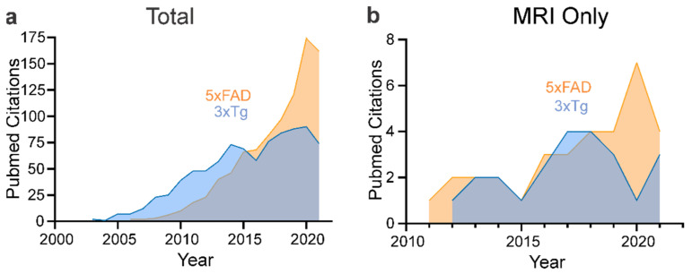
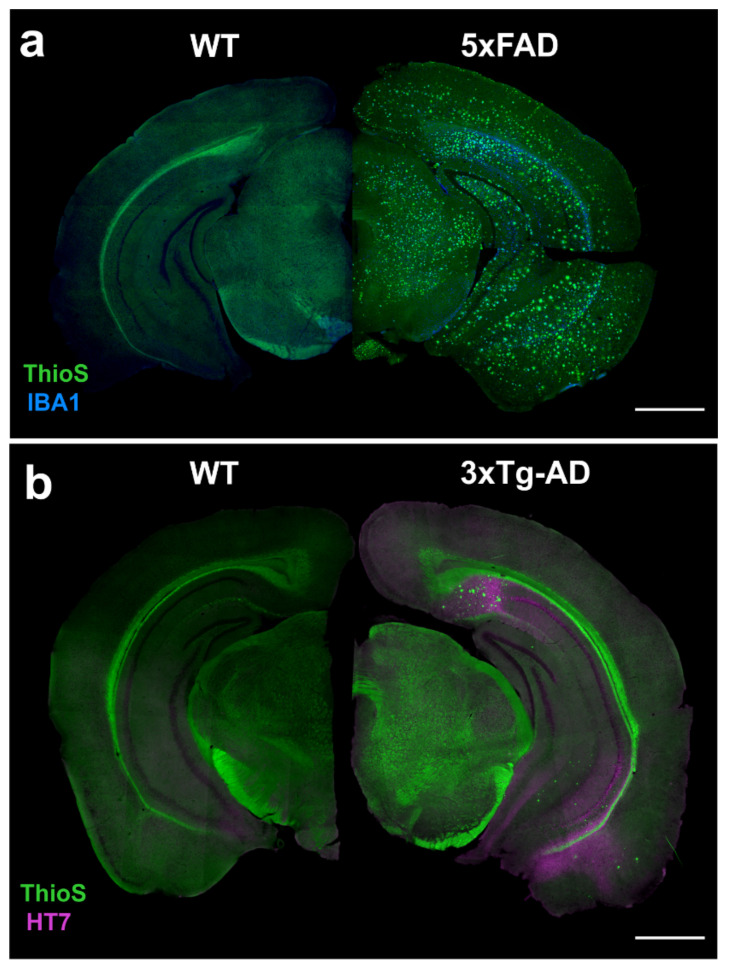
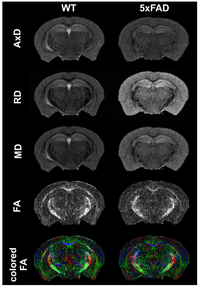
Magnetic resonance imaging (MRI) and positron emission tomography (PET) have made great strides in the diagnosis and our understanding of Alzheimer’s Disease (AD). Despite the knowledge gained from human studies, mouse models have and continue to play an important role in deciphering the cellular and molecular evolution of AD. MRI and PET are now being increasingly used to investigate neuroimaging features in mouse models and provide the basis for rapid translation to the clinical setting. Here, we provide an overview of the human MRI and PET imaging landscape as a prelude to an in-depth review of preclinical imaging in mice. A broad range of mouse models recapitulate certain aspects of the human AD, but no single model simulates the human disease spectrum. We focused on the two of the most popular mouse models, the 3xTg-AD and the 5xFAD models, and we summarized all known published MRI and PET imaging data, including contrasting findings. The goal of this review is to provide the reader with broad framework to guide future studies in existing and future mouse models of AD. We also highlight aspects of MRI and PET imaging that could be improved to increase rigor and reproducibility in future imaging studies.
Overview
The claims and statistics of this paper haven’t been checked yet.
Run the check — free, no account needed. It reads the open-access text and checks the sources it cites.
Integrity signals
Registry
Cites
Statistics
Figures
Verification
Claims the paper makes
The claims and statistics of this paper haven’t been checked yet.
Run the check — free, no account needed. It reads the open-access text and checks the sources it cites.
Claims other papers make about this one
Each is checked against this paper’s own text: does it say what the citing paper says it does?
The source backs part of the claim, not all of it — or backs it for a different population, condition or comparison.
In the citing paper
“Alongside the progression of AD pathology, age-related brain atrophy and myelin loss are observed on advanced high-field MRI (Ni, 2021; Jullienne et al., 2022; Bergamino et al., 2023).”
From Multimodal MRI combined with RNA sequencing reveals pathological signatures in the 9-month-old 3×Tg-AD mouse brain (2026) · cites this paper as Jullienne et al., 2022
Experimental — accuracy not yet measured
Self-check
Percentages, sums and reported differences re-computed from the paper’s own numbers.
The self-check needs the paper’s full text.
Citations
Cites
Items 201–229 of 229
Dynamic changes in vascular size and density in transgenic mice with Alzheimer’s disease
Xu, X., Meng, T., Wen, Q., Tao, M., Wang, P., Zhong, K. L., & Shen, Y. (2020) · Aging · doi:10.18632/aging.103672 · Cited by 21
Xu, W., Zhan, Y., Huang, W., Wang, X., Zhang, S., & Lei, H. C. (2010) · Journal of Neuroscience Research · doi:10.1002/jnr.22479 · Cited by 20
Brain Molecular Connectivity in Neurodegenerative Conditions
Carli, G., Tondo, G., Boccalini, C., & Perani, D. (2021) · Brain Sciences · doi:10.3390/brainsci11040433 · Cited by 20
Ishihara, Y., Itoh, K., Mitsuda, Y., Shimada, T., Kubota, T., Kato, C., Song, S. Y., Kobayashi, Y., Mori‐Yasumoto, K., Sekita, S., et al. (2013) · Free Radical Research · doi:10.3109/10715762.2013.818218 · Cited by 19
Jansen, D., Zerbi, V., Janssen, C. I. F., Dederen, P. J. W. C., Mutsaers, M. P. C., Hafkemeijer, A., Janssen, A.-L., Nobelen, C. L. M., Veltien, A. A., van Asten, J. J. A., et al. (2013) · PLoS ONE · doi:10.1371/journal.pone.0063643 · Cited by 19
Pan, G., King, A. E., Wu, F., Simpson, S., Woodhouse, A., Phipps, A., & Vickers, J. C. (2021) · Ageing Research Reviews · doi:10.1016/j.arr.2021.101402 · Cited by 19
Thiessen, J. D., Glazner, K. A. C., Nafez, S., Schellenberg, A. E., Buist, R. J., Martin, M., & Albensi, B. C. (2010) · Brain Structure and Function · doi:10.1007/s00429-010-0271-z · Cited by 19
Kara, F., Höfling, C., Roßner, S., Schliebs, R., der Linden, A. V., de Groot, H. J. M., & Alia, A. (2015) · Current Alzheimer Research · doi:10.2174/1567205012666151027123728 · Cited by 19
Distinct fiber-specific white matter reductions pattern in early- and late-onset Alzheimer’s disease
Luo, X., Wang, S., Jiaerken, Y., Li, K., Zeng, Q., Hong, H., Guan⁴, X., Guo, T., Wang, C., Zhang, R., et al. (2021) · Aging · doi:10.18632/aging.202702 · Cited by 19
Roy, U., Heredia-Muñoz, M. T., Stute, L., Höfling, C., Matysik, J., Meijer, J. H., Roßner, S., & Alia, A. (2019) · Journal of Alzheimer s Disease · doi:10.3233/jad-190037 · Cited by 18
Recent Advances in Imaging of Preclinical, Sporadic, and Autosomal Dominant Alzheimer's Disease
Buckley, R. F. (2021) · Neurotherapeutics · doi:10.1007/s13311-021-01026-5 · Cited by 18
Analysis of the vasculature by immunohistochemistry in paraffin-embedded brains
Decker, Y., Müller, A., Németh, E., Schulz‐Schaeffer, W., Fatar, M., Menger, M. D., Liu, Y., & Fassbender, K. C. (2017) · Brain Structure and Function · doi:10.1007/s00429-017-1595-8 · Cited by 17
van Duijn, S., Nabuurs, R. J. A., van Duinen, S. G., Natté, R., van Buchem, M. A., & Alia, A. (2013) · Journal of Alzheimer s Disease · doi:10.3233/jad-122188 · Cited by 16
Xie, Z., Yang, D., Stephenson, D. T., Morton, D. G., Hicks, C. D., Brown, T. M., & Bocan, T. M. A. (2010) · Lecture notes in computer science · doi:10.1007/978-3-642-15705-9_38 · Cited by 16
Altered brain development in an early-onset murine model of Alzheimer's disease
Allemang-Grand, R., Scholz, J., Ellegood, J., Cahill, L. S., Laliberté, C. L., Fraser, P. E., Josselyn, S. A., Sled, J. G., & Lerch, J. P. (2014) · Neurobiology of Aging · doi:10.1016/j.neurobiolaging.2014.08.032 · Cited by 16
Li, L., Wang, X., Gao, F., Wang, L., Xia, R., Li, Z., Wu, X., Tang, B., Zeng, Y., Zhou, G., et al. (2015) · Neuroscience Letters · doi:10.1016/j.neulet.2015.10.058 · Cited by 16
Kim, E., Di Censo, D., Baraldo, M., Simmons, C., Rosa, I., Randall, K., Ballard, C., Dickie, B. R., Williams, S. C. R., Killick, R., et al. (2021) · Scientific Reports · doi:10.1038/s41598-021-91899-5 · Cited by 15
Hinteregger, B., Loeffler, T., Flunkert, S., Neddens, J., Bayer, T. A., Madl, T., & Hutter‐Paier, B. (2021) · Journal of Alzheimer s Disease · doi:10.3233/jad-201204 · Cited by 15
Tataryn, N. M., Singh, V., Dyke, J. P., Berk-Rauch, H., Clausen, D. M., Aronowitz, E., Norris, E. H., Strickland, S., & Ahn, H. J. (2021) · Neurobiology of Aging · doi:10.1016/j.neurobiolaging.2021.07.015 · Cited by 15
Doert, A., Pilatus, U., Zanella, F., Müller, W. E. G., & Eckert, G. P. (2015) · Journal of Neural Transmission · doi:10.1007/s00702-015-1387-3 · Cited by 14
Cho, H., Baek, M. S., Lee, H. S., Lee, J., Ryu, Y. H., & Lyoo, C. H. (2020) · Alzheimer s Research & Therapy · doi:10.1186/s13195-020-00685-4 · Cited by 13
Chen, Q., Turnbull, A. G., Baran, T. M., & Lin, F. V. (2020) · eLife · doi:10.7554/elife.62114 · Cited by 13
Kim, H. J., Park, J., Jung, K. S., Kim, J., Jang, J. S., Kwon, S., Byun, M. S., Yi, D., Byeon, G., Jung, G., et al. (2021) · Experimental & Molecular Medicine · doi:10.1038/s12276-021-00638-3 · Cited by 13
Detrez, J. R., Ben-Nejma, I. R. H., Kolen, K. V., Dam, D. V., Deyn, P. P. D., Fransén, E., Verhoye, M., Timmermans, J., Nuydens, R., der Linden, A. V., et al. (2020) · Neurobiology of Disease · doi:10.1016/j.nbd.2020.105011 · Cited by 10
Mapping of microvascular architecture in the brain of an Alzheimer's disease mouse model using MRI
Chang, S., Kim, J., Lee, D.-K., Yoo, C. H., Jin, S., Rhee, H. Y., Ryu, C., Lee, J. K., Cho, H., & Jahng, G. (2021) · NMR in Biomedicine · doi:10.1002/nbm.4481 · Cited by 10
Bach, J. (2019) · Anais da Academia Brasileira de Ciências · doi:10.1590/0001-3765201920190339 · Cited by 7
Can MRI T1 be used to detect early changes in 5xFAD Alzheimer’s mouse brain?
Spencer, N. G., Lovell, D. P., Elderfield, K., Austen, B., & Howe, F. A. (2016) · Magnetic Resonance Materials in Physics Biology and Medicine · doi:10.1007/s10334-016-0593-9 · Cited by 5
Androuin, A., Abada, Y.-S. K., Ly, M., Santin, M. D., Petiet, A., Epelbaum, S., Bertrand, A., & Delatour, B. (2019) · Scientific Reports · doi:10.1038/s41598-018-37980-y · Cited by 5
Govaerts, K., Dresselaers, T., LEUVEN, F. V., & Himmelreich, U. (2019) · Molecular Imaging and Biology · doi:10.1007/s11307-019-01402-w · Cited by 2
Cited by
Sorted by citations
Alcohol Use Disorder and Dementia: A Review
Zahr, N. M. (2024) · Alcohol research · doi:10.35946/arcr.v44.1.03 · Cited by 42
Cortical cerebrovascular and metabolic perturbations in the 5xFAD mouse model of Alzheimer’s disease
Jullienne, A., Szu, J. I., Quan, R., Trinh, M. V., Norouzi, T., Noarbe, B. P., Bedwell, A. A., Eldridge, K., Persohn, S. A., Territo, P. R., et al. (2023) · Frontiers in Aging Neuroscience · doi:10.3389/fnagi.2023.1220036 · Cited by 30
Self-citationZeng, X., Cheung, S. K. K., Shi, M., Or, P. M. Y., Li, Z., Liu, J. Y. H., Ho, W. L.-H., Liu, T., Lu, K., Rudd, J. A., et al. (2023) · Journal of Neuroinflammation · doi:10.1186/s12974-023-02970-z · Cited by 28
Andrade-Guerrero, J., Rodríguez-Arellano, P., Barron-Leon, N., Orta‐Salazar, E., Ledesma-Alonso, C., Díaz-Cintra, S. Y., & Soto-Rojas, L. O. (2023) · Cells · doi:10.3390/cells12212531 · Cited by 28
Progressive Vascular Abnormalities in the Aging 3xTg-AD Mouse Model of Alzheimer’s Disease
Jullienne, A., Quan, R., Szu, J. I., Trinh, M. V., Behringer, E. J., & Obenaus, A. (2022) · Biomedicines · doi:10.3390/biomedicines10081967 · Cited by 23
Self-citation
Metadata
- Authors
- Jullienne, A., Trinh, M. V., & Obenaus, A.
- Affiliations
- University of California, Irvine (US)
- Department of Pediatrics, University of California, Irvine, CA 92697, USA
- Venue
- Biomedicines
- Publisher
- MDPI AG
- Published
- 2022-01-28
- Volume
- 10
- Issue
- 2
- Pages
- 305
- ISSN
- 2227-9059 (ISSN-L)
- PMC
- PMC8869427
- OpenAlex
- W4210462365
- Cited by
- 32
- References
- 232
- Citations by year
- 2026: 8 · 2025: 8 · 2024: 6 · 2023: 6 · 2022: 4
- FWCI
- 0.83
- Type
- journal article
- Language
- en
- Open access
- gold
- doi.org
- Licence
- Locations
- Biomedicines (publishedVersion, cc-by)
- PubMed Central (publishedVersion, cc-by)
- PubMed (publishedVersion)
- eScholarship (California Digital Library) (submittedVersion, cc-by)
- DOAJ (DOAJ: Directory of Open Access Journals) (submittedVersion, cc-by-sa)
- Funders
- National Institute on Aging (AG054345-01, AG054349-01)
- Topics
- Life Sciences › Neuroscience › Cellular and Molecular Neuroscience › Alzheimer's disease research and treatments
- Health Sciences › Medicine › Psychiatry and Mental health › Dementia and Cognitive Impairment Research
- Life Sciences › Neuroscience › Neurology › Neuroinflammation and Neurodegeneration Mechanisms
- Keywords
- Alzheimer's disease, mouse models of Alzheimer's disease, 3xTg-AD mice, 5xFAD mice, magnetic resonance imaging, positron emission tomography, imaging biomarkers
About these checks
Registry, retraction, Cites and statistics results are computed, not judged, with one exception: a printed reference that no index lists is read once by a model before it counts as a citation error. Claims are extracted by a model and kept only when their quote appears verbatim in the text; each cited source is then checked, with a model’s help, for whether it says what the claim relies on. Those verdicts can be wrong — read the claim’s quote and the cited source before relying on one.
Statistics and arithmetic are recomputed, not judged.
Confidence bands are shown only for fields calibrated on our annotated test set. None is calibrated yet, so no band is shown.
What counts as a claim ›How we measure confidence ›How we check this ›
Are you an author, or do you see a problem with this page? Request a review
