

The DOI matches its Crossref record; 174 of the 174 references it cites were found in a registry or an index.
Mechanisms of Neurodegeneration and Axonal Dysfunction in Progressive Multiple Sclerosis
Open accessAbstract
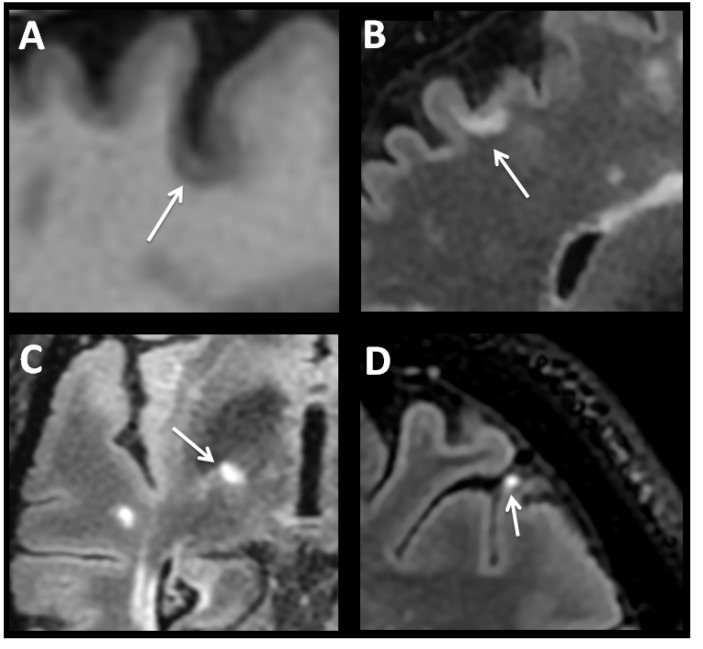
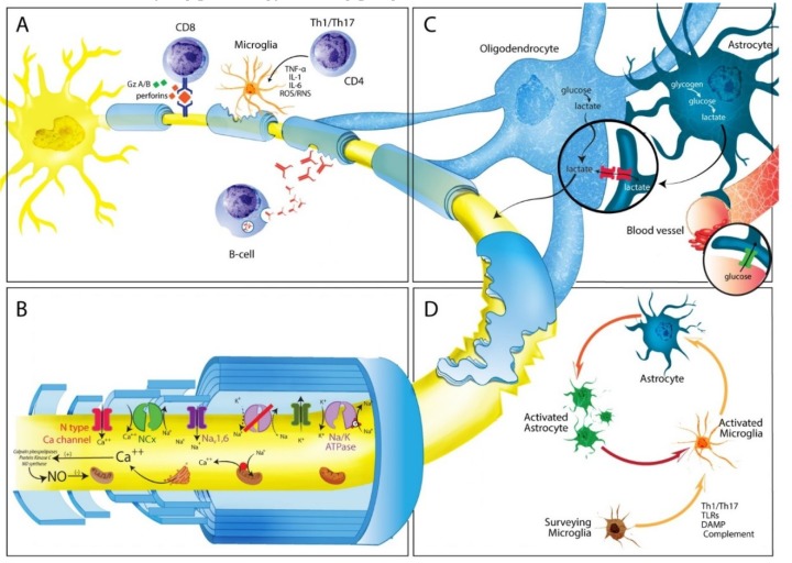
Multiple Sclerosis (MS) is a major cause of neurological disability, which increases predominantly during disease progression as a result of cortical and grey matter structures involvement. The gradual accumulation of disability characteristic of the disease seems to also result from a different set of mechanisms, including in particular immune reactions confined to the Central Nervous System such as: (a) B-cell dysregulation, (b) CD8+ T cells causing demyelination or axonal/neuronal damage, and (c) microglial cell activation associated with neuritic transection found in cortical demyelinating lesions. Other potential drivers of neurodegeneration are generation of oxygen and nitrogen reactive species, and mitochondrial damage, inducing impaired energy production, and intra-axonal accumulation of Ca2+, which in turn activates a variety of catabolic enzymes ultimately leading to progressive proteolytic degradation of cytoskeleton proteins. Loss of axon energy provided by oligodendrocytes determines further axonal degeneration and neuronal loss. Clearly, these different mechanisms are not mutually exclusive and could act in combination. Given the multifactorial pathophysiology of progressive MS, many potential therapeutic targets could be investigated in the future. This remains however, an objective that has yet to be undertaken.
Overview
The claims and statistics of this paper haven’t been checked yet.
Run the check — free, no account needed. It reads the open-access text and checks the sources it cites.
Integrity signals
Registry
Cites
Statistics
Figures
Verification
Claims the paper makes
The claims and statistics of this paper haven’t been checked yet.
Run the check — free, no account needed. It reads the open-access text and checks the sources it cites.
Claims other papers make about this one
Each is checked against this paper’s own text: does it say what the citing paper says it does?
In the citing paper
“Neurological deficits in MS patients result from two pathogenic events: acute inflammatory demyelination and axonal injury [150,151].”
From Beneficial Effects of Spirulina Consumption on Brain Health (2022) · cites this paper as [151]
Self-check
Percentages, sums and reported differences re-computed from the paper’s own numbers.
The self-check needs the paper’s full text.
Citations
Cites
How mitochondria produce reactive oxygen species
Murphy, M. P. (2009) · Biochemical Journal · doi:10.1042/bj20081386 · Cited by 8,207
New Multiple Sclerosis Phenotypic Classification
LUBLIN, F. D. (2014) · European Neurology · doi:10.1159/000367614 · Cited by 4,277
Axonal transection in the lesions of multiple sclerosis
Trapp, B. D., Peterson, J., Ransohoff, R. M., Rudick, R., Mörk, S., & Bö, L. (1998) · New England Journal of Medicine · doi:10.1056/nejm199801293380502 · Cited by 4,029
Defining the clinical course of multiple sclerosis
LUBLIN, F. D., & Reingold, S. C. (1996) · Neurology · doi:10.1212/wnl.46.4.907 · Cited by 3,762
Chondroitinase ABC promotes functional recovery after spinal cord injury
Bradbury, E. J., Moon, L. D. F., Popat, R. J., King, V. R., Bennett, G. S., Patel, P. N., Fawcett, J. W., & McMahon, S. B. (2002) · Nature · doi:10.1038/416636a · Cited by 2,357
Brain energy metabolism: focus on Astrocyte-neuron metabolic cooperation
Bélanger, M., Allaman, I., & Magistretti, P. J. (2011) · Cell Metabolism · doi:10.1016/j.cmet.2011.08.016 · Cited by 2,313
Neurofilaments as biomarkers in neurological disorders
Khalil, M., Teunissen, C. E., Otto, M., Piehl, F., Sormani, M. P., Gattringer, T., Barro, C., Kappos, L., Comabella, M., Fazekas, F., et al. (2018) · Nature Reviews Neurology · doi:10.1038/s41582-018-0058-z · Cited by 1,988
Thompson, A. J., Baranzini, S. E., Geurts, J. J. G., Hemmer, B., & Ciccarelli, O. (2018) · The Lancet · doi:10.1016/s0140-6736(18)30481-1 · Cited by 1,979
Cortical demyelination and diffuse white matter injury in multiple sclerosis
Kutzelnigg, A., Lucchinetti, C. F., Stadelmann, C., Brück, W., Rauschka, H., Bergmann, M., Schmidbauer, M., Parisi, J. E., & Lassmann, H. (2005) · Brain · doi:10.1093/brain/awh641 · Cited by 1,836
Oligodendroglia metabolically support axons and contribute to neurodegeneration
Lee, Y., Morrison, B. M., Li, Y., Lengacher, S., Farah, M. H., Hoffman, P. N., Liu, Y., Tsingalia, A., Jin, L., Zhang, P.-W., et al. (2012) · Nature · doi:10.1038/nature11314 · Cited by 1,664 · cited 1×
“Notably, both genetic and pharmacologic down-regulation of MCT1, which is present almost exclusively in oligodendrocytes, results in axon degeneration and neuronal loss both in vivo and in vitro, without obvious oligodendrocyte damage [165].”
Background
Cited by
Sorted by citations
Inflammatory Processes in Alzheimer’s Disease—Pathomechanism, Diagnosis and Treatment: A Review
Twarowski, B., & Herbet, M. (2023) · International Journal of Molecular Sciences · doi:10.3390/ijms24076518 · Cited by 489
Jamwal, S., Blackburn, J. K., & Elsworth, J. D. (2021) · Pharmacology & Therapeutics · doi:10.1016/j.pharmthera.2020.107705 · Cited by 236
Palanisamy, C. P., Pei, J., Alugoju, P., Anthikapalli, N. V. A., Jayaraman, S., Veeraraghavan, V. P., Gopathy, S., Roy, J. R., Janaki, C. S., Thalamati, D., et al. (2023) · Theranostics · doi:10.7150/thno.83066 · Cited by 165
Kamma, E., Lasisi, W. O., Libner, C. D., Ng, H. S., & Plemel, J. R. (2022) · Journal of Neuroinflammation · doi:10.1186/s12974-022-02408-y · Cited by 157
The Role of Vitamins in Neurodegenerative Disease: An Update
Nand, S., Singh, P., Steinbusch, H. W. M., Vamanu, E., Ashraf, G. M., & Singh, M. P. (2021) · Biomedicines · doi:10.3390/biomedicines9101284 · Cited by 126
Relationship between inflammation and oxidative stress and its effect on multiple sclerosis
Ramos-González, E. J., Bitzer-Quintero, O. K., Ortíz, G. G., Hernández-Cruz, J. J., & Ramírez-Jirano, L. J. (2021) · Neurología · doi:10.1016/j.nrl.2021.10.003 · Cited by 127
Praveenraj, S. S., Sonali, S., Anand, N., Hediyal, T. A., Vichitra, C., Kalyan, M., Kanna, P. V., Chandana, K. A., Paneyala, S., Mahalakshmi, A. M., et al. (2022) · Molecular Neurobiology · doi:10.1007/s12035-022-02990-5 · Cited by 116
Beneficial Effects of Spirulina Consumption on Brain Health
Trotta, T., Porro, C., Cianciulli, A., & Panaro, M. A. (2022) · Nutrients · doi:10.3390/nu14030676 · Cited by 98
Mitochondrial Behavior in Axon Degeneration and Regeneration
Wang, B., Huang, M., Shang, D., Yan, X., Zhao, B., & Zhang, X. (2021) · Frontiers in Aging Neuroscience · doi:10.3389/fnagi.2021.650038 · Cited by 98
Monitoring the Redox Status in Multiple Sclerosis
Tanaka, M., & Vécsei, L. (2020) · Biomedicines · doi:10.3390/biomedicines8100406 · Cited by 89
Microglial NLRP3 inflammasome activation in multiple sclerosis
(2020) · Advances in Protein Chemistry and Structural Biology · doi:10.1016/bs.apcsb.2019.08.007 · Cited by 83
Epilepsy in Neurodegenerative Diseases: Related Drugs and Molecular Pathways
Cano, A. C., Fonseca, E., Ettcheto, M., Sánchez‐López, E., de Rojas, I., Alonso-Lana, S., Morató, X., Souto, E. B., Toledo, M., Boada, M., et al. (2021) · Pharmaceuticals · doi:10.3390/ph14101057 · Cited by 90
Simkins, T., Duncan, G. J., & BOURDETTE, D. N. (2021) · Current Neurology and Neuroscience Reports · doi:10.1007/s11910-021-01110-5 · Cited by 79
CNS Border-Associated Macrophages: Ontogeny and Potential Implication in Disease
Dermitzakis, I., Theotokis, P., Evangelidis, P., Delilampou, E., Evangelidis, N., Chatzisavvidou, A., Avramidou, E., & Μanthou, M. E. (2023) · Current Issues in Molecular Biology · doi:10.3390/cimb45050272 · Cited by 74
Approved and Emerging Disease Modifying Therapies on Neurodegeneration in Multiple Sclerosis
Bross, M., Hackett, M., & Bernitsas, E. (2020) · International Journal of Molecular Sciences · doi:10.3390/ijms21124312 · Cited by 67
Alkhazaali-Ali, Z., Negah, S. S., Boroumand, A. R., & Tavakol‐Afshari, J. (2024) · Biomedicine & Pharmacotherapy · doi:10.1016/j.biopha.2024.116899 · Cited by 69
Functions and dysfunctions of oligodendrocytes in neurodegenerative diseases
Han, S.-W., Gim, Y., Jang, E., & Hur, E. (2022) · Frontiers in Cellular Neuroscience · doi:10.3389/fncel.2022.1083159 · Cited by 67
The SIRT-1/Nrf2/HO-1 axis: Guardians of neuronal health in neurological disorders
Sethi, P., Mehan, S., Khan, Z., Maurya, P. K., Kumar, B., Kumar, A., Tiwari, A., Sharma, T., Gupta, G. D., Narula, A. S., et al. (2024) · Behavioural Brain Research · doi:10.1016/j.bbr.2024.115280 · Cited by 59
Overview of Metformin and Neurodegeneration: A Comprehensive Review
Kruczkowska, W., Gałęziewska, J., Buczek, P., Płuciennik, E., Kciuk, M., & Śliwińska, A. (2025) · Pharmaceuticals · doi:10.3390/ph18040486 · Cited by 52
Hashemi, B., Abdollahi, M., Abbaspour‐Aghdam, S., Hazrati, A., Malekpour, K., Meshgi, S., Kafil, H. S., Ghazi, F., Yousefi, M., Roshangar, L., et al. (2023) · Biomedicine & Pharmacotherapy · doi:10.1016/j.biopha.2022.114195 · Cited by 49
Exosomes-Based Nanomedicine for Neurodegenerative Diseases: Current Insights and Future Challenges
Cano, A. C., Muñoz-Morales, Á., Sánchez‐López, E., Ettcheto, M., Souto, E. B., Camins, A., Boada, M., & Ruiz, A. (2023) · Pharmaceutics · doi:10.3390/pharmaceutics15010298 · Cited by 48
Dehghani, S., Ocakcı, O., Hatipoglu, P. T., Özalp, V. C., & Tevlek, A. (2025) · Molecular Neurobiology · doi:10.1007/s12035-025-04825-5 · Cited by 49
Roshanbakhsh, H., Salmani, M. E., Dehghan, S., Nazari, A., Javan, M., & Pourabdolhossein, F. (2020) · Life Sciences · doi:10.1016/j.lfs.2020.117671 · Cited by 43
Relationship between inflammation and oxidative stress and its effect on multiple sclerosis
Ramos-González, E. J., Bitzer-Quintero, O. K., Ortíz, G. G., Hernández-Cruz, J. J., & Ramírez-Jirano, L. J. (2024) · Neurología (English Edition) · doi:10.1016/j.nrleng.2021.10.010 · Cited by 48
Talebi, M., Talebi, M., Kakouri, E., Farkhondeh, T., Pourbagher‐Shahri, A. M., Tarantilis, P. Α., & Samarghandian, S. (2021) · International Journal of Biological Macromolecules · doi:10.1016/j.ijbiomac.2021.01.042 · Cited by 42
Chronic inflammation is associated with neural responses to faces in bangladeshi children
Xie, W., Kumar, S., Kakon, S. H., Haque, R., Petri, W. A., & Nelson, C. A. (2019) · NeuroImage · doi:10.1016/j.neuroimage.2019.116110 · Cited by 38
Liu, X., Zhang, X., Niu, X.-J., Zhang, P., Wang, Q., Xue, X.-H., Song, G., Yu, J.-Z., Xi, G.-P., Song, L.-J., et al. (2021) · Molecular Neurobiology · doi:10.1007/s12035-021-02552-1 · Cited by 37
Arnold, D. L., Piani-Meier, D., Bar‐Or, A., Benedict, R. H. B., Cree, B., Giovannoni, G., Gold, R., Vermersch, P., Arnould, S., Dahlke, F., et al. (2022) · Multiple Sclerosis Journal · doi:10.1177/13524585221076717 · Cited by 35
Lazzarotto, A., Hamzaoui, M., Tonietto, M., Dubessy, A.-L., Khalil, M., Pirpamer, L., Ropele, S., Enzinger, C., Battaglini, M., Stromillo, M. L., et al. (2024) · Brain · doi:10.1093/brain/awae024 · Cited by 33
Reduced TUBA1A Tubulin Causes Defects in Trafficking and Impaired Adult Motor Behavior
Buscaglia, G., Northington, K. R., Moore, J. K., & Bates, E. A. (2020) · eNeuro · doi:10.1523/eneuro.0045-20.2020 · Cited by 33
Effects of Palmitoylethanolamide on Neurodegenerative Diseases: A Review from Rodents to Humans
Landolfo, E., Cutuli, D., Petrosini, L., & Caltagirone, C. (2022) · Biomolecules · doi:10.3390/biom12050667 · Cited by 33
Delivanoglou, N., Boziki, M. K., Theotokis, P., Kesidou, E., Touloumi, O., Dafi, N., Nousiopoulou, E., Lagoudaki, R., Grigoriadis, N., Charalampopoulos, I., et al. (2020) · Journal of Neuroinflammation · doi:10.1186/s12974-020-1708-9 · Cited by 31
MRI biomarkers of disease progression and conversion to secondary-progressive multiple sclerosis
Tavazzi, E., Zivadinov, R., Dwyer, M. G., Jakimovski, D., Singhal, T., Weinstock‐Guttman, B., & Bergsland, N. (2020) · Expert Review of Neurotherapeutics · doi:10.1080/14737175.2020.1757435 · Cited by 33
Sen, M. K., Almuslehi, M. S. M., Shortland, P. J., Mahns, D. A., & Coorssen, J. R. (2021) · International Journal of Molecular Sciences · doi:10.3390/ijms22147377 · Cited by 31
Nogo-A and LINGO-1: Two Important Targets for Remyelination and Regeneration
Kalafatakis, I., Papagianni, F., Theodorakis, Κ., & Karagogeos, D. (2023) · International Journal of Molecular Sciences · doi:10.3390/ijms24054479 · Cited by 31
Corrales, A. V. G., Verberk, S. G. S., Haidar, M., Grajchen, E., Dehairs, J., Vanherle, S., Loix, M., Weytjens, T., Gervois, P., Matsuzaka, T., et al. (2023) · Proceedings of the National Academy of Sciences · doi:10.1073/pnas.2301030120 · Cited by 30
B-Cell Receptor Repertoire: Recent Advances in Autoimmune Diseases
Wang, Q., Feng, D., Jia, S., Lu, Q., & Zhao, M. (2024) · Clinical Reviews in Allergy & Immunology · doi:10.1007/s12016-024-08984-6 · Cited by 30
Clinical Use of Schistosoma mansoni Antigens as Novel Immunotherapies for Autoimmune Disorders
Cleenewerk, L., Garssen, J., & Hogenkamp, A. (2020) · Frontiers in Immunology · doi:10.3389/fimmu.2020.01821 · Cited by 27
Fettig, N. M., & Osborne, L. C. (2021) · Microbes and Infection · doi:10.1016/j.micinf.2021.104814 · Cited by 26
Matrine Inhibits CNS Autoimmunity Through an IFN-β-Dependent Mechanism
Chu, Y., Ma, W., Thomé, R., Ping, J.-D., Liu, F., Wang, M., Zhang, M., Zhang, G., & Zhu, L. D. (2020) · Frontiers in Immunology · doi:10.3389/fimmu.2020.569530 · Cited by 26
Hasan, A. R., Tasnim, F., Aktaruzzaman, M., Islam, M. T., Rayhan, R., Brishti, A., Hur, J., Porter, J. E., & Raihan, M. O. (2024) · Neuroglia · doi:10.3390/neuroglia5040027 · Cited by 25
How do neurons age? A focused review on the aging of the microtubular cytoskeleton
Richardson, B., Goedert, T., Quraishe, S., Deinhardt, K., & Mudher, A. K. (2023) · Neural Regeneration Research · doi:10.4103/1673-5374.390974 · Cited by 27
The Na+/Ca2+ exchangers in demyelinating diseases
Boscia, F., de Rosa, V., Cammarota, M., Secondo, A., Pannaccione, A., & Annunziato, L. (2019) · Cell Calcium · doi:10.1016/j.ceca.2019.102130 · Cited by 22
Dziedzic, A., Maciak, K., Miller, E. D., Starosta, M., & Saluk, J. (2024) · International Journal of Molecular Sciences · doi:10.3390/ijms25073858 · Cited by 25
Analysis of shared underlying mechanism in neurodegenerative disease
Butler, R., Bradford, D. R., & Rodgers, K. E. (2022) · Frontiers in Aging Neuroscience · doi:10.3389/fnagi.2022.1006089 · Cited by 24
Fu, L., Luo, T., Hao, Z., Pan, Y., Xin, W., Zhang, L., Lai, Z., Zhang, H., Liu, H., & Wei, W. (2025) · Frontiers in Neuroscience · doi:10.3389/fnins.2025.1603292 · Cited by 21
Bernstein, H., Keilhoff, G., Dobrowolny, H., & Steiner, J. H. (2019) · Medical Hypotheses · doi:10.1016/j.mehy.2019.109443 · Cited by 20
Using Redox Proteomics to Gain New Insights into Neurodegenerative Disease and Protein Modification
Cadenas-Garrido, P., Schonvandt-Alarcos, A., Herrera‐Quintana, L., Vázquez‐Lorente, H., Santamaría-Quiles, A., de Francisco, J. R., Moya-Escudero, M., Martín‐Oliva, D., Martín-Guerrero, S. M., Rodríguez-Santana, C., et al. (2024) · Antioxidants · doi:10.3390/antiox13010127 · Cited by 20
Wigner, P., Dziedzic, A., Synowiec, E., Miller, E. D., Bijak, M., & Saluk, J. (2022) · Scientific Reports · doi:10.1038/s41598-022-14795-6 · Cited by 20
Identifying miRNAs in multiple sclerosis gray matter lesions that correlate with atrophy measures
Tripathi, A. K., Pandit, I., Perles, A., Zhou, Y., Cheng, F., & Dutta, R. (2021) · Annals of Clinical and Translational Neurology · doi:10.1002/acn3.51365 · Cited by 18
Nazari, N., Aligholipour, A., & Sadeghi, M. (2020) · BMC Women s Health · doi:10.1186/s12905-020-01109-z · Cited by 23
Cognitive Decline in Older People with Multiple Sclerosis—A Narrative Review of the Literature
Chiang, H., Khera, A., Stopschinski, B. E., Stüve, O., Hart, J., Kelley, B. J., & Nguyen, T. P. (2022) · Geriatrics · doi:10.3390/geriatrics7030061 · Cited by 16
Modulators of ASIC1a and its potential as a therapeutic target for age-related diseases
Zhou, R., Liang, H., Hu, W., Ding, J., Li, S., Chen, Y., Zhao, Y., Lü, C., Chen, F., & Hu, W. (2022) · Ageing Research Reviews · doi:10.1016/j.arr.2022.101785 · Cited by 16
Astroglial connexin 43 is a novel therapeutic target for chronic multiple sclerosis model
Takase, E. O., Yamasaki, R., Nagata, S., Watanabe, M., Masaki, K., Yamaguchi, H., Kira, J., Takeuchi, H., & Isobe, N. (2024) · Scientific Reports · doi:10.1038/s41598-024-61508-2 · Cited by 15
Tremolanti, C., Cavallini, C., Meyer, L., Klein, C., Pozzo, E. D., Costa, B., Germelli, L., Taliani, S., Patte‐Mensah, C., & Mensah‐Nyagan, A. G. (2022) · Molecular Neurobiology · doi:10.1007/s12035-022-02737-2 · Cited by 17
Che, J., Li, D., Hong, W., Wang, L., Guo, Y. L., Wu, M., Lu, J., Tong, L., Weng, Q., Wang, J., et al. (2022) · European Journal of Medicinal Chemistry · doi:10.1016/j.ejmech.2022.114732 · Cited by 15
Hemmati, S., Sadeghi, M. A., Yousefi‐Manesh, H., Eslamiyeh, M., Vafaei, A., Foroutani, L., Donyadideh, G., Dehpour, A. R., & Rezaei, N. (2020) · Journal of Inflammation Research · doi:10.2147/jir.s258991 · Cited by 15
Rahmani, S., Noorolyai, S., Ayromlou, H., Khaze, V., Shanehbandi, D., Baghbani, E., Shadbad, M. A., Jigari‐Asl, F., Khamiriyan, M., Safaralizadeh, R., et al. (2020) · Gene · doi:10.1016/j.gene.2020.145236 · Cited by 15
Risk of secondary progressive multiple sclerosis after early worsening of disability
Dzau, W., Sharmin, S., Patti, F., Izquierdo, G., Eichau, S., Prat, A., Girard, M., Duquette, P., ONOFRJ, M. C., Lugaresi, A., et al. (2023) · Journal of Neurology Neurosurgery & Psychiatry · doi:10.1136/jnnp-2023-331748 · Cited by 15
Farzan, M., Rounkian, M. S., Rizi, A. A., Heidari, Z., Farzan, M., Fathi, M., Aghaei, A., Azadegan‐Dehkordi, F., & Bagheri, N. (2024) · Experimental Neurology · doi:10.1016/j.expneurol.2024.115071 · Cited by 15
Sutiwisesak, R., Burns, T. C., Rodriguez, M., & Warrington, A. E. (2021) · Expert Opinion on Investigational Drugs · doi:10.1080/13543784.2021.1942840 · Cited by 14
Liu, F., Alfarhan, M., Baker, L., Shenoy, N., Liao, Y., Henry-Ojo, H. O., Somanath, P. R., & Narayanan, S. P. (2022) · Cells · doi:10.3390/cells11244100 · Cited by 14
Li, W., Wu, M., Li, Y., & Shen, J. (2023) · Free Radical Biology and Medicine · doi:10.1016/j.freeradbiomed.2023.07.032 · Cited by 16
Multiple Sclerosis and Related Challenges to Young women’s Health: Canadian Expert Review
Vorobeychik, G., Black, D., Cooper, P. E., & Cox, A. R. (2020) · Neurodegenerative Disease Management · doi:10.2217/nmt-2020-0010 · Cited by 14
An Interpretable Machine Learning Model to Predict Cortical Atrophy in Multiple Sclerosis
Conti, A., Treaba, C. A., Mehndiratta, A., Barletta, V. T., Mainero, C., & Toschi, N. (2023) · Brain Sciences · doi:10.3390/brainsci13020198 · Cited by 12
Turano, E., Scambi, I., Bonafede, R., Dusi, S., Angelini, G., Lopez, N., Marostica, G., Rossi, B., Furlan, R., Constantin, G., et al. (2024) · Cytotherapy · doi:10.1016/j.jcyt.2023.12.007 · Cited by 11
Esmaeilnejad, S., Semnanian, S., & Javan, M. (2021) · PubMed Central · doi:10.22074/cellj.2021.7174 · Cited by 12
Pearse, D. D., Otero, P. A., Diaz, A., Pan, X., & Ghosh, M. (2020) · Neuroscience · doi:10.1016/j.neuroscience.2020.11.034 · Cited by 10
Schröder, L.-J., Thiesler, H., Gretenkort, L., Möllenkamp, T. M., Stangel, M., Gudi, V., & Hildebrandt, H. (2023) · Frontiers in Cellular Neuroscience · doi:10.3389/fncel.2023.1207540 · Cited by 10
Mohammadi, S., Ghaderi, S., & Fatehi, F. (2024) · Brain and Behavior · doi:10.1002/brb3.70093 · Cited by 9
Jalilian, M., Elhaie, M., Sharifi, M., & Abedi, I. (2024) · BMJ Neurology Open · doi:10.1136/bmjno-2024-000788 · Cited by 9
Zhang, J., Ma, W., Liu, R., Li, X., Yuan, Z., & Cheng, J. (2025) · Neural Regeneration Research · doi:10.4103/nrr.nrr-d-24-01648 · Cited by 10
Astrocytes as Therapeutic Targets in Neurodegenerative Disorders
Salas, I. H., Alfonso-Triguero, M., Castro, B. D., Elazar, N., Escartin, C., Akl, C. F., Hill, S. A., Quintana, F. J., & Arranz, A. M. (2025) · Journal of Neuroscience · doi:10.1523/jneurosci.1379-25.2025 · Cited by 10
Gräfe, E. L., Reid, H. M. O., Shkolnikov, I., Conway, K., Kit, A., Acosta, C., & Christie, B. R. (2023) · Frontiers in Neuroendocrinology · doi:10.1016/j.yfrne.2023.101076 · Cited by 10
Razi, O., Tartibian, B., Teixeira, A. M., Zamani, N., Govindasamy, K., Suzuki, K., Laher, I., & Zouhal, H. (2022) · Multiple Sclerosis and Related Disorders · doi:10.1016/j.msard.2022.103557 · Cited by 10
Beneficial Effects of Spirulina on Brain Health: A Systematic Review
Kumar, S., Saha, S., Singh, K., Singh, T., Miahra, A. K., Dubey, B. N., & Singh, S. (2024) · Current Functional Foods · doi:10.2174/0126668629269256231222092721 · Cited by 17
Exosome-based platforms for treatment of multiple sclerosis
Zonouz, A. M., Rahbardar, M. G., & Alibolandi, M. (2025) · Brain Research Bulletin · doi:10.1016/j.brainresbull.2025.111256 · Cited by 9
Cladribine tablets in the new multiple sclerosis era
Buttari, F., Dolcetti, E., Rizzo, F. R., Rizzi, C., Lauritano, G., Borrelli, A., Azzolini, F., Fantozzi, R., Assogna, F., Conte, A., et al. (2025) · Therapeutic Advances in Neurological Disorders · doi:10.1177/17562864251342855 · Cited by 9
Chisari, C. G., Aguglia, U., Amato, M. P., Bergamaschi, R. C., Bertolotto, A., Bonavita, S., Morra, V. B., Cavalla, P., Cocco, E. E., Conte, A., et al. (2024) · Neurotherapeutics · doi:10.1016/j.neurot.2024.e00363 · Cited by 8
Multiple sclerosis from onset to secondary progression: a 30-year Italian register study
Zanghì, A., Copetti, M., Avolio, C., Paolicelli, D., Romeo, M. A. L., Patti, F., Luca, G. D., Amato, M. P., Galgani, S., Sola, P., et al. (2025) · Journal of Neurology Neurosurgery & Psychiatry · doi:10.1136/jnnp-2025-335958 · Cited by 7
Thümmler, K., Wrzos, C., Franz, J., McElroy, D., Cole, J. J., Hayden, L., Arseni, D., Schwarz, F., Junker, A., Edgar, J. M., et al. (2023) · Neuropathology and Applied Neurobiology · doi:10.1111/nan.12935 · Cited by 9
Stojsavljević, A., Jagodić, J., Pavlović, S. Z., Dinčić, E., Kuveljić, J., Manojlović, D., & Živković, M. (2024) · Journal of Trace Elements in Medicine and Biology · doi:10.1016/j.jtemb.2024.127421 · Cited by 8
Sokratous, D., Charalambous, C. C., Zamba‐Papanicolaou, E., Michailidou, K., & Konstantinou, N. (2024) · PLoS ONE · doi:10.1371/journal.pone.0299611 · Cited by 7
Exploring non-canonical targets in Alzheimer’s disease: a departure from the norm
Jain, S., & Chauhan, N. (2024) · The Egyptian Journal of Neurology Psychiatry and Neurosurgery · doi:10.1186/s41983-024-00908-7 · Cited by 7
Ad'hiah, A. H., Atiyah, N. S., & Fadhil, H. Y. (2023) · Iraqi Journal of Science · doi:10.24996/ijs.2023.64.1.13 · Cited by 7
Pathogenic Microglia Orchestrate Neurotoxic Properties of Eomes-Expressing Helper T Cells
Zhang, C., Raveney, B. J. E., Takahashi, F., Yeh, T.-W., Hohjoh, H., Yamamura, T., & Oki, S. (2023) · Cells · doi:10.3390/cells12060868 · Cited by 10
OVERVIEW OF MULTIPLE SCLEROSIS
Al-Dawani, A. A. A., Albakhite, H. A. A., Albunayyan, R. F., Almutiri, T. M. M., Nasrallah, L. Z. A., Aljehani, S. T., Alrashidi, S. S. S., Althobaiti, M. A. A., Alotaibi, F. M. M., Aljuaid, M. M., et al. (2023) · Journal of Population Therapeutics and Clinical Pharmacology · doi:10.53555//jptcp.v30i17.5823 · Cited by 6
Ghafoor, D. D., Ahmad, D. O., Najmuldeen, H. H. R., Mohammed, S. J., Jalal, P. J., Mustafa, F. S., Alghofaili, F., Zangana, B. A., & Al-Bajalan, S. J. (2025) · Inflammopharmacology · doi:10.1007/s10787-025-02012-5 · Cited by 5
Yuan, B., Jin, X., Li, C., Shen, Y., Tian, J., & Duan, S. (2026) · Frontiers in Pharmacology · doi:10.3389/fphar.2026.1806176 · Cited by 6
Shenoy, N., Liu, F., & Narayanan, S. P. (2024) · Journal of Integrative Neuroscience · doi:10.31083/j.jin2307129 · Cited by 5
Miscioscia, A., Mainero, C., Treaba, C. A., Silvestri, E., Scialpi, G., Berardi, A., Causin, F., Anglani, M. G., Rinaldi, F., Perini, P., et al. (2024) · Journal of Neurology · doi:10.1007/s00415-024-12622-8 · Cited by 6
Sharma, K., Dixon, K. M., Münch, G. W., Chang, D., & Zhou, X. (2024) · Frontiers in Public Health · doi:10.3389/fpubh.2024.1505904 · Cited by 5
Hurtado, J. C., González, L. M., Ospina, L., Sperling, L. E., & Chaparro, O. (2021) · Current Stem Cell Research & Therapy · doi:10.2174/1574888x16666211209155333 · Cited by 5
The evaluation of small fibers in multiple sclerosis
Bekircan‐Kurt, C. E., Jahanroshan, J., Tuncer, A., Ergul-Ulger, Z., Güneş, G., Erdem‐Özdamar, S., & Tan, E. (2023) · Multiple Sclerosis and Related Disorders · doi:10.1016/j.msard.2023.104602 · Cited by 5
Raghib, M. F., & Bernitsas, E. (2023) · Biomedicines · doi:10.3390/biomedicines11113069 · Cited by 5
Breaking boundaries: the transformative role of exercise in managing multiple sclerosis
Saedmocheshi, S., Yousfi, N., & Chamari, K. (2023) · EXCLI journal · doi:10.17179/excli2024-6932 · Cited by 5
Stojsavljević, A., Jagodić, J., Perović, T., Manojlović, D., & Pavlović, S. Z. (2024) · Biomedicines · doi:10.3390/biomedicines12071589 · Cited by 6
Brain macrophages and pial fibroblasts promote inflammation in a hypomyelination model
Okuno, N., Yamamoto, S., Hamashima, T., Tung, D. S., Okita, N., Fujikawa, M., Kunisawa, T., Takakura, N., Fujimori, T., Mori, H., et al. (2025) · Acta Neuropathologica Communications · doi:10.1186/s40478-025-02063-3 · Cited by 6
Myelin–Proteinoids Interactions in Neural Signaling
Mougkogiannis, P., & Adamatzky, A. (2025) · Langmuir · doi:10.1021/acs.langmuir.5c03469 · Cited by 4
Pathological and protective effects of tumor necrosis factor-alpha in multiple sclerosis
Смагина, И. В., Elchaninova, S. A., Palashchenko, A. S., & Галактионова, Л. П. (2019) · S S Korsakov Journal of Neurology and Psychiatry · doi:10.17116/jnevro201911910214 · Cited by 4
Metadata
- Authors
- Correale, J., Marrodan, M., & Ysrraelit, M. C.
- Affiliations
- Fundación para la Lucha contra las Enfermedades Neurológicas de la Infancia (AR)
- Department of Neurology, FLENI, Buenos Aires 1428, Argentina
- Corresponding author
- Correale, Jorge
- Venue
- Biomedicines
- Publisher
- MDPI AG
- Published
- 2019-02-20
- Volume
- 7
- Issue
- 1
- Pages
- 14
- ISSN
- 2227-9059 (ISSN-L)
- PMC
- PMC6466454
- OpenAlex
- W2917933180
- Cited by
- 167
- References
- 174
- Citations by year
- 2026: 15 · 2025: 25 · 2024: 39 · 2023: 27 · 2022: 20 · 2021: 15 · 2020: 19 · 2019: 7
- FWCI
- 2.27
- Type
- journal article
- Language
- en
- Open access
- gold
- doi.org
- Licence
- Locations
- Biomedicines (publishedVersion, cc-by)
- PubMed Central (publishedVersion, cc-by)
- PubMed (publishedVersion)
- DOAJ (DOAJ: Directory of Open Access Journals) (submittedVersion)
- Funders
- Fleni
- Topics
- Health Sciences › Medicine › Clinical Neurology › Multiple Sclerosis Research Studies
- Life Sciences › Neuroscience › Neurology › Neuroinflammation and Neurodegeneration Mechanisms
- Health Sciences › Medicine › Clinical Neurology › Peripheral Neuropathies and Disorders
- Keywords
- multiple sclerosis, progressive multiple sclerosis, neurodegeneration, axonal dysfunction, axonal degeneration, B cell dysregulation, CD8+ T cells, demyelination, microglial activation, reactive oxygen species, mitochondrial damage, calcium accumulation, neuronal loss, disability progression, therapeutic target
About these checks
Registry, retraction, Cites and statistics results are computed, not judged, with one exception: a printed reference that no index lists is read once by a model before it counts as a citation error. Claims are extracted by a model and kept only when their quote appears verbatim in the text; each cited source is then checked, with a model’s help, for whether it says what the claim relies on. Those verdicts can be wrong — read the claim’s quote and the cited source before relying on one.
Statistics and arithmetic are recomputed, not judged.
Confidence bands are shown only for fields calibrated on our annotated test set. None is calibrated yet, so no band is shown.
What counts as a claim ›How we measure confidence ›How we check this ›
Are you an author, or do you see a problem with this page? Request a review
