

The DOI matches its Crossref record; 51 of the 51 references it cites were found in a registry or an index.
Ovarian Cancer Relies on Glucose Transporter 1 to Fuel Glycolysis and Growth: Anti-Tumor Activity of BAY-876
Open accessAbstract
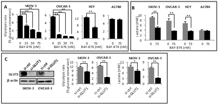
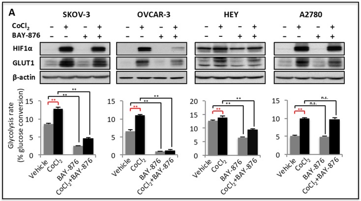
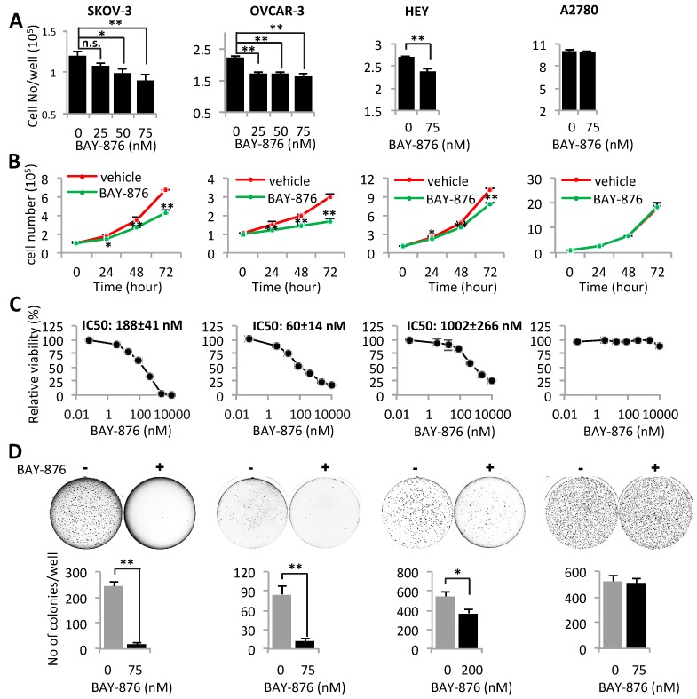
The recent progresses in understanding of cancer glycolytic phenotype have offered new strategies to manage ovarian cancer and other malignancies. However, therapeutic targeting of glycolysis to treat cancer remains unsuccessful due to complex mechanisms of tumor glycolysis and the lack of selective, potent and safe glycolytic inhibitors. Recently, BAY-876 was identified as a new-generation inhibitor of glucose transporter 1 (GLUT1), a GLUT isoform commonly overexpressed but functionally poorly defined in ovarian cancer. Notably, BAY-876 has not been evaluated in any cell or preclinical animal models since its discovery. We herein took advantage of BAY-876 and molecular approaches to study GLUT1 regulation, targetability, and functional relevance to cancer glycolysis. The anti-tumor activity of BAY-876 was evaluated with ovarian cancer cell line- and patient-derived xenograft (PDX) models. Our results show that inhibition of GLUT1 is sufficient to block basal and stress-regulated glycolysis, and anchorage-dependent and independent growth of ovarian cancer cells. BAY-876 dramatically inhibits tumorigenicity of both cell line-derived xenografts and PDXs. These studies provide direct evidence that GLUT1 is causally linked to the glycolytic phenotype in ovarian cancer. BAY-876 is a potent blocker of GLUT1 activity, glycolytic metabolism and ovarian cancer growth, holding promise as a novel glycolysis-targeted anti-cancer agent.
Overview
The claims and statistics of this paper haven’t been checked yet.
Run the check — free, no account needed. It reads the open-access text and checks the sources it cites.
Integrity signals
Registry
Cites
Statistics
Figures
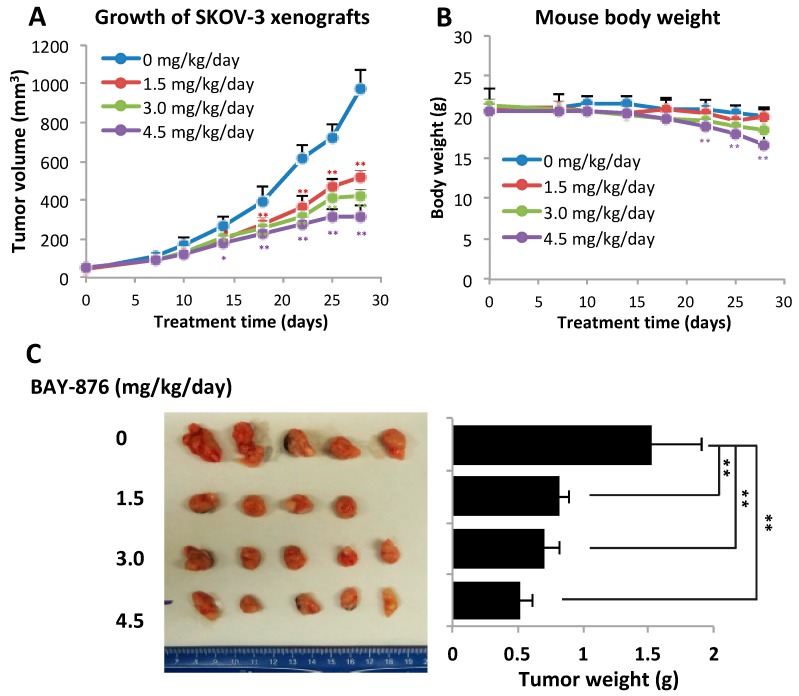
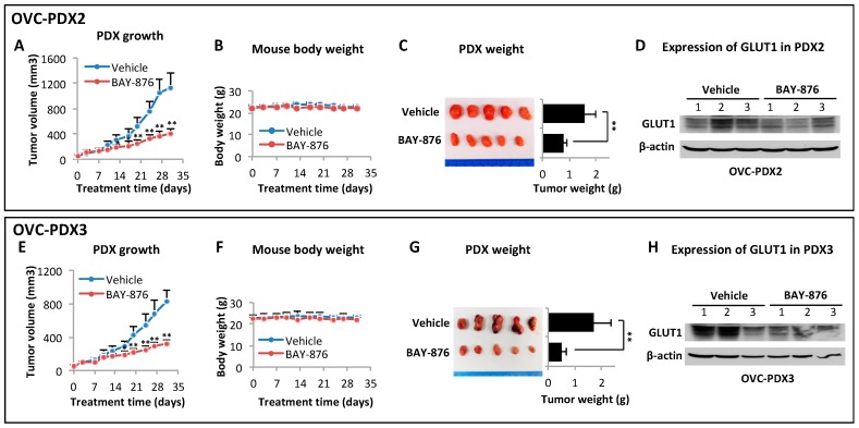
Verification
Claims the paper makes
The claims and statistics of this paper haven’t been checked yet.
Run the check — free, no account needed. It reads the open-access text and checks the sources it cites.
Claims other papers make about this one
Each is checked against this paper’s own text: does it say what the citing paper says it does?
In the citing paper
“BAY-876, a potent GLUT1 inhibitor49, was separately shown as an effective candidate in pre-clinical setting 50.”
From The metabolic landscape of RAS-driven cancers from biology to therapy (2021) · cites this paper as [50]
Self-check
Percentages, sums and reported differences re-computed from the paper’s own numbers.
The self-check needs the paper’s full text.
Citations
Cites
Warburg, O. (1956) · Science · doi:10.1126/science.123.3191.309 · Cited by 13,425
The Warburg Effect: How Does it Benefit Cancer Cells?
Liberti, M. V., & Locasale, J. W. (2016) · Trends in Biochemical Sciences · doi:10.1016/j.tibs.2015.12.001 · Cited by 5,150
Regulation of cancer cell metabolism
Cairns, R. A., Harris, I. S., & Mak, T. W. (2011) · Nature Reviews Cancer · doi:10.1038/nrc2981 · Cited by 4,744
ONCOMINE: A Cancer Microarray Database and Integrated Data-Mining Platform
Rhodes, D. R., Yu, J., Sundaram, S. K., Deshpande, N., Varambally, R., Ghosh, D., Barrette, T. R., Pander, A., & Chinnaiyan, A. M. (2004) · Neoplasia · doi:10.1016/s1476-5586(04)80047-2 · Cited by 3,719
Fundamentals of Cancer Metabolism
DeBerardinis, R. J., & Chandel, N. S. (2016) · Science Advances · doi:10.1126/sciadv.1600200 · Cited by 3,013
Inhibition of lactate dehydrogenase A induces oxidative stress and inhibits tumor progression
Le, A., Cooper, C. R., Gouw, A. M., Dinavahi, R., Maitra, A., Deck, L. M., Royer, R. E., Jagt, D. L. V., Semenza, G. L., & Dang, C. V. (2010) · Proceedings of the National Academy of Sciences · doi:10.1073/pnas.0914433107 · Cited by 1,528
Rethinking ovarian cancer: recommendations for improving outcomes
Vaughan, S., Coward, J. I. G., Bast, R. C., Berchuck, A., Berek, J. S., Brenton, J. D., Coukos, G., Crum, C. P., Drapkin, R., Etemadmoghadam, D., et al. (2011) · Nature reviews. Cancer · doi:10.1038/nrc3144 · Cited by 1,484
The Control of the Metabolic Switch in Cancers by Oncogenes and Tumor Suppressor Genes
Levine, A. J., & Puzio-Kuter, A. M. (2010) · Science · doi:10.1126/science.1193494 · Cited by 1,243
The hypoxic tumour microenvironment
Petrova, V., Annicchiarico‐Petruzzelli, M., Melino, G., & Amelio, I. (2018) · Oncogenesis · doi:10.1038/s41389-017-0011-9 · Cited by 1,077
Glucose transporters in the 21st Century
Thorens, B., & Mueckler, M. (2010) · American Journal of Physiology-Endocrinology and Metabolism · doi:10.1152/ajpendo.00712.2009 · Cited by 1,013
Research works printed but not linked: 1
- Warburg, 1924, üeber den Stoffwechsel der Tumoren, Biochem. Z., 152, 319
Cited by
Sorted by citations
Metabolomics in cancer research and emerging applications in clinical oncology
Schmidt, D. R., Patel, R., Kirsch, D. G., Lewis, C. A., Heiden, M. G. V., & Locasale, J. W. (2021) · CA A Cancer Journal for Clinicians · doi:10.3322/caac.21670 · Cited by 675
The metabolic landscape of RAS-driven cancers from biology to therapy
Mukhopadhyay, S., Vander Heiden, M. G., & McCormick, F. (2021) · Nature Cancer · doi:10.1038/s43018-021-00184-x · Cited by 315
GLUT1 inhibition blocks growth of RB1-positive triple negative breast cancer
Wu, Q., Ba-Alawi, W., Deblois, G., Cruickshank, J., Duan, S., Lima‐Fernandes, E., Haight, J., Tonekaboni, S. A. M., Fortier, A.-M., Kuasne, H., et al. (2020) · Nature Communications · doi:10.1038/s41467-020-18020-8 · Cited by 305
TGFβ-induced metabolic reprogramming during epithelial-to-mesenchymal transition in cancer
Hua, W., ten Dijke, P., Kostidis, S., Giera, M. A., & Hornsveld, M. (2019) · Cellular and Molecular Life Sciences · doi:10.1007/s00018-019-03398-6 · Cited by 245
Gao, T., Zhang, X., Zhao, J., Zhou, F., Wang, Y., Zhao, Z., Xing, J., Chen, B., Li, J., & Liu, S. (2019) · Cancer Letters · doi:10.1016/j.canlet.2019.10.029 · Cited by 158
Targeting glucose metabolism enzymes in cancer treatment: current and emerging strategies
Zhang, Y., Li, Q., Huang, Z., Li, B., Nice, E. C., Huang, C., Wei, L., & Zou, B. (2022) · Cancers · doi:10.3390/cancers14194568 · Cited by 144
Liu, Y., Zhao, Y., Song, H., Li, Y., Liu, Z., Ye, Z., Zhao, J., Wu, Y. C., Tang, J., & Yao, M. (2024) · Cancer Letters · doi:10.1016/j.canlet.2024.217076 · Cited by 146
Ren, X., Cheng, Z., He, J., Yao, X., Liu, Y., Cai, K., Li, M., Hu, Y., & Luo, Z. Y. (2023) · Nature Communications · doi:10.1038/s41467-023-42883-2 · Cited by 119
The Druggability of Solute Carriers
Wang, W., Gallo, L. H., Jadhav, A. M., Hawkins, R., & Parker, C. G. (2019) · Journal of Medicinal Chemistry · doi:10.1021/acs.jmedchem.9b01237 · Cited by 113
Self-citationLi, Y., Song, Z., Han, Q., Zhao, H., Pan, Z., Lei, Z., & Zhang, J. (2022) · Molecular Oncology · doi:10.1002/1878-0261.13263 · Cited by 97
Small‐Molecule Inhibition of Glucose Transporters GLUT‐1–4
Reckzeh, E. S., & Waldmann, H. (2019) · ChemBioChem · doi:10.1002/cbic.201900544 · Cited by 101
GLUT and HK: Two primary and essential key players in tumor glycolysis
Yadav, D., Yadav, A., Bhattacharya, S., Dagar, A., Kumar, V., & Rani, R. (2024) · Seminars in Cancer Biology · doi:10.1016/j.semcancer.2024.03.001 · Cited by 87
The regulatory roles and clinical significance of glycolysis in tumor
Qiao, Q., Hu, S., & Wang, X. (2024) · 癌症:英文版 · doi:10.1002/cac2.12549 · Cited by 90
Self-citationJin, M., Shi, C., Yang, C., Liu, J., & Huang, G. (2019) · Molecular Therapy — Nucleic Acids · doi:10.1016/j.omtn.2019.08.016 · Cited by 80
RetractedMa, Y., Wang, W., Devarakonda, T., Zhou, H., Wang, X., Salloum, F. N., Spiegel, S., & Fang, X. (2020) · Scientific Reports · doi:10.1038/s41598-020-58334-7 · Cited by 74
Self-citationYao, J., Tang, S., Shi, C., Lin, Y., Ge, L., Chen, Q., Ou, B., Liu, D., Miao, Y., Xie, Q., et al. (2022) · Autophagy · doi:10.1080/15548627.2022.2119353 · Cited by 69
Shriwas, P., Roberts, D. A., Li, Y., Wang, L., Qian, Y., Bergmeier, S. C., Hines, J. V., Adhicary, S., Nielsen, C. M., & Chen, X. (2021) · Cancer & Metabolism · doi:10.1186/s40170-021-00248-7 · Cited by 61
Metabolic dependencies and targets in ovarian cancer
Zhang, Y., Wang, Y., Zhao, G., Oršulić, S., & Matei, D. E. (2023) · Pharmacology & Therapeutics · doi:10.1016/j.pharmthera.2023.108413 · Cited by 65
GLUT1 expression in high-risk prostate cancer: correlation with 18F-FDG-PET/CT and clinical outcome
Meziou, S., Goulet, C. R., Hovington, H., Lefebvre, V., Lavallée, É., Bergeron, M., Brisson, H., Champagne, A., Neveu, B., Lacombe, D., et al. (2020) · Prostate Cancer and Prostatic Diseases · doi:10.1038/s41391-020-0202-x · Cited by 57
Development of Glucose Transporter (GLUT) Inhibitors
Reckzeh, E. S., & Waldmann, H. (2019) · European Journal of Organic Chemistry · doi:10.1002/ejoc.201901353 · Cited by 58
Tyagi, K., Mandal, S., & Roy, A. (2021) · Biochimica et Biophysica Acta (BBA) - Reviews on Cancer · doi:10.1016/j.bbcan.2021.188563 · Cited by 57
Henry, Ó. C., & O'Neill, L. A. J. (2025) · European Journal of Immunology · doi:10.1002/eji.202451381 · Cited by 62
Resveratrol Action on Lipid Metabolism in Cancer
Kisková, T., & Kassayová, M. (2019) · International Journal of Molecular Sciences · doi:10.3390/ijms20112704 · Cited by 52
RelB acts as a molecular switch driving chronic inflammation in glioblastoma multiforme
Waters, M. R., Gupta, A. S., Mockenhaupt, K., Brown, L. N., Biswas, D. D., & Kordula, T. (2019) · Oncogenesis · doi:10.1038/s41389-019-0146-y · Cited by 52
Pharmacological Targeting of GLUT1 to Control Autoreactive T Cell Responses
Dedda, C. D., Vignali, D., Piemonti, L., & Monti, P. (2019) · International Journal of Molecular Sciences · doi:10.3390/ijms20194962 · Cited by 47
Disulfidptosis, A Novel Cell Death Pathway: Molecular Landscape and Therapeutic Implications
Gu, Q., An, Y., Xu, M., Huang, X., Chen, X., Li, X., Shan, H., & Zhang, M. (2024) · Aging and Disease · doi:10.14336/ad.2024.0083 · Cited by 49
Rashid, K., Ahmad, A., Li, L., Liu, M., Cui, Y., & Liu, T. (2021) · Drug Discovery Today · doi:10.1016/j.drudis.2021.03.004 · Cited by 43
Yu, J., Li, Q., Wei, Z., Fan, G., Wan, F., & Tian, L. (2023) · Acta Biomaterialia · doi:10.1016/j.actbio.2023.08.025 · Cited by 41
Extracellular Citrate Fuels Cancer Cell Metabolism and Growth
Haferkamp, S., Drexler, K., Federlin, M., Schlitt, H. J., Berneburg, M., Adamski, J., Gaumann, A. K. A., Geissler, E. K., Ganapathy, V., Parkinson, E. K., et al. (2020) · Frontiers in Cell and Developmental Biology · doi:10.3389/fcell.2020.602476 · Cited by 44
Glycolysis: A fork in the path of normal and pathological pregnancy
Gou, R., & Zhang, X. (2023) · The FASEB Journal · doi:10.1096/fj.202301230r · Cited by 43
Metabolic Regulation of Epithelial to Mesenchymal Transition: Implications for Endocrine Cancer
Bhattacharya, D., & Scimè, A. (2019) · Frontiers in Endocrinology · doi:10.3389/fendo.2019.00773 · Cited by 39
Gauthier-Coles, G., Bröer, A., McLeod, M. D., George, A. J., Hannan, R. D., & Bröer, S. (2022) · Frontiers in Pharmacology · doi:10.3389/fphar.2022.963066 · Cited by 38
Targeting RAS mutants in malignancies: successes, failures, and reasons for hope
Yang, H. S., Zhou, X., Fu, D., Le, C., Wang, J., Zhou, Q., Liu, X., Yuan, Y., Ding, K., & Xiao, Q. (2022) · 癌症:英文版 · doi:10.1002/cac2.12377 · Cited by 38
Oncogenic KRAS triggers metabolic reprogramming in pancreatic ductal adenocarcinoma
Shen, X., Niu, N., & Xue, J. (2022) · Journal of Translational Internal Medicine · doi:10.2478/jtim-2022-0022 · Cited by 39
In Vitro and In Vivo Efficacy of a Novel Glucose–Methotrexate Conjugate in Targeted Cancer Treatment
Woźniak, M., Pastuch-Gawołek, G., Makuch, S., Wiśniewski, J. Ł., Krenács, T., Hamar, P., Gamian, A., Szeja, W., Szkudlarek, D., Krawczyk, M., et al. (2021) · International Journal of Molecular Sciences · doi:10.3390/ijms22041748 · Cited by 34
Identification of a novel GLUT1 inhibitor with in vitro and in vivo anti-tumor activity
Chen, X., Zhao, Y., He, C., Gao, G., Li, J., Qiu, L., Wang, X., Gao, Y., Qi, Y., Sun, K., et al. (2022) · International Journal of Biological Macromolecules · doi:10.1016/j.ijbiomac.2022.07.123 · Cited by 36
Self-citationYang, H., Zhang, M.-Z.-H., Sun, H., Chai, Y., Li, X., Jiang, Q., & Hou, J. (2021) · Frontiers in Oncology · doi:10.3389/fonc.2021.783194 · Cited by 34
García‐Cañaveras, J. C., & Lahoz, A. (2021) · Cancers · doi:10.3390/cancers13133230 · Cited by 33
Lin, M., Fang, Y., Li, Z., Li, Y., Feng, X., Zhan, Y., Xie, Y., Liu, Y. S., Liu, Z., Li, G., et al. (2021) · British Journal of Cancer · doi:10.1038/s41416-021-01306-z · Cited by 31
Luo, F., Zhao, J., Liu, S., Xue, Y., Tang, D., Yang, J., Mei, Y., Li, G., & Xie, Y. (2022) · Biochemical Pharmacology · doi:10.1016/j.bcp.2022.115278 · Cited by 34
Characterization of the effect of the GLUT-1 inhibitor BAY-876 on T cells and macrophages
Chen, Z., Vaeth, M., Eckstein, M., Delgobo, M., Ramos, G. C., Frantz, S., Hofmann, U., & Gladow, N. (2023) · European Journal of Pharmacology · doi:10.1016/j.ejphar.2023.175552 · Cited by 34
Deregulated Metabolic Pathways in Ovarian Cancer: Cause and Consequence
Murali, R., Balasubramaniam, V., Srinivas, S., Sundaram, S., Venkatraman, G., Warrier, S. R., Dharmarajan, A., & Gandhirajan, R. K. (2023) · Metabolites · doi:10.3390/metabo13040560 · Cited by 34
Pharmacological inhibition of GLUT1 as a new immunotherapeutic approach after myocardial infarction
Chen, Z., Dudek, J., Maack, C., & Hofmann, U. (2021) · Biochemical Pharmacology · doi:10.1016/j.bcp.2021.114597 · Cited by 31
Dong, P., Wang, F., Taheri, M. N., Xiong, Y., Ihira, K., Kobayashi, N., Konno, Y., Yue, J., & Watari, H. (2022) · Frontiers in Oncology · doi:10.3389/fonc.2022.912935 · Cited by 30
The “Sweet Spot” of Targeting Tumor Metabolism in Ovarian Cancers
Tondo-Steele, K., & McLean, K. (2022) · Cancers · doi:10.3390/cancers14194696 · Cited by 29
Rodriguez‐Zabala, M., Ramakrishnan, R., Reinbach, K., Ghosh, S., Oburoglu, L., Falqués, T., Bellamkonda, K., Ehinger, M., Peña‐Martínez, P., Puente‐Moncada, N., et al. (2023) · Blood Advances · doi:10.1182/bloodadvances.2023009967 · Cited by 28
Liu, M., Zhang, Y., Xu, Q., Liu, G., Sun, N., Che, H., & He, T. (2021) · Frontiers in Oncology · doi:10.3389/fonc.2021.682917 · Cited by 28
Guo, L., Zhang, W., Xie, Y., Chen, X., Olmstead, E. E., Lian, M., Zhang, B., Zaytseva, Y. Y., Evers, B. M., Spielmann, H. P., et al. (2022) · Molecular Cancer Therapeutics · doi:10.1158/1535-7163.mct-21-0925 · Cited by 30
The peculiarities of cancer cell metabolism: A route to metastasization and a target for therapy
Carvalho, T. M. A., Cardoso, H. J., Figueira, M. I., Vaz, C. V., & Socorro, S. (2019) · European Journal of Medicinal Chemistry · doi:10.1016/j.ejmech.2019.03.053 · Cited by 27
Ferroptosis, Metabolic Rewiring, and Endometrial Cancer
Žalytė, E. (2023) · International Journal of Molecular Sciences · doi:10.3390/ijms25010075 · Cited by 29
Manipulation of Glucose Availability to Boost Cancer Immunotherapies
Marchesi, F., Vignali, D., Manini, B., Rigamonti, A., & Monti, P. (2020) · Cancers · doi:10.3390/cancers12102940 · Cited by 27
Wang, W., Wu, J., Mukherjee, A., He, T., Wang, X., Ma, Y., & Fang, X. (2020) · The FASEB Journal · doi:10.1096/fj.202001136r · Cited by 26
Self-citationMetabolomics and cellular altered pathways in cancer biology: A review
Bhardwaj, J. K., Siwach, A., & Sachdeva, S. N. (2024) · Journal of Biochemical and Molecular Toxicology · doi:10.1002/jbt.23807 · Cited by 27
Hayashi, M., Nakamura, K., Harada, S., Tanaka, M. H., Kobayashi, A., Saito, H., Tsuji, T., Yamamoto, D., Moriyama, H., Kinoshita, J., et al. (2025) · BMC Cancer · doi:10.1186/s12885-025-14141-9 · Cited by 32
Silva, C., Andrade, N., Guimarães, J. T., Patrício, E., & Martel, F. (2021) · European Journal of Pharmacology · doi:10.1016/j.ejphar.2021.173980 · Cited by 25
DLAT activates EMT to promote HCC metastasis by regulating GLUT1-mediated aerobic glycolysis
Yin, Q., Yao, Y., Ni, J., Zhang, Y., Wu, J., Zeng, H., Wu, W., Zhuo, W., Ying, J., & Li, J. (2025) · Molecular Medicine · doi:10.1186/s10020-025-01125-5 · Cited by 26
The Application of Patient-Derived Xenograft Models in Gynecologic Cancers
Jiang, W., Xie, S., Liu, Y., Zou, S., & Zhu, X. (2020) · Journal of Cancer · doi:10.7150/jca.46145 · Cited by 24
Baczewska, M., Supruniuk, E., Bojczuk, K., Guzik, P. W., Milewska, P., Konończuk, K., Dobroch, J., Chabowski, A., & Knapp, P. (2022) · International Journal of Molecular Sciences · doi:10.3390/ijms23168968 · Cited by 24
Tumor metabolism rewiring in epithelial ovarian cancer
Wang, M., Zhang, J., & Wu, Y. (2023) · Journal of Ovarian Research · doi:10.1186/s13048-023-01196-0 · Cited by 27
Liu, Y., Li, S., Wu, Y., Zhang, P., Yu, Y., Chen, X., Yu, L., Yang, X., Li, Y., Wu, C., et al. (2025) · Cellular & Molecular Biology Letters · doi:10.1186/s11658-025-00743-5 · Cited by 34
Temre, M. K., Yadav, S., Goel, Y., Pandey, S. K., Kumar, A., & Singh, S. M. (2022) · Frontiers in Oncology · doi:10.3389/fonc.2022.925666 · Cited by 22
Active-targeting long-acting protein-glycopolymer conjugates for selective cancer therapy
Sun, J., Guo, J., Zhang, L., Gong, L., Sun, Y., Deng, X., & Gao, W. (2023) · Journal of Controlled Release · doi:10.1016/j.jconrel.2023.02.046 · Cited by 20
Babuka, D., Kolouchová, K., Hrubý, M., Groborz, O., Tošner, Z., Zhigunov, A., & Štěpánek, P. (2019) · European Polymer Journal · doi:10.1016/j.eurpolymj.2019.109306 · Cited by 20
Santana‐Rivera, Y., Rabelo-Fernández, R. J., Quiñones‐Díaz, B. I., Grafals‐Ruiz, N., Santiago-Sánchez, G. S., Lozada-Delgado, E. L., Echevarría-Vargas, I. M., Apiz, J., Soto, D., Rosado, A., et al. (2020) · PubMed Central · Cited by 20
The emerging role of glycolysis and immune evasion in ovarian cancer
Jin, B., Miao, Z., Pan, J., Zhang, Z., Yang, Y., Zhou, Y., Jin, Y., Niu, Z., & Xu, Q. (2025) · Cancer Cell International · doi:10.1186/s12935-025-03698-x · Cited by 19
Zhong, J., Tian, L., Gou, Y., Zhao, P., Dong, X., Guo, M., Zhao, G., Li, A., Hao, A., He, T., et al. (2023) · Cancer & Metabolism · doi:10.1186/s40170-023-00310-6 · Cited by 18
Zhang, S., Wang, X., Zhang, R., Cui, Y., Zhang, H., Song, W., Hou, X., Fu, S., Gao, Q., & Liu, S. (2021) · Analytica Chimica Acta · doi:10.1016/j.aca.2021.338593 · Cited by 18
Self-citationQin, Q., Yang, B. V., Liu, J., Song, E., & Song, Y. (2021) · Journal of Hazardous Materials · doi:10.1016/j.jhazmat.2021.127512 · Cited by 18
New Developments in T Cell Immunometabolism and Therapeutic Implications for Type 1 Diabetes
Zhang, M., Zhou, Y., Xie, Z., Luo, S., Zhou, Z., Huang, J., & Zhao, B. (2022) · Frontiers in Endocrinology · doi:10.3389/fendo.2022.914136 · Cited by 19
Target Therapy in Malignant Pleural Mesothelioma: Hope or Mirage?
Borea, F., Frańczak, M. A., Garcia, M., Perrino, M., Cordua, N., Smoleński, R., Peters, G., Dziadziuszko, R., Santoro, A., Zucali, P., et al. (2023) · International Journal of Molecular Sciences · doi:10.3390/ijms24119165 · Cited by 15
Chen, L., Wang, Y., Hu, Q., Liu, Y., Qi, X., Tang, Z., Hu, H., Lin, N., Zeng, S., & Yu, L. (2023) · Frontiers in Immunology · doi:10.3389/fimmu.2023.1225948 · Cited by 18
Therapeutic implications of the metabolic changes associated with BRAF inhibition in melanoma
Loftus, A. W., Zarei, M., Kakish, H. H., Hajihassani, O., Hue, J. J., Boutros, C., Graor, H. J., Nakazzi, F., Bahlibi, T., Winter, J. M., et al. (2024) · Cancer Treatment Reviews · doi:10.1016/j.ctrv.2024.102795 · Cited by 20
Possible Role of Metformin as an Immune Modulator in the Tumor Microenvironment of Ovarian Cancer
Tsogas, F. K., Majerczyk, D., & Hart, P. C. (2021) · International Journal of Molecular Sciences · doi:10.3390/ijms22020867 · Cited by 17
Liu, D., Wang, Y., Li, X., Wang, Y., Zhang, Z., Wang, Z., & Zhang, X. (2023) · Amino Acids · doi:10.1007/s00726-023-03316-y · Cited by 17
Guo, L., Zhang, B., Zhang, W., Xie, Y., Chen, X., Sun, X., Watt, D. S., Liu, C., Spielmann, H. P., & Liu, X. (2024) · Cancers · doi:10.3390/cancers16071399 · Cited by 16
Glucose metabolism reprogramming in gynecologic malignant tumors
Ma, J., Yao, Z., Ma, L., Zhu, Q., Zhang, J., Li, L., & Liu, C. (2024) · Journal of Cancer · doi:10.7150/jca.91131 · Cited by 15
Obesity and Energy Substrate Transporters in Ovarian Cancer—Review
Baczewska, M., Bojczuk, K., Kołakowski, A., Dobroch, J., Guzik, P. W., & Knapp, P. (2021) · Molecules · doi:10.3390/molecules26061659 · Cited by 14
GLUT1 mediates the release of HMGB1 from airway epithelial cells in mixed granulocytic asthma
Tang, H., Guo, Y., Gan, S., Chen, Z., Dong, M., Lin, L., Chen, H., Ji, X., Xian, M., Shi, X., et al. (2024) · Biochimica et Biophysica Acta (BBA) - Molecular Basis of Disease · doi:10.1016/j.bbadis.2024.167040 · Cited by 14
Yu, J., Tang, M., Zhou, Z., Wei, Z., Wan, F., Hou, S., Li, Q., Li, Y., & Tian, L. (2024) · Acta Biomaterialia · doi:10.1016/j.actbio.2024.02.030 · Cited by 14
Iron promotes glycolysis to drive colon tumorigenesis
Liu, Z., Villareal, L., Goodla, L., Kim, H., Falcon, D. M., Haneef, M., Martin, D. R., Zhang, L., Lee, H., Kremer, D. M., et al. (2023) · Biochimica et Biophysica Acta (BBA) - Molecular Basis of Disease · doi:10.1016/j.bbadis.2023.166846 · Cited by 11
Choi, H., & Na, K. J. (2021) · Frontiers in Oncology · doi:10.3389/fonc.2021.769393 · Cited by 12
Deng, Z., Li, B., Wang, W., Xia, W., Zhang, L., Chen, L., & Jin, W. (2024) · European journal of medical research · doi:10.1186/s40001-024-02050-9 · Cited by 12
Self-citationLong noncoding RNAs: glycolysis regulators in gynaecologic cancers
Lv, N., Shen, S., Chen, Q., & Tong, J. (2023) · Cancer Cell International · doi:10.1186/s12935-023-02849-2 · Cited by 12
Zhu, Z. Y., Sun, J., Xu, W., Zeng, Q., Feng, H., Zang, L., He, Y., He, X. H., Sheng, N., Ren, X., et al. (2024) · Advanced Science · doi:10.1002/advs.202409764 · Cited by 11
Hypoxia Boosts Aerobic Glycolysis in Carcinoma: A Complex Process for Tumour Development
Zheng, X., Fan, H. J., Liu, Y., Wei, Z., Li, X., Wang, A., Chen, W., & Lu, Y. (2021) · Current Molecular Pharmacology · doi:10.2174/1874467214666210811145752 · Cited by 11
Hung, H.-C., Li, L., Guh, J., Kung, F., & Hsu, L. (2022) · Molecules · doi:10.3390/molecules27228106 · Cited by 12
Insights into the tumor-stromal-immune cell metabolism cross talk in ovarian cancer
Tang, P. W., Frisbie, L. G., Hempel, N., & Coffman, L. G. (2023) · American Journal of Physiology-Cell Physiology · doi:10.1152/ajpcell.00588.2022 · Cited by 10
GLUT1 contributes to impaired epithelial tight junction in the late phase of acute lung injury
Tang, H., Chen, Z., Gan, S., Liang, Y., Zhang, H., Yang, C., Lin, L., Guo, Y., Li, S., Li, J., et al. (2023) · European Journal of Pharmacology · doi:10.1016/j.ejphar.2023.176185 · Cited by 10
Identification of novel inhibitors of GLUT1 by virtual screening and cell-based assays
Chen, X., Zhao, Y., Lyu, S., Gao, G., Gao, Y., Qi, Y., & Du, J. (2021) · Investigational New Drugs · doi:10.1007/s10637-021-01109-2 · Cited by 10
Park, G. B., Jeong, J.-Y., Choi, S., Yoon, Y. S., & Kim, D. (2021) · Anti-Cancer Drugs · doi:10.1097/cad.0000000000001215 · Cited by 10
Liang, M., Li, J. W., Luo, H., Lulu, S., Calbay, O., Shenoy, A. K., Tan, M., Law, B. K., Huang, S., Xiao, T. S., et al. (2022) · Cells · doi:10.3390/cells11142208 · Cited by 9
Amahong, K., Yan, M., Li, J., Yang, N., Liu, H., Bi, X., Vuitton, D. A., Lin, R., & Lü, G. (2021) · Frontiers in Cellular and Infection Microbiology · doi:10.3389/fcimb.2021.747739 · Cited by 9
Ubiquitin-like protein 5 is a novel player in the UPR–PERK arm and ER stress–induced cell death
Wang, W., Hawkridge, A. M., Ma, Y., Zhang, B., Mangrum, J. B., Hassan, Z. H., He, T., Blat, S., Guo, C., Zhou, H., et al. (2023) · Journal of Biological Chemistry · doi:10.1016/j.jbc.2023.104915 · Cited by 8
Self-citationMoskovich, D., Beilinson, D., Rosemarin, A., Cohen, A., Fabian, I., Lifschytz, T., Lerer, B., Mugesh, G., Gottfried, M., & Ashur‐Fabian, O. (2025) · Molecular Metabolism · doi:10.1016/j.molmet.2025.102225 · Cited by 8
Czegle, I., Huang, C., Soria, P., Purkiss, D., Shields, A., & Wappler-Guzzetta, E. A. (2023) · Life · doi:10.3390/life13040996 · Cited by 10
Sheng, M., Zhang, Y., Wang, Y., Liu, W., Wang, X., Ke, T.-Y., Liu, P., Wang, S., & Shao, W. (2023) · Journal of Cancer Research and Clinical Oncology · doi:10.1007/s00432-023-05474-8 · Cited by 9
Self-citationLin, F., Ma, L., Yu, S., Lin, J., Xu, Z., Xia, H., Song, Y., Wang, H., Wu, Y., Chen, Y., et al. (2024) · Biochimica et Biophysica Acta (BBA) - Molecular Cell Research · doi:10.1016/j.bbamcr.2024.119715 · Cited by 8
Alteration of glucose metabolism and expression of glucose transporters in ovarian cancer
Ali, F. B., Qmichou, Z., Oukabli, M., Dakka, N., Bakri, Y., Eddouks, M., & Hassani, R. A. E. (2024) · Exploration of Targeted Anti-tumor Therapy · doi:10.37349/etat.2024.00224 · Cited by 11
GLUT1: a promising drug target for cancer treatment
Chen, X., Zhao, Y., Gao, Y., Qi, Y., & Du, J. (2021) · Anti-Cancer Drugs · doi:10.1097/cad.0000000000001029 · Cited by 8
A Dichotomized Rendezvous Algorithm for Mesh Bluetooth Scatternets
Wang, Q., & Agrawal, D. P. (2005) · Ad Hoc & Sensor Wireless Networks · Cited by 8
Metadata
- Authors
- Ma, Y., Wang, W., Idowu, M. O., Oh, U., Wang, X.-Y., Temkin, S. M., & Fang, X.
- Affiliations
- Virginia Commonwealth University (US)
- Corresponding author
- Fang, Xianjun
- Venue
- Cancers
- Publisher
- MDPI AG
- Published
- 2018-12-31
- Volume
- 11
- Issue
- 1
- Pages
- 33
- ISSN
- 2072-6694 (ISSN-L)
- PMC
- PMC6356953
- OpenAlex
- W2907710771
- Cited by
- 145
- References
- 51
- Citations by year
- 2026: 14 · 2025: 15 · 2024: 21 · 2023: 25 · 2022: 18 · 2021: 27 · 2020: 9 · 2019: 14
- FWCI
- 3.85
- Type
- journal article
- Language
- en
- Open access
- gold
- dx.doi.org
- Licence
- Locations
- Cancers (publishedVersion, cc-by)
- Europe PMC (PubMed Central) (publishedVersion, cc-by)
- PubMed Central (publishedVersion, cc-by)
- PubMed (publishedVersion)
- DOAJ (DOAJ: Directory of Open Access Journals) (submittedVersion, cc-by-sa)
- Funders
- National Institutes of Health (P30 CA16059)
- Astar Biotechnology Research award (N/A)
- Topics
- Life Sciences › Biochemistry, Genetics and Molecular Biology › Cancer Research › Cancer, Hypoxia, and Metabolism
- Life Sciences › Biochemistry, Genetics and Molecular Biology › Molecular Biology › Metabolism, Diabetes, and Cancer
- Health Sciences › Medicine › Oncology › Ovarian cancer diagnosis and treatment
- Keywords
- ovarian cancer, glucose transporter 1, GLUT1, BAY-876, glycolysis, cancer metabolism, glycolysis inhibitors, tumor growth, patient-derived xenografts, anchorage-independent growth, anticancer activity
About these checks
Registry, retraction, Cites and statistics results are computed, not judged, with one exception: a printed reference that no index lists is read once by a model before it counts as a citation error. Claims are extracted by a model and kept only when their quote appears verbatim in the text; each cited source is then checked, with a model’s help, for whether it says what the claim relies on. Those verdicts can be wrong — read the claim’s quote and the cited source before relying on one.
Statistics and arithmetic are recomputed, not judged.
Confidence bands are shown only for fields calibrated on our annotated test set. None is calibrated yet, so no band is shown.
What counts as a claim ›How we measure confidence ›How we check this ›
Are you an author, or do you see a problem with this page? Request a review
