

The DOI matches its Crossref record; 118 of the 118 references it cites were found in a registry or an index.
Chaperone-Mediated Autophagy and Its Emerging Role in Hematological Malignancies
Abstract
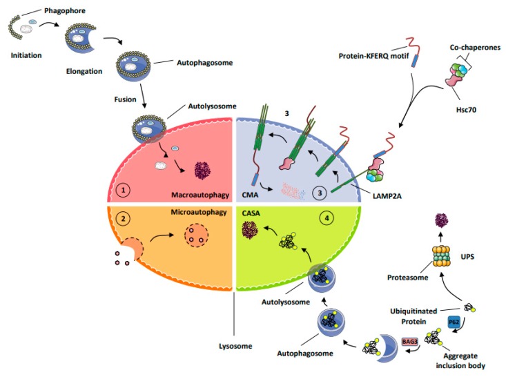
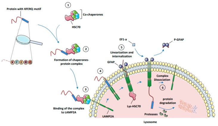
Chaperone-mediated autophagy (CMA) ensures the selective degradation of cellular proteins endowed with a KFERQ-like motif by lysosomes. It is estimated that 30% of all cellular proteins can be directed to the lysosome for CMA degradation, but only a few substrates have been formally identified so far. Mechanistically, the KFERQ-like motifs present in substrate proteins are recognized by the molecular chaperone Hsc70c (Heat shock cognate 71 kDa protein cytosolic), also known as HSPA8, and directed to LAMP2A, which acts as the CMA receptor at the lysosomal surface. Following linearization, the protein substrate is next transported to the lumen of the lysosomes, where it is degraded by resident proteases, mainly cathepsins and eventually recycled to sustain cellular homeostasis. CMA is induced by different stress conditions, including energy deprivation that also activates macro-autophagy (MA), that may make it difficult to decipher the relative impact of both pathways on cellular homeostasis. Besides common inducing triggers, CMA and MA might be induced as compensatory mechanisms when either mechanism is altered, as it is the often the case in different pathological settings. Therefore, CMA activation can compensate for alterations of MA and vice versa. In this context, these compensatory mechanisms, when occurring, may be targeted for therapeutic purposes. Both processes have received particular attention from scientists and clinicians, since modulation of MA and CMA may have a profound impact on cellular proteostasis, metabolism, death, differentiation, and survival and, as such, could be targeted for therapeutic intervention in degenerative and immune diseases, as well as in cancer, including hematopoietic malignancies. The role of MA in cancer initiation and progression is now well established, but whether and how CMA is involved in tumorigenesis has been only sparsely explored. In the present review, we encompass the description of the mechanisms involved in CMA, its function in the physiology and pathogenesis of hematopoietic cells, its emerging role in cancer initiation and development, and, finally, the potential therapeutic opportunity to target CMA or CMA-mediated compensatory mechanisms in hematological malignancies.
Overview
The claims and statistics of this paper haven’t been checked yet.
Run the check — free, no account needed. It reads the open-access text and checks the sources it cites.
Integrity signals
Registry
Cites
Statistics
Figures
Verification
Claims the paper makes
The claims and statistics of this paper haven’t been checked yet.
Run the check — free, no account needed. It reads the open-access text and checks the sources it cites.
Claims other papers make about this one
Each is checked against this paper’s own text: does it say what the citing paper says it does?
In the citing paper
“This approach can be applied to RhoH as the presence of the inserted domain, LFSINE, at the C-terminal was shown to act as a recognition signal for lysosomal uptake and CMA-mediated degradation, while not affecting its function on both T cells and B cells [26].”
From The Role of RhoH in TCR Signalling and Its Involvement in Diseases (2021) · cites this paper as [112]
In the citing paper
“Additionally, deregulated RhoH protein expression can be corrected by using the current approach that relies on the potential therapeutic opportunity to target CMA [112].”
From The Role of RhoH in TCR Signalling and Its Involvement in Diseases (2021) · cites this paper as [112]
Self-check
Percentages, sums and reported differences re-computed from the paper’s own numbers.
The self-check needs the paper’s full text.
Citations
Cites
1 retracted
RETRACTED ARTICLE: LAMP2 expression dictates azacytidine response and prognosis in MDS/AML
Dubois, A., Furstoss, N., Calleja, A., Zerhouni, M., Cluzeau, T., Savy, C., Marchetti, S., Hamouda, M. A., Boulakirba, S., Orange, F., et al. (2019) · Leukemia · doi:10.1038/s41375-018-0336-1 · Cited by 24
Self-citationRetractedRepertoires of Autophagy in the Pathogenesis of Ocular Diseases
Li, Y.-J., Jiang, Q., Cao, G.-F., Yao, J., & Yan, B. (2015) · Cellular Physiology and Biochemistry · doi:10.1159/000373980 · Cited by 16,304
Genomic classification and prognosis in acute myeloid leukemia
Papaemmanuil, E., Gerstung, M., Bullinger, L., Gaidzik, V. I., Paschka, P., Roberts, N. D., Potter, N. E., Heuser, M., Thol, F., Bolli, N., et al. (2016) · New England Journal of Medicine · doi:10.1056/nejmoa1516192 · Cited by 4,350
Tissue fractionation studies. 6. Intracellular distribution patterns of enzymes in rat-liver tissue
de Duve, C., Pressman, B. C., Gianetto, R., Wattiaux, R., & Appelmans, F. (1955) · Biochemical Journal · doi:10.1042/bj0600604 · Cited by 4,174
de Duve, C., & Wattiaux, R. (1966) · Annual Review of Physiology · doi:10.1146/annurev.ph.28.030166.002251 · Cited by 3,033 · cited 1×
“The term micro-autophagy was introduced by de Duve and Wattiaux at the end of the sixties to describe a process involving the invagination of the lysosomal membrane to sequestrate small cytoplasmic constituents for degradation [7].”
BackgroundAdapting Proteostasis for Disease Intervention
Balch, W. E., Morimoto, R. I., Dillin, A., & Kelly, J. W. (2008) · Science · doi:10.1126/science.1141448 · Cited by 2,495
Rel/NF-kappa B/I kappa B family: intimate tales of association and dissociation.
Verma, I. M., Stevenson, J. K., Schwarz, E. M., Van Antwerp, D., & Miyamoto, S. (1995) · Genes & Development · doi:10.1101/gad.9.22.2723 · Cited by 1,724
Lysosome biogenesis and lysosomal membrane proteins: trafficking meets function
Saftig, P., & Klumperman, J. (2009) · Nature Reviews Molecular Cell Biology · doi:10.1038/nrm2745 · Cited by 1,648
Accumulation of autophagic vacuoles and cardiomyopathy in LAMP-2-deficient mice
Tanaka, Y., Guhde, G., Suter, A., Eskelinen, E.-L., Hartmann, D., Lüllmann-Rauch, R., Janssen, P. M. L., Blanz, J., von Figura, K., & Saftig, P. (2000) · Nature · doi:10.1038/35022595 · Cited by 925
Microautophagy of Cytosolic Proteins by Late Endosomes
Sahu, R., Kaushik, S., Clement, C. C., Cannizzo, E. S., Scharf, B., Follenzi, A., Potolicchio, I., Nieves, E., Cuervo, A. M., & Santambrogio, L. (2011) · Developmental Cell · doi:10.1016/j.devcel.2010.12.003 · Cited by 902
Cited by
The Role of RhoH in TCR Signalling and Its Involvement in Diseases
Ahmad Mokhtar, A. M., Hashim, I. F., Mohd Zaini Makhtar, M., Salikin, N. H., & Amin-Nordin, S. (2021) · Cells · doi:10.3390/cells10040950 · Cited by 25
Metadata
- Authors
- Robert, G., Jacquel, A., & Auberger, P.
- Affiliations
- Mediterranean Center for Molecular Medicine ,Université Nice Côte d’Azur, C3M/Inserm1065, 06100 Nice, France
- Venue
- Cells
- Publisher
- MDPI AG
- Published
- 2019-10-16
- Volume
- 8
- Issue
- 10
- Pages
- 1260
- ISSN
- 2073-4409
- PMC
- PMC6830112
- Cited by
- 45
- References
- 118
- Language
- en
- Funders
- Fondation ARC pour la Recherche sur le Cancer (Equipe labellisée 2017-19-PGA1 RF20170205389)
- Institut National Du Cancer (PRTK 2017-2019)
About these checks
Registry, retraction, Cites and statistics results are computed, not judged, with one exception: a printed reference that no index lists is read once by a model before it counts as a citation error. Claims are extracted by a model and kept only when their quote appears verbatim in the text; each cited source is then checked, with a model’s help, for whether it says what the claim relies on. Those verdicts can be wrong — read the claim’s quote and the cited source before relying on one.
Statistics and arithmetic are recomputed, not judged.
Confidence bands are shown only for fields calibrated on our annotated test set. None is calibrated yet, so no band is shown.
What counts as a claim ›How we measure confidence ›How we check this ›
Are you an author, or do you see a problem with this page? Request a review
Data sources
Bibliographic data: Semantic Scholar, OpenAlex, Crossref.
Contains information from the Semantic Scholar Academic Graph Datasets, made available under the ODC Attribution License (ODC-BY 1.0).
Semantic Scholar’s data is described in: Kinney, R., et al. (2023). The Semantic Scholar Open Data Platform. arXiv:2301.10140.
