

The DOI matches its Crossref record; 249 of the 249 references it cites were found in a registry or an index.
Changes in Regenerative Capacity through Lifespan
Open accessAbstract
Most organisms experience changes in regenerative abilities through their lifespan. During aging, numerous tissues exhibit a progressive decline in homeostasis and regeneration that results in tissue degeneration, malfunction and pathology. The mechanisms responsible for this decay are both cell intrinsic, such as cellular senescence, as well as cell-extrinsic, such as changes in the regenerative environment. Understanding how these mechanisms impact on regenerative processes is essential to devise therapeutic approaches to improve tissue regeneration and extend healthspan. This review offers an overview of how regenerative abilities change through lifespan in various organisms, the factors that underlie such changes and the avenues for therapeutic intervention. It focuses on established models of mammalian regeneration as well as on models in which regenerative abilities do not decline with age, as these can deliver valuable insights for our understanding of the interplay between regeneration and aging.
Overview
The claims and statistics of this paper haven’t been checked yet.
Free: it reads the open-access text and checks the first 5 claims that cite a source we can check. Create a free account to check every claim.
Integrity signals
Registry
Cites
Statistics
Figures



Verification
Claims the paper makes
The claims and statistics of this paper haven’t been checked yet.
Free: it reads the open-access text and checks the first 5 claims that cite a source we can check. Create a free account to check every claim.
Claims other papers make about this one
Each is checked against this paper’s own text: does it say what the citing paper says it does?
The source backs part of the claim, not all of it — or backs it for a different population, condition or comparison.
In the citing paper
“In mammals, one of the first systems to lose regenerative potential is the CNS5–7.”
From Reprogramming to recover youthful epigenetic information and restore vision (2020) · cites this paper as [7]
Experimental — accuracy not yet measuredThe cited source says what the claim attributes to it.
In the citing paper
“Over time, the central nervous system (CNS) loses function and regenerative capacity5–7.”
From Reprogramming to recover youthful epigenetic information and restore vision (2020) · cites this paper as [7]
Self-check
Percentages, sums and reported differences re-computed from the paper’s own numbers.
The self-check needs the paper’s full text.
Citations
Cites
Items 201–249 of 249
Regulation of p53 is critical for vertebrate limb regeneration
Yun, M. H., Gates, P. B., & Brockes, J. P. (2013) · Proceedings of the National Academy of Sciences · doi:10.1073/pnas.1310519110 · Cited by 121 · cited 2×
“regeneration of full limbs in salamanders, by promoting the dedifferentiation of adult cells to generate progenitors for regeneration [50].”
BackgroundSelf-citationEpithelial cell senescence impairs repair process and exacerbates inflammation after airway injury
Zhou, F. J., Onizawa, S., Nagai, A., & Aoshiba, K. (2011) · Respiratory Research · doi:10.1186/1465-9921-12-78 · Cited by 119 · cited 1×
“repair and regenerative capacities [231].”
Spinal cord regeneration: Lessons for mammals from non‐mammalian vertebrates
Lee‐Liu, D., Edwards‐Faret, G., Tapia, V. S., & Larraín, J. (2013) · genesis · doi:10.1002/dvg.22406 · Cited by 117 · cited 1×
“In humans and mice, adult neurogenesis occurs in the hippocampus and the olfactory bulb [25].”
BackgroundInvolvement of the INK4a/Arf gene locus in senescence
Collins, C. J., & Sedivy, J. M. (2003) · Aging Cell · doi:10.1046/j.1474-9728.2003.00048.x · Cited by 113 · cited 1×
“arrest [215–219].”
Epigenetic regulation of aging stem cells
Pollina, E. A., & Brunet, A. (2011) · Oncogene · doi:10.1038/onc.2011.45 · Cited by 111 · cited 1×
“are frequent during aging and have recently emerged as potentially critical factors contributing to ageassociated decline in regenerative abilities [120].”
BackgroundThe Hydra model: disclosing an apoptosis-driven generator of Wnt-based regeneration
Galliot, B., & Chera, S. (2010) · Trends in Cell Biology · doi:10.1016/j.tcb.2010.05.006 · Cited by 110 · cited 1×
“Two remarkable organisms, the freshwater cnidarian hydra and the planarian flatworm represent the most extreme case, as they are able to regenerate whole bodies from a single fragment [37–39].”
BackgroundImmortality and the base of multicellular life: Lessons from cnidarian stem cells
Watanabe, H., Hoang, V. T., Mättner, R. A., & Holstein, T. W. (2009) · Seminars in Cell and Developmental Biology · doi:10.1016/j.semcdb.2009.09.008 · Cited by 108 · cited 1×
“Two remarkable organisms, the freshwater cnidarian hydra and the planarian flatworm represent the most extreme case, as they are able to regenerate whole bodies from a single fragment [37–39].”
BackgroundAging and Regeneration in Vertebrates
Sousounis, K., Baddour, J., & Tsonis, P. A. (2014) · Current topics in developmental biology/Current Topics in Developmental Biology · doi:10.1016/b978-0-12-391498-9.00008-5 · Cited by 107 · cited 2×
“Interestingly, recent gene expression analysis during lens regeneration in adult newts, a process unaffected by aging, revealed an upregulation of DNA repair genes during regeneration [14,67].”
BackgroundWahlestedt, M., Norddahl, G. L., Sten, G., Ugale, A., Frisk, M.-A. M., Mattsson, R., Deierborg, T., Sigvardsson, M., & Bryder, D. (2013) · Blood · doi:10.1182/blood-2012-11-469080 · Cited by 106 · cited 1×
“This strategy has been successfully used to reprogramme aged haematopoietic progenitors [240] and T cells [241,242], and the resulting iPSC were differentiated following expansion into the respective cell types, leading to “rejuvenated””
MethodTelomerase Protects Werner Syndrome Lineage-Specific Stem Cells from Premature Aging
Cheung, H.-H., Liu, X., Canterel-Thouennon, L., Li, L., Edmonson, C., & Rennert, O. M. (2014) · Stem Cell Reports · doi:10.1016/j.stemcr.2014.02.006 · Cited by 102
Hematopoietic Stem Cell Aging: Wrinkles In Stem Cell Potential
Chambers, S. M., & Goodell, M. A. (2007) · Stem Cell Reviews and Reports · doi:10.1007/s12015-007-0027-1 · Cited by 100 · cited 1×
“kinetics and skewed differentiation towards myeloid lineages [18–21].”
BackgroundThe molecular basis of amphibian limb regeneration: integrating the old with the new
Gardiner, D. M., Endo, T., & Bryant, S. V. (2002) · Seminars in Cell and Developmental Biology · doi:10.1016/s1084952102000903 · Cited by 95 · cited 1×
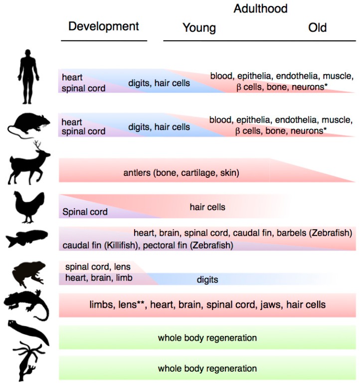
“Among vertebrates, these organisms are considered the champions of regeneration, as they are able to regrow a wide range of structures including parts of their heart and brain, jaws, entire tails (including the spinal cord), lens and hair cells, and they are the only vertebrate able to regenerate full limbs in the adu…”
BackgroundSehm, T., Sachse, C., Frenzel, C., & Echeverri, K. (2009) · Developmental Biology · doi:10.1016/j.ydbio.2009.08.008 · Cited by 95
Fülöp, T., Franceschi, C., Hirokawa, K., & Pawelec, G. (2009) · Springer eBooks · doi:10.1007/978-1-4020-9063-9 · Cited by 92
Lens regeneration in axolotl: new evidence of developmental plasticity
Suetsugu‐Maki, R., Maki, N., Nakamura, K., Sumanas, S., Zhu, J., Rio‐Tsonis, K. D., & Tsonis, P. A. (2012) · BMC Biology · doi:10.1186/1741-7007-10-103 · Cited by 88 · cited 1×
“However, there are other examples among amphibians that do not seem to be related to metamorphosis-induced changes, such as the case of the axolotl lens, which can only be regenerated for a period of two weeks after hatching [12].”
BackgroundExperimentally induced metamorphosis in axolotls reduces regenerative rate and fidelity
Monaghan, J. R., Stier, A. C., Michonneau, F., Smith, M. D., Pasch, B. S., Maden, M., & Seifert, A. W. (2014) · Regeneration · doi:10.1002/reg2.8 · Cited by 87
Gannon, H. S., Donehower, L. A., Lyle, S., & Jones, S. N. (2011) · Developmental Biology · doi:10.1016/j.ydbio.2011.02.007 · Cited by 87 · cited 1×
“suggest that an increase in p53 expression or activity levels can lead to aging and reduced lifespan, and lead to stem cell death in response to telomere attrition [74–76].”
BackgroundImpact of Aging on Rat Bone Marrow-Derived Stem Cell Chondrogenesis
Zheng, H., Martin, J. A., Duwayri, Y. M., Falcon, G., & Buckwalter, J. A. (2007) · The Journals of Gerontology Series A · doi:10.1093/gerona/62.2.136 · Cited by 85 · cited 1×
“Alterations include a loss in chondrogenic potential leading to impaired chondrocyte formation, which results in decreased cartilage repair in aged mammals [22].”
BackgroundFlores, I., & Blasco, M. A. (2009) · PLoS ONE · doi:10.1371/journal.pone.0004934 · Cited by 83 · cited 1×
“and improved wound healing responses and functionality in various tissues such as intestines, skin and the haematopoietic system [95,96].”
BackgroundHair cell regeneration in sensory epithelia from the inner ear of a urodele amphibian
Taylor, R., & Forge, A. (2005) · The Journal of Comparative Neurology · doi:10.1002/cne.20450 · Cited by 82 · cited 1×
“In contrast to salamanders, birds and fish, which can regenerate their hair cells through adulthood [7], in mammals this is only possible during development [8].”
BackgroundMacrophages decide between regeneration and fibrosis in muscle
Muñoz‐Cánoves, P., & Serrano, A. L. (2015) · Trends in Endocrinology and Metabolism · doi:10.1016/j.tem.2015.07.005 · Cited by 81 · cited 1×
“regeneration and tissue repair contexts, as demonstrated in various systems including the mammalian muscle [198,199], the zebrafish brain [200] and fin [201], and the axolotl limb [202].”
BackgroundLife-long preservation of the regenerative capacity in the fin and heart in zebrafish
Itou, J., Kawakami, H., Burgoyne, T., & Kawakami, Y. (2012) · Biology Open · doi:10.1242/bio.20121057 · Cited by 79 · cited 1×
“Notably, detailed studies in young and old individuals have revealed that the ability to regenerate the caudal fin, the barbels and the heart does not decrease with age [43,44] or repetitive regeneration cycles [44–46] although age and sex-related differences do affect regeneration of the pectoral fin [47].”
BackgroundLeClair, E. E., & Topczewski, J. (2010) · PLoS ONE · doi:10.1371/journal.pone.0008737 · Cited by 74 · cited 1×
“Notably, detailed studies in young and old individuals have revealed that the ability to regenerate the caudal fin, the barbels and the heart does not decrease with age [43,44] or repetitive regeneration cycles [44–46] although age and sex-related differences do affect regeneration of the pectoral fin [47].”
BackgroundAlterations in the TGFβ signaling pathway in myogenic progenitors with age
Beggs, M. L., Nagarajan, R., Taylor‐Jones, J. M., Nolen, G., MacNicol, M., & Peterson, C. A. (2004) · Aging Cell · doi:10.1111/j.1474-9728.2004.00135.x · Cited by 74 · cited 1×
“tissue upon muscle injury [13,15].”
Revisiting the relationship between regenerative ability and aging
Seifert, A. W., & Voss, S. R. (2013) · BMC Biology · doi:10.1186/1741-7007-11-2 · Cited by 73 · cited 2×
“First, tissue regeneration tends to be high during early life stages, and various regenerative time-windows have been defined through ontogenesis [4].”
BackgroundWalters, B. J., & Zuo, J. (2012) · Hearing Research · doi:10.1016/j.heares.2012.11.009 · Cited by 56 · cited 2×
“regulators such as retinoic acid and growth factors underlie changes in hair cell regenerative capacity [8].”
BackgroundLack of age‐associated telomere shortening in long‐ and short‐lived species of sea urchins
Francis, N. L., Gregg, T., Owen, R., Ebert, T., & Bodnar, A. G. (2006) · FEBS Letters · doi:10.1016/j.febslet.2006.07.049 · Cited by 56 · cited 1×
“vertebrate species such as sea urchins and planarians, species in which tumour formation is rarely observed [99,100], suggesting that active telomere extension could contribute to extend regenerative capacities upon aging, and do so in a tumour-free manner.”
BackgroundSexually Dimorphic Fin Regeneration in Zebrafish Controlled by Androgen/GSK3 Signaling
Nachtrab, G. T., Czerwinski, M. J., & Poss, K. D. (2011) · Current Biology · doi:10.1016/j.cub.2011.09.050 · Cited by 56 · cited 1×
“Notably, detailed studies in young and old individuals have revealed that the ability to regenerate the caudal fin, the barbels and the heart does not decrease with age [43,44] or repetitive regeneration cycles [44–46] although age and sex-related differences do affect regeneration of the pectoral fin [47].”
BackgroundYun, M. H., Gates, P. B., & Brockes, J. P. (2014) · Stem Cell Reports · doi:10.1016/j.stemcr.2014.05.009 · Cited by 55 · cited 1×
“This process involves the reversion of certain epigenetic modifications [246,247], but whether”
BackgroundSelf-citationTissue regeneration after injury in adult zebrafish: The regenerative potential of the caudal fin
Shao, J., Chen, D., Ye, Q., Cui, J., Li, Y., & Li, L. (2011) · Developmental Dynamics · doi:10.1002/dvdy.22603 · Cited by 52 · cited 1×
“Notably, detailed studies in young and old individuals have revealed that the ability to regenerate the caudal fin, the barbels and the heart does not decrease with age [43,44] or repetitive regeneration cycles [44–46] although age and sex-related differences do affect regeneration of the pectoral fin [47].”
BackgroundPrice, J. S., Oyajobi, B. O., Oreffo, R. O. C., & Russell, R. G. G. (1994) · Journal of Endocrinology · doi:10.1677/joe.0.143r009 · Cited by 51 · cited 1×
“The role of hormones as systemic regulators of regeneration is elegantly exemplified by the phenomenon of deer antler regeneration, one of the fastest organogenesis processes in the natural world, which is linked to the annual breeding cycle and hence controlled by fluctuations in the concentration of sex hormones suc…”
BackgroundDNA methylation in adult stem cells: New insights into self-renewal
Trowbridge, J. J., & Orkin, S. H. (2010) · Epigenetics · doi:10.4161/epi.5.3.11374 · Cited by 42 · cited 1×
“In support of this idea, modulators of DNA methylation are required for the function of many adult stem cells [126–130].”
BackgroundSousounis, K., Looso, M., Maki, N., Ivester, C. J., Braun, T. W., & Tsonis, P. A. (2013) · PLoS ONE · doi:10.1371/journal.pone.0061445 · Cited by 42 · cited 1×
“Interestingly, recent gene expression analysis during lens regeneration in adult newts, a process unaffected by aging, revealed an upregulation of DNA repair genes during regeneration [14,67].”
BackgroundGéraudie, J., & Singer, M. (1985) · Journal of Experimental Zoology · doi:10.1002/jez.1402340306 · Cited by 40 · cited 1×
“annelid segments [178,179].”
Hamada, Y., Bando, T., Nakamura, T., Ishimaru, Y., Mito, T., Noji, S., Tomioka, K., & Ohuchi, H. (2015) · Development · doi:10.1242/dev.122598 · Cited by 40
Carrasco‐García, E., Arrizabalaga, O., Serrano, M., Lovell-Badge, R. H., & Matheu, A. (2015) · Aging Cell · doi:10.1111/acel.12343 · Cited by 37 · cited 1×
“Surprisingly, these mice show a delayed onset of aging, extended lifespan and normal regeneration and wound healing phenotypes [79,80].”
BackgroundWitman, N., Heigwer, J., Thaler, B., Lui, W., & Morrison, J. I. (2013) · Developmental Biology · doi:10.1016/j.ydbio.2013.09.011 · Cited by 33 · cited 1×
“Furthermore, miRNA regulation is important for regeneration in species that do not exhibit an age-associated decline in this process such zebrafish [144,145] and salamanders [146,147].”
BackgroundDissecting the role of mTOR complexes in cellular senescence
Serrano, M. (2012) · Cell Cycle · doi:10.4161/cc.21065 · Cited by 30 · cited 1×
“In this connection, it is worth noting that mTOR activity is regulated by nutrient availability, and has also been implicated in the regulation of cellular senescence [157,158].”
BackgroundFletcher, T. (1978) · Hormones and Behavior · doi:10.1016/0018-506x(78)90059-4 · Cited by 28 · cited 1×
“The role of hormones as systemic regulators of regeneration is elegantly exemplified by the phenomenon of deer antler regeneration, one of the fastest organogenesis processes in the natural world, which is linked to the annual breeding cycle and hence controlled by fluctuations in the concentration of sex hormones suc…”
BackgroundAging profoundly delays functional recovery from gustatory nerve injury
He, L., Yadgarov, A., Sharif, S., & McCluskey, L. P. (2012) · Neuroscience · doi:10.1016/j.neuroscience.2012.02.012 · Cited by 27 · cited 2×
“Indeed, a decline in gustatory nerve regeneration is associated with the impairment in taste bud regeneration in aging mammals [27].”
Background(2013) · Wiley eBooks · doi:10.1002/9781118300312 · Cited by 21
Plasticity of Mammalian Myotubes Upon Stimulation with a Thrombin-activated Serum Factor
Lööf, S., Straube, W. L., Drechsel, D. N., Tanaka, E. M., & Simon, A. (2007) · Cell Cycle · doi:10.4161/cc.6.9.4141 · Cited by 20 · cited 1×
“This process involves the reversion of certain epigenetic modifications [246,247], but whether”
BackgroundMemoirs of a Reincarnated T Cell
Crompton, J. G., Rao, M. S., & Restifo, N. P. (2013) · Cell stem cell · doi:10.1016/j.stem.2012.12.009 · Cited by 12 · cited 1×
“Likewise, it is not clear if reprogrammed cells are indeed rejuvenated [243].”
BackgroundSchmuck, E. G., Mulligan, J. D., & Saupe, K. W. (2010) · AGE · doi:10.1007/s11357-010-9166-4 · Cited by 12 · cited 1×
“For example, multiple cycles of fasting have been shown to improve HSC self-renewal and regenerative capacities, in an IGF1-dependent manner [165], while short-term caloric restriction can revert age-associated increases in adipose-derived stem cells [166] and leads to enhancements in muscle stem cell function and rep…”
BackgroundCurrent advances in tissue repair and regeneration: the future is bright
Ninov, N., & Yun, M. H. (2015) · Regeneration · doi:10.1002/reg2.30 · Cited by 11 · cited 1×
“important signals for regenerative responses in highly regenerative organisms, such as hydra, drosophila, planarians, zebrafish and amphibians [105,106].”
BackgroundSelf-citationMargotta, V., Filoni, S., Merante, A., & Chimenti, C. A. (2002) · PubMed · Cited by 11
microRNA biogenesis and senescence
Gómez-Cabello, D., Adrados, I., & Palmero, I. (2013) · Aging · doi:10.18632/aging.100607 · Cited by 10 · cited 1×
“cellular senescence [143].”
Stem Cells and Aging: What’s Next?
Artandi, S. E., Blau, H. M., de Haan, G., Geiger, H., Goodell, M. A., Jones, D. L., Levine, R. L., Muñoz‐Cánoves, P., Rodewald, H.-R., Wagers, A. J., et al. (2015) · Cell stem cell · doi:10.1016/j.stem.2015.05.005 · Cited by 8 · cited 1×
“terms, and whether any of the modifications discussed herein can actually induce cells to revert to a more youthful status [244].”
BackgroundThe Emergence of GeroMIRs: A Group of MicroRNAs Implicated in Aging
Ugalde, A. P., Kwarciak, A., Caravia, X. M., López-Otín, C., & Ramsay, A. J. (2013) · Wiley eBooks · doi:10.1002/9781118300312.ch5 · Cited by 5 · cited 1×
“These age-associated epigenetic modifications could underlie some of the transcriptional signatures associated with aging, such as the dysregulation of gero-miRNAs [140], a miRNA class which has been shown to affect longevity in Caenorhabditis elegans [141] and Drosophila melanogaster [142] as”
Background
Research works printed but not linked: 1
- Finch, C.E. (1994). Longevity, Senescence and the Genome, The University of Chicago Press.
Cited by
Reprogramming to recover youthful epigenetic information and restore vision
Lu, Y., Brommer, B., Tian, X., Krishnan, A., Meer, M., Wang, C., Vera, D. L., Zeng, Q., Yu, D., Bonkowski, M. S., et al. (2020) · Nature · doi:10.1038/s41586-020-2975-4 · Cited by 820
Model systems for regeneration: salamanders
Joven, A., Elewa, A., & Simon, A. (2019) · Development · doi:10.1242/dev.167700 · Cited by 191
“In fact, among tetrapods, salamanders exhibit the widest range of regenerative capacity, with an impressive ability to regrow tissues, organs and entire body parts (Tanaka, 2003; Yun, 2015).”
BackgroundBiomaterials to enhance stem cell transplantation
Kharbikar, B. N., Mohindra, P., & Desai, T. A. (2022) · Cell Stem Cell · doi:10.1016/j.stem.2022.04.002 · Cited by 172
Scarring vs. functional healing: Matrix-based strategies to regulate tissue repair
Keane, T. J., Horejs, C., & Stevens, M. M. (2018) · Advanced Drug Delivery Reviews · doi:10.1016/j.addr.2018.02.002 · Cited by 149
“In general, regenerative potential decreases with gestation time and continues to decline through lifespan, and it's clear that both cell-intrinsic (regenerative capacity) and cellextrinsic (regenerative environment) factors play a role in this decline [32].”
BackgroundMechanical state transitions in the regulation of tissue form and function
Mao, Y., & Wickström, S. A. (2024) · Nature Reviews Molecular Cell Biology · doi:10.1038/s41580-024-00719-x · Cited by 162
“Ageing is associated with changes in the mechanical properties of tissues, such as reduced elasticity, leading to altered mechanotransduction and mechanosensitivity, which compromises tissue form and function (one example being a decline in repair capacity) 203 .”
Metadata
- Authors
- Yun, M.
- Affiliations
- Institute of Structural and Molecular Biology (GB)
- Corresponding author
- Yun, Maximina
- ORCID
- Yun, Maximina
- Venue
- International Journal of Molecular Sciences
- Publisher
- MDPI AG
- Published
- 2015-10-23
- Volume
- 16
- Issue
- 10
- Pages
- 25392-25432
- ISSN
- 1422-0067 (ISSN-L)
- PMC
- PMC4632807
- OpenAlex
- W1834172848
- Cited by
- 216
- References
- 249
- Citations by year
- 2026: 19 · 2025: 26 · 2024: 28 · 2023: 13 · 2022: 28 · 2021: 35 · 2020: 19 · 2019: 15 · 2018: 20 · 2017: 7 · 2016: 5
- FWCI
- 0.94
- Type
- journal article
- Language
- en
- Open access
- gold
- dx.doi.org
- Licence
- Locations
- International Journal of Molecular Sciences (publishedVersion, cc-by)
- PubMed Central (publishedVersion, cc-by)
- PubMed (publishedVersion)
- DOAJ (DOAJ: Directory of Open Access Journals) (submittedVersion, cc-by-sa)
- UCL Discovery (University College London) (submittedVersion)
- Topics
- Life Sciences › Biochemistry, Genetics and Molecular Biology › Aging › Genetics, Aging, and Longevity in Model Organisms
- Health Sciences › Medicine › Surgery › Tissue Engineering and Regenerative Medicine
- Life Sciences › Biochemistry, Genetics and Molecular Biology › Molecular Biology › Planarian Biology and Electrostimulation
- Keywords
- regenerative capacity, aging, tissue regeneration, cellular senescence, healthspan extension, tissue homeostasis, regenerative therapy
- MeSH terms
- Aging, Animals, Humans, Regeneration, Stem Cells, Cellular Senescence, Tissue Engineering, Cell Proliferation
About these checks
Registry, retraction, Cites and statistics results are computed, not judged, with one exception: a printed reference that no index lists is read once by a model before it counts as a citation error. Claims are extracted by a model and kept only when their quote appears verbatim in the text; each cited source is then checked, with a model’s help, for whether it says what the claim relies on. Those verdicts can be wrong — read the claim’s quote and the cited source before relying on one.
Statistics and arithmetic are recomputed, not judged.
Confidence bands are shown only for fields calibrated on our annotated test set. None is calibrated yet, so no band is shown.
What counts as a claim ›How we measure confidence ›How we check this ›
Are you an author, or do you see a problem with this page? Request a review
Data sources
Bibliographic data: Semantic Scholar, OpenAlex, Crossref.
Contains information from the Semantic Scholar Academic Graph Datasets, made available under the ODC Attribution License (ODC-BY 1.0).
Semantic Scholar’s data is described in: Kinney, R., et al. (2023). The Semantic Scholar Open Data Platform. arXiv:2301.10140.
