

A p53-Dependent Response Limits Epidermal Stem Cell Functionality and Organismal Size in Mice with Short Telomeres
Open accessThe DOI matches its Crossref record; 46 of the 46 references it cites were found in a registry or an index.
Abstract
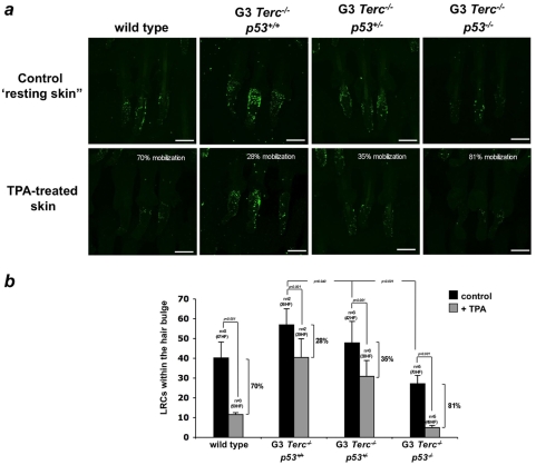
Telomere maintenance is essential to ensure proper size and function of organs with a high turnover. In particular, a dwarf phenotype as well as phenotypes associated to premature loss of tissue regeneration, including the skin (hair loss, hair graying, decreased wound healing), are found in mice deficient for telomerase, the enzyme responsible for maintaining telomere length. Coincidental with the appearance of these phenotypes, p53 is found activated in several tissues from these mice, where is thought to trigger cellular senescence and/or apoptotic responses. Here, we show that p53 abrogation rescues both the small size phenotype and restitutes the functionality of epidermal stem cells (ESC) of telomerase-deficient mice with dysfunctional telomeres. In particular, p53 ablation restores hair growth, skin renewal and wound healing responses upon mitogenic induction, as well as rescues ESCmobilization defects in vivo and defective ESC clonogenic activity in vitro. This recovery of ESC functions is accompanied by a downregulation of senescence markers and an increased proliferation in the skin and kidney of telomerase-deficient mice with critically short telomeres without changes in apoptosis rates. Together, these findings indicate the existence of a p53-dependent senescence response acting on stem/progenitor cells with dysfunctional telomeres that is actively limiting their contribution to tissue regeneration, thereby impinging on tissue fitness.
Overview
The claims and statistics of this paper haven’t been checked yet.
Free: it reads the open-access text and checks the first 5 claims that cite a source we can check. Create a free account to check every claim.
Integrity signals
Registry
Cites
Statistics
Figures




Verification
Claims the paper makes
The claims and statistics of this paper haven’t been checked yet.
Free: it reads the open-access text and checks the first 5 claims that cite a source we can check. Create a free account to check every claim.
Claims other papers make about this one
Each is checked against this paper’s own text: does it say what the citing paper says it does?
The source backs part of the claim, not all of it — or backs it for a different population, condition or comparison.
In the citing paper
“Skin aging associated with ROS accumulation, thus, was found to be rescued in the skin of late-generation p53 knockout mice [120].”
From Ultraviolet Radiation‐Induced Skin Aging: The Role of DNA Damage and Oxidative Stress in Epidermal Stem Cell Damage Mediated Skin Aging (2016) · cites this paper as [120]
Experimental — accuracy not yet measuredThe source backs part of the claim, not all of it — or backs it for a different population, condition or comparison.
In the citing paper
“In these mice, there were improved wound healing, hair growth, and skin renewal to levels comparable to those of wild-type mice, as well as increased ESC numbers and improved mobilization capacity [120].”
From Ultraviolet Radiation‐Induced Skin Aging: The Role of DNA Damage and Oxidative Stress in Epidermal Stem Cell Damage Mediated Skin Aging (2016) · cites this paper as [120]
Experimental — accuracy not yet measured
Self-check
Percentages, sums and reported differences re-computed from the paper’s own numbers.
The self-check needs the paper’s full text.
Citations
Cites
Shelterin: the protein complex that shapes and safeguards human telomeres
de Lange, T. (2005) · Genes & Development · doi:10.1101/gad.1346005 · Cited by 3,056
A DNA damage checkpoint response in telomere-initiated senescence
Fagagna, F. D. D., Reaper, P. M., Clay-Farrace, L., Fiegler, H., Carr, P., von Zglinicki, T., Saretzki, G., Carter, N. P., & Jackson, S. P. (2003) · Nature · doi:10.1038/nature02118 · Cited by 2,917
Cotsarelis, G., Sun, T.-T., & Lavker, R. M. (1990) · Cell · doi:10.1016/0092-8674(90)90696-c · Cited by 2,276
Defining the Epithelial Stem Cell Niche in Skin
Tumbar, T., Guasch, G., Greco, V., Blanpain, C., Lowry, W. E., Rendl, M., & Fuchs, E. (2004) · Science · doi:10.1126/science.1092436 · Cited by 2,031
Cellular senescence in cancer and aging.
Collado, M., Blasco, M. A., & Serrano, M. (2007) · Cell · doi:10.1016/j.cell.2007.07.003 · Cited by 1,708
Self-citationBlanpain, C., Lowry, W. E., Geoghegan, A., Polak, L., & Fuchs, E. (2004) · Cell · doi:10.1016/j.cell.2004.08.012 · Cited by 1,428
DNA Damage Foci at Dysfunctional Telomeres
Takai, H., Smogorzewska, A., & de Lange, T. (2003) · Current Biology · doi:10.1016/s0960-9822(03)00542-6 · Cited by 1,409
Longevity, Stress Response, and Cancer in Aging Telomerase-Deficient Mice
Rudolph, K. L., Chang, S., Lee, H.-W., Blasco, M., Gottlieb, G. J., Greider, C., & DePinho, R. A. (1999) · Cell · doi:10.1016/s0092-8674(00)80580-2 · Cited by 1,349 · cited 5×
“, all mice were dead with in the first 40 weeks of life; not shown) prevented the analysis of the role of p53 in additional skin defects associated to critically short telomeres, such as hair graying and alopecia [27,28].”
BackgroundMethodInfluentialSelf-citationTelomeric Repeat–Containing RNA and RNA Surveillance Factors at Mammalian Chromosome Ends
Azzalin, C. M., Reichenbach, P., Khoriauli, L., Giulotto, E., & Lingner, J. (2007) · Science · doi:10.1126/science.1147182 · Cited by 1,329
Three clonal types of keratinocyte with different capacities for multiplication.
Barrandon, Y., & Green, H. (1987) · Proceedings of the National Academy of Sciences · doi:10.1073/pnas.84.8.2302 · Cited by 1,294
Cited by
Targeting p53 pathways: mechanisms, structures, and advances in therapy
Wang, H., Guo, M., Wei, H., & Chen, Y. (2023) · Signal Transduction and Targeted Therapy · doi:10.1038/s41392-023-01347-1 · Cited by 1,016
Linking functional decline of telomeres, mitochondria and stem cells during ageing
Sahin, E., & DePinho, R. A. (2010) · Nature · doi:10.1038/nature08982 · Cited by 759
Telomeric and extra-telomeric roles for telomerase and the telomere-binding proteins
Martínez, P., & Blasco, M. A. (2011) · Nature Reviews Cancer · doi:10.1038/nrc3025 · Cited by 468
Self-citationAxis of ageing: telomeres, p53 and mitochondria
Sahin, E., & DePinho, R. A. (2012) · Nature Reviews Molecular Cell Biology · doi:10.1038/nrm3352 · Cited by 345
Panich, U., Sittithumcharee, G., Rathviboon, N., & Jirawatnotai, S. (2016) · Stem Cells International · doi:10.1155/2016/7370642 · Cited by 374
“In these mice, there were improved wound healing, hair growth, and skin renewal to levels comparable to those of wild-type mice, as well as increased ESC numbers and improved mobilization capacity [120].”
Metadata
- Authors
- Flores, I., & Blasco, M. A.
- Affiliations
- Spanish National Cancer Research Centre (ES)
- ORCID
- Flores, Ignacio
- Venue
- PLoS ONE
- Publisher
- Public Library of Science (PLoS)
- Published
- 2009-03-19
- Volume
- 4
- Issue
- 3
- Pages
- e4934
- ISSN
- 1932-6203 (ISSN-L)
- PMC
- PMC2654505
- OpenAlex
- W1983400015
- Cited by
- 83
- References
- 46
- Citations by year
- 2026: 2 · 2025: 1 · 2024: 2 · 2023: 2 · 2022: 4 · 2021: 3 · 2020: 2 · 2019: 2 · 2018: 1 · 2016: 4 · 2015: 5 · 2014: 3 · 2013: 9 · 2012: 14
- FWCI
- 9.53
- Type
- journal article
- Language
- en
- Open access
- gold
- doi.org
- Locations
- PLoS ONE (publishedVersion, cc-by)
- PubMed Central (publishedVersion, cc-by)
- PubMed (publishedVersion)
- DOAJ (DOAJ: Directory of Open Access Journals) (submittedVersion, cc-by-sa)
- Figshare (submittedVersion, cc-by)
- Funders
- European Commission (FI6R-CT-2003 508842, RISC-RAD FI6R-CT-2003-508842, LSHC-CT-2003-506803, LSHC-CT-2004-502943, FIGH-CT-2002-00217, FOOD-CT-2003-506850)
- Topics
- Life Sciences › Biochemistry, Genetics and Molecular Biology › Cell Biology › Telomeres, Telomerase, and Senescence
- Health Sciences › Medicine › Dermatology › Hair Growth and Disorders
- Life Sciences › Biochemistry, Genetics and Molecular Biology › Aging › Genetics, Aging, and Longevity in Model Organisms
- Keywords
- p53, telomere shortening, epidermal stem cells, cellular senescence, tissue regeneration, hair growth, wound healing, stem cell mobilization, clonogenic activity, stem cell proliferation
- MeSH terms
- Epidermal Cells, Animals, Epidermis, Kidney, Phenotype, Stem Cells, Wound Healing, Tumor Suppressor Protein p53, Telomere, Cellular Senescence, Mice, Knockout, Hair Follicle, Telomerase, Cell Proliferation, Mice
About these checks
Registry, retraction, Cites and statistics results are computed, not judged, with one exception: a printed reference that no index lists is read once by a model before it counts as a citation error. Claims are extracted by a model and kept only when their quote appears verbatim in the text; each cited source is then checked, with a model’s help, for whether it says what the claim relies on. Those verdicts can be wrong — read the claim’s quote and the cited source before relying on one.
Statistics and arithmetic are recomputed, not judged.
Confidence bands are shown only for fields calibrated on our annotated test set. None is calibrated yet, so no band is shown.
What counts as a claim ›How we measure confidence ›How we check this ›
Are you an author, or do you see a problem with this page? Request a review
How to cite
Cite CoChat for the checks on this page, and the paper for its own findings.
- This page
- CoChat. (2026). Citation check: A p53-Dependent Response Limits Epidermal Stem Cell Functionality and Organismal Size in Mice with Short Telomeres. https://app.cochat.ai/papers/10.1371/journal.pone.0004934
- The paper
- Flores, I., & Blasco, M. A. (2009). A p53-Dependent Response Limits Epidermal Stem Cell Functionality and Organismal Size in Mice with Short Telomeres. PLoS ONE. https://doi.org/10.1371/journal.pone.0004934
Data sources
Bibliographic data: Semantic Scholar, OpenAlex, Crossref.
Contains information from the Semantic Scholar Academic Graph Datasets, made available under the ODC Attribution License (ODC-BY 1.0).
Semantic Scholar’s data is described in: Kinney, R., et al. (2023). The Semantic Scholar Open Data Platform. arXiv:2301.10140.
