

The DOI matches its Crossref record; 249 of the 249 references it cites were found in a registry or an index.
Changes in Regenerative Capacity through Lifespan
Open accessAbstract
Most organisms experience changes in regenerative abilities through their lifespan. During aging, numerous tissues exhibit a progressive decline in homeostasis and regeneration that results in tissue degeneration, malfunction and pathology. The mechanisms responsible for this decay are both cell intrinsic, such as cellular senescence, as well as cell-extrinsic, such as changes in the regenerative environment. Understanding how these mechanisms impact on regenerative processes is essential to devise therapeutic approaches to improve tissue regeneration and extend healthspan. This review offers an overview of how regenerative abilities change through lifespan in various organisms, the factors that underlie such changes and the avenues for therapeutic intervention. It focuses on established models of mammalian regeneration as well as on models in which regenerative abilities do not decline with age, as these can deliver valuable insights for our understanding of the interplay between regeneration and aging.
Overview
The claims and statistics of this paper haven’t been checked yet.
Free: it reads the open-access text and checks the first 5 claims that cite a source we can check. Create a free account to check every claim.
Integrity signals
Registry
Cites
Statistics
Figures
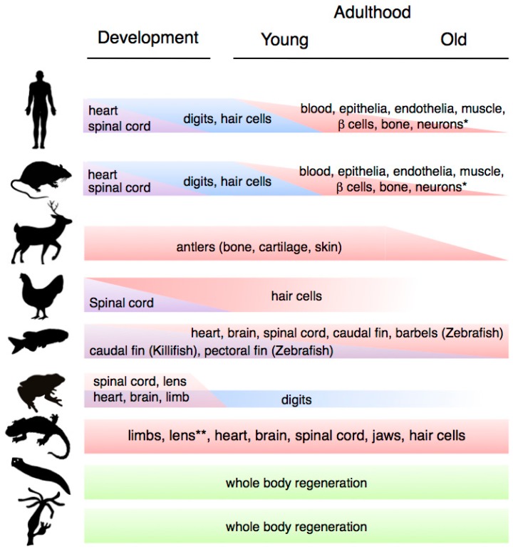


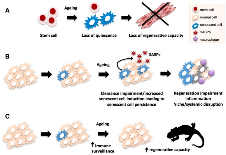

Verification
Claims the paper makes
The claims and statistics of this paper haven’t been checked yet.
Free: it reads the open-access text and checks the first 5 claims that cite a source we can check. Create a free account to check every claim.
Claims other papers make about this one
Each is checked against this paper’s own text: does it say what the citing paper says it does?
The source backs part of the claim, not all of it — or backs it for a different population, condition or comparison.
In the citing paper
“In mammals, one of the first systems to lose regenerative potential is the CNS5–7.”
From Reprogramming to recover youthful epigenetic information and restore vision (2020) · cites this paper as [7]
Experimental — accuracy not yet measuredThe cited source says what the claim attributes to it.
In the citing paper
“Over time, the central nervous system (CNS) loses function and regenerative capacity5–7.”
From Reprogramming to recover youthful epigenetic information and restore vision (2020) · cites this paper as [7]
Self-check
Percentages, sums and reported differences re-computed from the paper’s own numbers.
The self-check needs the paper’s full text.
Citations
Cites
López-Otín, C., Blasco, M. A., Partridge, L., Serrano, M., & Kroemer, G. (2013) · Cell · doi:10.1016/j.cell.2013.05.039 · Cited by 14,982
The serial cultivation of human diploid cell strains
Hayflick, L., & Moorhead, P. S. (1961) · Experimental Cell Research · doi:10.1016/0014-4827(61)90192-6 · Cited by 8,508 · cited 1×
“Telomere shortening, initially associated with the triggering of replicative senescence in proliferating cells in vitro [81], has been also observed in vivo during normal mammalian aging [82], in a number of regenerative cell types such as hair follicle [83] and hematopoietic stem cells [84].”
BackgroundA biomarker that identifies senescent human cells in culture and in aging skin in vivo.
Dimri, G. P., Lee, X., Basile, G., Acosta, M., Scott, G., Roskelley, C., Medrano, E. E., Linskens, M., Rubelj, I., & Pereira-Smith, O. (1995) · Proceedings of the National Academy of Sciences · doi:10.1073/pnas.92.20.9363 · Cited by 7,477 · cited 1×
“which include the expansion of mitochondrial and lysosomal networks (leading to high levels of senescence-associated-β-galactosidase (SAβgal) activity, the best-known senescent cell marker [220],”
BackgroundLane, D. P. (1992) · Nature · doi:10.1038/358015a0 · Cited by 5,138
Clearance of p16Ink4a-positive senescent cells delays ageing-associated disorders
Baker, D. J., Wijshake, T., Tchkonia, T., LeBrasseur, N. K., Childs, B. G., van de Sluis, B., Kirkland, J. L., & van Deursen, J. M. (2011) · Nature · doi:10.1038/nature10600 · Cited by 3,670 · cited 4×
“Studies in mouse models have shown its potential as a means to enhance regenerative abilities [59,232] as well as to improve a number of age-associated pathologies [228].”
BackgroundInfluentialMitochondrial membrane permeabilization in cell death
Kroemer, G., Galluzzi, L., & Brenner, C. (2007) · Physiological Reviews · doi:10.1152/physrev.00013.2006 · Cited by 3,598 · cited 1×
“Alternatively, mitochondrial mutations could impact on regenerative abilities independently of changes in the oxidative environment, through affecting proteins involved in apoptotic or intracellular signalling in stem cells or their niches [3,118,119].”
BackgroundLive or let die: the cell's response to p53
Vousden, K. H., & Lu, X. (2002) · Nature Reviews Cancer · doi:10.1038/nrc864 · Cited by 3,192 · cited 1×
“Named “the guardian of the genome” [72], p53 is activated by DNA damage including telomere erosion (see below), reactive oxygen species and oncogene activation, leading to a transient cell cycle arrest, apoptosis or senescence depending on the severity of the stress incurred [73].”
BackgroundThe role of senescent cells in ageing
van Deursen, J. M. (2014) · Nature · doi:10.1038/nature13193 · Cited by 2,623
The Achilles' heel of senescent cells: from transcriptome to senolytic drugs
Zhu, Y., Tchkonia, T., Pirtskhalava, T., Gower, A. C., Ding, H., Giorgadze, N., Palmer, A. K., Ikeno, Y., Hubbard, G. B., Lenburg, M., et al. (2015) · Aging Cell · doi:10.1111/acel.12344 · Cited by 2,584 · cited 1×
“Progress has been made with the recent finding that a combination of such compounds, dasatinib and quercetin, leads to improved senescent cell elimination in a mouse model [239].”
BackgroundTransient Regenerative Potential of the Neonatal Mouse Heart
Porrello, E. R., Mahmoud, A. I., Simpson, E., Hill, J. A., Richardson, J. A., Olson, E. N., & Sadek, H. A. (2011) · Science · doi:10.1126/science.1200708 · Cited by 2,545 · cited 1×
“For example, mammals have the ability to regenerate their hearts until shortly after birth, when this ability is lost [5].”
BackgroundRejuvenation of aged progenitor cells by exposure to a young systemic environment
Conboy, I. M., Conboy, M. J., Wagers, A. J., Girma, E. R., Weissman, I. L., & Rando, T. A. (2005) · Nature · doi:10.1038/nature03260 · Cited by 2,287 · cited 1×
“Together, this suggests that age-related alterations in Notch signalling in the satellite cell niche are responsible for the decrease in muscle regenerative potential [176].”
BackgroundA complex secretory program orchestrated by the inflammasome controls paracrine senescence
Acosta, J. C., Banito, A., Wuestefeld, T., Georgilis, A., Janich, P., Morton, J. P., Athineos, D., Kang, T.-W., Lasitschka, F., Andrulis, M., et al. (2013) · Nature Cell Biology · doi:10.1038/ncb2784 · Cited by 2,278 · cited 1×
“These effects can be cell intrinsic, as in the case of the direct abrogation of proliferative capacity or alterations in cell function, or cell extrinsic, such as the paracrine induction of senescence in neighbouring cells [224,225] and the promotion of inflammation [206], which are mediated by the SASP.”
BackgroundAdult pancreatic β-cells are formed by self-duplication rather than stem-cell differentiation
Dor, Y., Brown, J., Martinez, O. I., & Melton, D. A. (2004) · Nature · doi:10.1038/nature02520 · Cited by 2,219
Rubinsztein, D. C., Mariño, G., & Kroemer, G. (2011) · Cell · doi:10.1016/j.cell.2011.07.030 · Cited by 2,107 · cited 1×
“In addition, most proteostasis mechanisms have been shown to decline with age in human and model organisms, including authopagy [152], the endoplasmic reticulum stress response [153], the ubiquitin-proteasome degradation system [154], and the mitochondrial unfolded protein stress response [64].”
BackgroundOncogene-Induced Senescence Relayed by an Interleukin-Dependent Inflammatory Network
Kuilman, T., Michaloglou, C., Vredeveld, L. C. W., Douma, S., van Doorn, R., Desmet, C. J., Aarden, L. A., Mooi, W. J., & Peeper, D. S. (2008) · Cell · doi:10.1016/j.cell.2008.03.039 · Cited by 2,086
An Essential Role for Senescent Cells in Optimal Wound Healing through Secretion of PDGF-AA
Demaria, M., Ohtani, N., Youssef, S. A., Rodier, F., Toussaint, W., Mitchell, J. R., Laberge, R.-M., Vijg, J., Van Steeg, H., Dollé, M. E. T., et al. (2014) · Developmental Cell · doi:10.1016/j.devcel.2014.11.012 · Cited by 2,045 · cited 1×
“In support of this view, the transient induction senescent cells have recently been found to contribute to tissue remodelling during mouse embryonic development [233,234], wound healing [235,236], and fibrosis restriction in organs such as liver [237] and heart [238].”
BackgroundSenescence of Activated Stellate Cells Limits Liver Fibrosis
Krizhanovsky, V., Yon, M., Dickins, R. A., Hearn, S., Simon, J., Miething, C., Yee, H., Zender, L., & Lowe, S. W. (2008) · Cell · doi:10.1016/j.cell.2008.09.015 · Cited by 2,027 · cited 1×
“In support of this view, the transient induction senescent cells have recently been found to contribute to tissue remodelling during mouse embryonic development [233,234], wound healing [235,236], and fibrosis restriction in organs such as liver [237] and heart [238].”
BackgroundArnold, L., Henry, A., Poron, F., Baba-Amer, Y., van Rooijen, N., Plonquet, A., Gherardi, R. K., & Chazaud, B. (2007) · The Journal of Experimental Medicine · doi:10.1084/jem.20070075 · Cited by 2,002 · cited 1×
“regeneration and tissue repair contexts, as demonstrated in various systems including the mammalian muscle [198,199], the zebrafish brain [200] and fin [201], and the axolotl limb [202].”
BackgroundThe Biologic Clock: The Mitochondria?
HARMAN, D. (1972) · Journal of the American Geriatrics Society · doi:10.1111/j.1532-5415.1972.tb00787.x · Cited by 1,991 · cited 1×
“dysfunction of the respiratory chain results in increase production of ROS, which causes further deterioration, damage of the mitochondrial genome, and global cellular impairment [102].”
BackgroundBmi-1 is required for maintenance of adult self-renewing haematopoietic stem cells.
Park, I.-K., Qian, D., Kiel, M., Becker, M. W., Pihalja, M., Weissman, I. L., Morrison, S. J., & Clarke, M. F. (2003) · Nature · doi:10.1038/nature01587 · Cited by 1,871 · cited 1×
“Bmi1, a component of the Polycomb complex I that is required for the generation of self-renewing adult HSC and neural stem cells [137], can prevent premature senescence of neural stem cells by repressing p16ink4a and ARF [138].”
BackgroundControl of Stem Cell Fate by Physical Interactions with the Extracellular Matrix
Guilak, F., Cohen, D. M., Estes, B. T., Gimble, J. M., Liedtke, W., & Chen, C. S. (2009) · Cell Stem Cell · doi:10.1016/j.stem.2009.06.016 · Cited by 1,850 · cited 1×
“In various contexts, ageing leads to alterations in the composition of the extracellular matrix (ECM) which can affect stem/progenitor cell functions such as self-renewal, quiescence and differentiation [184].”
BackgroundChemokine signaling via the CXCR2 receptor reinforces senescence
Acosta, J. C., O'Loghlen, A., Banito, A., Guijarro, M. V., Augert, A., Raguz, S., Fumagalli, M., Da Costa, M., Brown, C., Popov, N., et al. (2008) · Cell · doi:10.1016/j.cell.2008.03.038 · Cited by 1,801 · cited 1×
“up-regulation of DNA checkpoint proteins such as p53, p21 and p16ink4a, rearrangements in nuclear architecture and heterochromatin organization, and the acquisition of a senescence-associated secretory phenotype (SASP) which comprises growth factors, proinflammatory cytokines, chemokines and matrix-remodelling protein…”
BackgroundThe ageing systemic milieu negatively regulates neurogenesis and cognitive function
Villeda, S. A., Luo, J., Mosher, K. I., Zou, B., Britschgi, M., Bieri, G., Stan, T. M., Fainberg, N., Ding, Z., Eggel, A., et al. (2011) · Nature · doi:10.1038/nature10357 · Cited by 1,763
Senescence surveillance of pre-malignant hepatocytes limits liver cancer development
Kang, T.-W., Yevsa, T., Woller, N., Hoenicke, L., Wuestefeld, T., Dauch, D., Hohmeyer, A., Gereke, M., Rudalska, R., Potapova, A., et al. (2011) · Nature · doi:10.1038/nature10599 · Cited by 1,621 · cited 1×
“Furthermore, this accumulation of senescent cells is likely the result of failures in clearance mechanisms that depend on the immune system [206,207], which are thought to decline with”
BackgroundThe interplay between the intestinal microbiota and the brain
Collins, S. M., Surette, M., & Bercik, P. (2012) · Nature Reviews Microbiology · doi:10.1038/nrmicro2876 · Cited by 1,611 · cited 1×
“This has that revealed numerous aspects of an organism’s physiology are affected (at the local or systemic levels) by the composition of its gut microbiota, ranging from pathogen protection, metabolite synthesis, and detoxification, to the modulation of nervous, immunological and developmental processes [210,211].”
BackgroundSubstrate Elasticity Regulates Skeletal Muscle Stem Cell Self-Renewal in Culture
Gilbert, P. M., Havenstrite, K. L., Magnusson, K. E. G., Sacco, A., Leonardi, N. A., Kraft, P., Nguyen, N. K., Thrun, S., Lutolf, M. P., & Blau, H. M. (2010) · Science · doi:10.1126/science.1191035 · Cited by 1,587 · cited 1×
“Changes in substrate elasticity regulate skeletal muscle stem cell self-renewal in culture [186].”
BackgroundIncreased Wnt signaling during aging alters muscle stem cell fate and increases fibrosis
Brack, A. S., Conboy, M. J., Roy, S., Lee, M., Kuo, C. J., Keller, C., & Rando, T. A. (2007) · Science · doi:10.1126/science.1144090 · Cited by 1,564 · cited 1×
“To date, a handful of putative regulators exhibiting age-related alterations have been identified, including members of the Wnt [193] and TGF-β families (such as GDF11 [190]), cytokines such as CCL11 [192], and hormones such as oxytocin [194].”
BackgroundA tissue-scale gradient of hydrogen peroxide mediates rapid wound detection in zebrafish
Niethammer, P., Grabher, C., Look, A. T., & Mitchison, T. J. (2009) · Nature · doi:10.1038/nature08119 · Cited by 1,541
p53 mutant mice that display early ageing-associated phenotypes
Tyner, S. D., Venkatachalam, S., Choi, J., Jones, S., Ghebranious, N., Igelmann, H., Lu, X., Soron, G., Cooper, B., Brayton, C., et al. (2002) · Nature · doi:10.1038/415045a · Cited by 1,459 · cited 1×
“compartment [77,78].”
Reversal of human cellular senescence: roles of the p53 and p16 pathways
Beauséjour, C. M., Krtolica, A., Galimi, F., Narita, M., Lowe, S. W., Yaswen, P., & Campisi, J. (2003) · The EMBO Journal · doi:10.1093/emboj/cdg417 · Cited by 1,457 · cited 1×
“arrest [215–219].”
Programmed cell senescence during mammalian embryonic development
Muñoz-Espín, D., Cañamero, M., Maraver, A., Gómez-López, G., Contreras, J., Murillo-Cuesta, S., Rodríguez-Baeza, A., Varela-Nieto, I., Ruberte, J., Collado, M., et al. (2013) · Cell · doi:10.1016/j.cell.2013.10.019 · Cited by 1,428 · cited 1×
“In support of this view, the transient induction senescent cells have recently been found to contribute to tissue remodelling during mouse embryonic development [233,234], wound healing [235,236], and fibrosis restriction in organs such as liver [237] and heart [238].”
BackgroundLongevity, Stress Response, and Cancer in Aging Telomerase-Deficient Mice
Rudolph, K. L., Chang, S., Lee, H.-W., Blasco, M., Gottlieb, G. J., Greider, C., & DePinho, R. A. (1999) · Cell · doi:10.1016/s0092-8674(00)80580-2 · Cited by 1,349 · cited 1×
“Defects in proliferation are found in epidermal stem cells, loss of self-renewal and differentiation impairments are observed in neural stem cells, and increases in apoptosis lead to depletion of intestinal stem cells [92–94].”
BackgroundmiR-34a repression of SIRT1 regulates apoptosis
Yamakuchi, M., Ferlito, M., & Lowenstein, C. J. (2008) · Proceedings of the National Academy of Sciences · doi:10.1073/pnas.0801613105 · Cited by 1,342 · cited 1×
“case of miRNA-217, which can affect SIRT1 expression [148,149].”
BackgroundEssential role of mouse telomerase in highly proliferative organs
Lee, H.-W., Blasco, M. A., Gottlieb, G. J., Horner, J. W., Greider, C. W., & DePinho, R. A. (1998) · Nature · doi:10.1038/33345 · Cited by 1,270 · cited 1×
“Genetic disruption of the gene encoding the RNA component of telomerase (Tertc) leads a severe degenerative phenotype characterised by stem cell depletion, impaired responses to tissue injury and functional damage in a variety of tissues [91].”
BackgroundPromoting Health and Longevity through Diet: From Model Organisms to Humans
Fontana, L., & Partridge, L. (2015) · Cell · doi:10.1016/j.cell.2015.02.020 · Cited by 1,245 · cited 1×
“Although the molecular mechanisms that mediate the beneficial effects of caloric restriction are not fully understood, it is clear that the IGF1 and mTOR networks play important roles [168].”
BackgroundStenderup, K. (2003) · Bone · doi:10.1016/j.bone.2003.07.005 · Cited by 1,206 · cited 2×
“Furthermore, studies in human-derived bone marrow MSC revealed age-dependent decreases in their capacity to differentiate to osteoblasts, which are related to increases in the level of MSC apoptosis and senescence upon aging [23,24].”
BackgroundMolecular inflammation: Underpinnings of aging and age-related diseases
Chung, H. Y., Cesari, M., Anton, S., Marzetti, E., Giovannini, S., Seo, A. Y., Carter, C., Yu, B. P., & Leeuwenburgh, C. (2009) · Ageing Research Reviews · doi:10.1016/j.arr.2008.07.002 · Cited by 1,203 · cited 1×
“This is highly relevant to mammalian aging, as this process is characterised by an increase in inflammatory responses [203] as well as a decrease in the functionality of the adaptive immune system [204].”
BackgroundAubert, G., & Lansdorp, P. M. (2008) · Physiological Reviews · doi:10.1152/physrev.00026.2007 · Cited by 1,188
Senescence is a developmental mechanism that contributes to embryonic growth and patterning
Storer, M., Mas, A., Robert-Moreno, A., Pecoraro, M., Ortells, M. C., Di Giacomo, V., Yosef, R., Pilpel, N., Krizhanovsky, V., Sharpe, J., et al. (2013) · Cell · doi:10.1016/j.cell.2013.10.041 · Cited by 1,186 · cited 1×
“In support of this view, the transient induction senescent cells have recently been found to contribute to tissue remodelling during mouse embryonic development [233,234], wound healing [235,236], and fibrosis restriction in organs such as liver [237] and heart [238].”
BackgroundA telomerase component is defective in the human disease dyskeratosis congenita
Mitchell, J. R., Wood, E., & Collins, K. (1999) · Nature · doi:10.1038/990141 · Cited by 1,168 · cited 1×
“originates from a mutation in a key component of the telomerase complex, DKC1, and results in the inability to reconstitute the bone marrow [90].”
BackgroundLaberge, R.-M., Sun, Y., Orjalo, A. V., Patil, C. K., Freund, A., Zhou, L., Curran, S. C., Davalos, A. R., Wilson-Edell, K. A., Liu, S., et al. (2015) · Nature Cell Biology · doi:10.1038/ncb3195 · Cited by 1,160 · cited 1×
“Indeed, rapamycin treatment can lead to considerable inhibition of cell senescence in various contexts [159,160].”
BackgroundConversion of adult pancreatic α-cells to β-cells after extreme β-cell loss
Thorel, F., Népote, V., Avril, I., Kohno, K., Desgraz, R., Chera, S., & Herrera, P. L. (2010) · Nature · doi:10.1038/nature08894 · Cited by 1,136
Evidence for a mitotic clock in human hematopoietic stem cells: loss of telomeric DNA with age.
Vaziri, H., Dragowska, W., Allsopp, R. C., Thomas, T. E., Harley, C. B., & Lansdorp, P. M. (1994) · Proceedings of the National Academy of Sciences · doi:10.1073/pnas.91.21.9857 · Cited by 1,132 · cited 1×
“Telomere shortening, initially associated with the triggering of replicative senescence in proliferating cells in vitro [81], has been also observed in vivo during normal mammalian aging [82], in a number of regenerative cell types such as hair follicle [83] and hematopoietic stem cells [84].”
BackgroundNotch-Mediated Restoration of Regenerative Potential to Aged Muscle
Conboy, I. M., Conboy, M. J., Smythe, G. M., & Rando, T. A. (2003) · Science · doi:10.1126/science.1087573 · Cited by 1,121 · cited 2×
“In agreement with this, forced activation of Notch signalling in aged mice muscle restores its regenerative potential following injury [173].”
BackgroundDeficiencies in DNA damage repair limit the function of haematopoietic stem cells with age
Rossi, D. J., Bryder, D., Seita, J., Nussenzweig, A., Hoeijmakers, J., & Weissman, I. L. (2007) · Nature · doi:10.1038/nature05862 · Cited by 1,115 · cited 1×
“repair show diminished self-renewal, increased apoptosis and functional exhaustion with age [21,58].”
BackgroundStem-cell ageing modified by the cyclin-dependent kinase inhibitor p16INK4a
Janzen, V., Forkert, R., Fleming, H. E., Saito, Y., Waring, M. T., Dombkowski, D. M., Cheng, T., DePinho, R. A., Sharpless, N. E., & Scadden, D. T. (2006) · Nature · doi:10.1038/nature05159 · Cited by 1,106 · cited 1×
“Its overexpression has been shown to contribute to the replicative failure of many regenerative cell types [69,70], while its downregulation or genetic deletion has been shown to ameliorate the ageassociated functional and proliferative impairment in stem and progenitor cells [68,230].”
BackgroundA Senescence Program Controlled by p53 and p16INK4a Contributes to the Outcome of Cancer Therapy
Schmitt, C. A., Fridman, J. S., Yang, M., Lee, S., Baranov, E., Hoffman, R. M., & Lowe, S. W. (2002) · Cell · doi:10.1016/s0092-8674(02)00734-1 · Cited by 1,050 · cited 1×
“arrest [215–219].”
Vascular and Neurogenic Rejuvenation of the Aging Mouse Brain by Young Systemic Factors
Katsimpardi, L., Litterman, N. K., Schein, P. A., Miller, C. M., Loffredo, F. S., Wojtkiewicz, G. R., Chen, J. W., Lee, R. T., Wagers, A. J., & Rubin, L. L. (2014) · Science · doi:10.1126/science.1251141 · Cited by 1,037 · cited 1×
“Heterochronic pairing of young and old mice results in improved regenerative capacities in skeletal and cardiac muscle, enhanced neurogenesis and remyelination in the old parabiont [176,189–192].”
BackgroundMetabolic requirements for the maintenance of self-renewing stem cells
Ito, K., & Suda, T. (2014) · Nature Reviews Molecular Cell Biology · doi:10.1038/nrm3772 · Cited by 1,014 · cited 1×
“mammals, ROS can recruit stem cells to proliferate or differentiate [112].”
BackgroundGeriatric muscle stem cells switch reversible quiescence into senescence
Sousa-Victor, P., Gutarra, S., García-Prat, L., Rodriguez-Ubreva, J., Ortet, L., Ruiz-Bonilla, V., Jardí, M., Ballestar, E., González, S., Serrano, A. L., et al. (2014) · Nature · doi:10.1038/nature13013 · Cited by 1,010 · cited 5×
“In this connection, it is worth noting that p38 activation has also been linked to the promotion of cellular senescence, a process that is prevalent among aged muscle stem cells [59].”
BackgroundInfluentialp16INK4a induces an age-dependent decline in islet regenerative potential
Krishnamurthy, J., Ramsey, M. R., Ligon, K. L., Torrice, C., Koh, A., Bonner-Weir, S., & Sharpless, N. E. (2006) · Nature · doi:10.1038/nature05092 · Cited by 1,003 · cited 1×
“Its overexpression has been shown to contribute to the replicative failure of many regenerative cell types [69,70], while its downregulation or genetic deletion has been shown to ameliorate the ageassociated functional and proliferative impairment in stem and progenitor cells [68,230].”
BackgroundChin, L., Artandi, S. E., Shen, Q., Tam, A., Lee, S.-L., Gottlieb, G. J., Greider, C. W., & DePinho, R. A. (1999) · Cell · doi:10.1016/s0092-8674(00)80762-x · Cited by 979 · cited 1×
“and improved wound healing responses and functionality in various tissues such as intestines, skin and the haematopoietic system [95,96].”
BackgroundIncreasing p16INK4a expression decreases forebrain progenitors and neurogenesis during ageing
Molofsky, A. V., Slutsky, S. G., Joseph, N. M., He, S., Pardal, R., Krishnamurthy, J., Sharpless, N. E., & Morrison, S. J. (2006) · Nature · doi:10.1038/nature05091 · Cited by 972 · cited 1×
“Its overexpression has been shown to contribute to the replicative failure of many regenerative cell types [69,70], while its downregulation or genetic deletion has been shown to ameliorate the ageassociated functional and proliferative impairment in stem and progenitor cells [68,230].”
BackgroundAgeing and neuronal vulnerability
Mattson, M. P., & Magnus, T. (2006) · Nature Reviews Neuroscience · doi:10.1038/nrn1886 · Cited by 957 · cited 1×
“Factors that underlie such defects include reduced stem cell proliferation and increased apoptosis in the regenerated neuronal progeny [26].”
BackgroundLoffredo, F. S., Steinhauser, M. L., Jay, S. M., Gannon, J., Pancoast, J. R., Yalamanchi, P., Sinha, M., Dall’Osso, C., Khong, D., Shadrach, J. L., et al. (2013) · Cell · doi:10.1016/j.cell.2013.04.015 · Cited by 948 · cited 2×
“To date, a handful of putative regulators exhibiting age-related alterations have been identified, including members of the Wnt [193] and TGF-β families (such as GDF11 [190]), cytokines such as CCL11 [192], and hormones such as oxytocin [194].”
BackgroundNeuronal Activity–Induced Gadd45b Promotes Epigenetic DNA Demethylation and Adult Neurogenesis
Ma, D. K., Jang, M.-H., Guo, J. U., Kitabatake, Y., Chang, M.-L., Pow-anpongkul, N., Flavell, R. A., Lu, B., Ming, G.-L., & Song, H. (2009) · Science · doi:10.1126/science.1166859 · Cited by 924 · cited 1×
“In support of this idea, modulators of DNA methylation are required for the function of many adult stem cells [126–130].”
BackgroundHuman Telomerase and Its Regulation
Cong, Y.-S., Wright, W. E., & Shay, J. W. (2002) · Microbiology and Molecular Biology Reviews · doi:10.1128/mmbr.66.3.407-425.2002 · Cited by 898 · cited 1×
“Telomere length is commonly maintained by the cellular retrotranscriptase telomerase, an RNA-dependent DNA polymerase capable of replicating telomeres and thus overcoming the end-replication problem posed by linear telomeric sequences [89].”
BackgroundProteotoxic stress and inducible chaperone networks in neurodegenerative disease and aging
Morimoto, R. I. (2008) · Genes & Development · doi:10.1101/gad.1657108 · Cited by 872 · cited 1×
“Indeed, age is the principal risk factor for most protein misfolding disorders [151].”
BackgroundTelomerase reactivation reverses tissue degeneration in aged telomerase-deficient mice
Jaskelioff, M., Muller, F. L., Paik, J.-H., Thomas, E., Jiang, S., Adams, A. C., Sahin, E., Kost-Alimova, M., Protopopov, A., Cadiñanos, J., et al. (2011) · Nature · doi:10.1038/nature09603 · Cited by 857 · cited 1×
“Indeed, recent studies show that telomerase reactivation can reverse tissue degeneration in mice with shortened telomeres [97], and improve wound healing as well as”
BackgroundInfluence of aging on peripheral nerve function and regeneration
Verdu, E., Ceballos, D., Vilches, J. J., & Navarro, X. (2000) · Journal of the Peripheral Nervous System · doi:10.1046/j.1529-8027.2000.00026.x · Cited by 855 · cited 1×
“These are important considerations in the light of the general decline in nerve regeneration observed during aging [183].”
BackgroundThe aged niche disrupts muscle stem cell quiescence
Chakkalakal, J. V., Jones, K. M., Basson, M. A., & Brack, A. S. (2012) · Nature · doi:10.1038/nature11438 · Cited by 851 · cited 1×
“FGF2 signalling, which drives satellite cells out of quiescence, promoting stem-cell depletion and loss of regenerative capacity [172].”
BackgroundConservation of the human telomere sequence (TTAGGG)n among vertebrates.
Meyne, J., Ratliff, R. L., & Moyzis, R. K. (1989) · Proceedings of the National Academy of Sciences · doi:10.1073/pnas.86.18.7049 · Cited by 843 · cited 1×
“Telomeres are tracts of multiple nucleotide repeats, highly conserved through evolution, and located at chromosome ends [85,86].”
BackgroundMacrophages are required for adult salamander limb regeneration
Godwin, J. W., Pinto, A. R., & Rosenthal, N. A. (2013) · Proceedings of the National Academy of Sciences · doi:10.1073/pnas.1300290110 · Cited by 829 · cited 1×
“regeneration and tissue repair contexts, as demonstrated in various systems including the mammalian muscle [198,199], the zebrafish brain [200] and fin [201], and the axolotl limb [202].”
BackgroundHow Telomeres Solve the End-Protection Problem
de Lange, T. (2009) · Science · doi:10.1126/science.1170633 · Cited by 826 · cited 1×
“They associate with a number of proteins, such as the shelterin complex [87], forming a cap that prevents the activation of the DNA damage response, which could otherwise lead to widespread genomic instability driven by chromosome breakage-fusion events [88].”
BackgroundChronic inflammation induces telomere dysfunction and accelerates ageing in mice
Jurk, D., Wilson, C., Passos, J. F., Oakley, F., Correia-Melo, C., Greaves, L., Saretzki, G., Fox, C., Lawless, C., Anderson, R., et al. (2014) · Nature Communications · doi:10.1038/ncomms5172 · Cited by 819 · cited 1×
“The threat posed by inflammatory responses during aging is further aggravated by the age-related accumulation of senescent cells (see discussion below), which can itself be promoted by chronic inflammation [205].”
BackgroundStem cell aging: mechanisms, regulators and therapeutic opportunities
Oh, J., Lee, Y. D., & Wagers, A. J. (2014) · Nature Medicine · doi:10.1038/nm.3651 · Cited by 796 · cited 1×
“These include mutations in the DNA damage sensor ATM and in the RecQ DNA helicases, which lead to the Ataxia Telangiectasia and the Werner, Blood and Rothmund-Thompson progeroid syndromes respectively (human diseases that recapitulate some aspects of chronological aging) [61].”
BackgroundNonmyelinating Schwann Cells Maintain Hematopoietic Stem Cell Hibernation in the Bone Marrow Niche
Yamazaki, S., Ema, H., Karlsson, G., Yamaguchi, T., Miyoshi, H., Shioda, S., Taketo, M. M., Karlsson, S., Iwama, A., & Nakauchi, H. (2011) · Cell · doi:10.1016/j.cell.2011.09.053 · Cited by 795
Zhou, S., Greenberger, J. S., Epperly, M. W., Goff, J. P., Adler, C., LeBoff, M. S., & Glowacki, J. (2008) · Aging Cell · doi:10.1111/j.1474-9726.2008.00377.x · Cited by 785 · cited 1×
“Furthermore, studies in human-derived bone marrow MSC revealed age-dependent decreases in their capacity to differentiate to osteoblasts, which are related to increases in the level of MSC apoptosis and senescence upon aging [23,24].”
BackgroundLinking functional decline of telomeres, mitochondria and stem cells during ageing
Sahin, E., & DePinho, R. A. (2010) · Nature · doi:10.1038/nature08982 · Cited by 759 · cited 3×
“Furthermore, cells undergoing telomere stress due to lack of telomerase exhibit a decrease in gluconeogenesis, impaired glucose tolerance and mitochondrial compromise, which are reversed by telomerase overexpression [13,170].”
BackgroundA role for both RB and p53 in the regulation of human cellular senescence*1
SHAY, J. (1991) · Experimental Cell Research · doi:10.1016/0014-4827(91)90453-2 · Cited by 757 · cited 1×
“arrest [215–219].”
A senescent cell bystander effect: senescence‐induced senescence
Nelson, G., Wordsworth, J., Wang, C., Jurk, D., Lawless, C., Martin‐Ruiz, C., & von Zglinicki, T. (2012) · Aging Cell · doi:10.1111/j.1474-9726.2012.00795.x · Cited by 755 · cited 1×
“These effects can be cell intrinsic, as in the case of the direct abrogation of proliferative capacity or alterations in cell function, or cell extrinsic, such as the paracrine induction of senescence in neighbouring cells [224,225] and the promotion of inflammation [206], which are mediated by the SASP.”
BackgroundmTORC1 in the Paneth cell niche couples intestinal stem-cell function to calorie intake
Yilmaz, Ö. H., Katajisto, P., Lamming, D. W., Gültekin, Y., Bauer-Rowe, K. E., Sengupta, S., Birsoy, K., Dursun, A., Yilmaz, V. O., Selig, M., et al. (2012) · Nature · doi:10.1038/nature11163 · Cited by 753 · cited 1×
“HSC self-renewal during aging [162] and promotes maintenance of intestinal stem cells in mouse through non-autonomous effects on Paneth cells [164].”
BackgroundTelomere length, stem cells and aging
Blasco, M. A. (2007) · Nature Chemical Biology · doi:10.1038/nchembio.2007.38 · Cited by 747 · cited 1×
“Telomere shortening, initially associated with the triggering of replicative senescence in proliferating cells in vitro [81], has been also observed in vivo during normal mammalian aging [82], in a number of regenerative cell types such as hair follicle [83] and hematopoietic stem cells [84].”
BackgroundAging Hematopoietic Stem Cells Decline in Function and Exhibit Epigenetic Dysregulation
Chambers, S. M., Shaw, C. A., Gatza, C., Fisk, C. J., Donehower, L. A., & Goodell, M. A. (2007) · PLoS Biology · doi:10.1371/journal.pbio.0050201 · Cited by 731 · cited 1×
“kinetics and skewed differentiation towards myeloid lineages [18–21].”
BackgroundBuchon, N., Broderick, N. A., Chakrabarti, S., & Lemaitre, B. (2009) · Genes & Development · doi:10.1101/gad.1827009 · Cited by 721 · cited 1×
“melanogaster have uncovered a role for the gut microbiota in the modulation of regenerative responses, by showing that gut bacteria promote intestinal epithelium renewal through the induction of intestinal stem cell proliferation [212].”
BackgroundDNA damage response and cellular senescence in tissues of aging mice
Wang, C., Jurk, D., Maddick, M., Nelson, G., Martin‐Ruiz, C., & Von Zglinicki, T. (2009) · Aging Cell · doi:10.1111/j.1474-9726.2009.00481.x · Cited by 696 · cited 1×
“The negative functions of cellular senescence are of particular relevance to aging, as senescent cells have been shown to accumulate in various tissues (including skin, lung, liver, spleen and kidney) in mammals as they age [226,227].”
BackgroundSun, D., Luo, M., Jeong, M., Rodriguez, B., Xia, Z., Hannah, R., Wang, H., Le, T., Faull, K. F., Chen, R., et al. (2014) · Cell Stem Cell · doi:10.1016/j.stem.2014.03.002 · Cited by 686 · cited 1×
“could affect their self-renewal and differentiation abilities [65,125].”
BackgroundChen, C., Liu, Y., Liu, R., Ikenoue, T., Guan, K.-L., Liu, Y., & Zheng, P. (2008) · The Journal of Experimental Medicine · doi:10.1084/jem.20081297 · Cited by 682 · cited 2×
“HSC self-renewal during aging [162] and promotes maintenance of intestinal stem cells in mouse through non-autonomous effects on Paneth cells [164].”
BackgroundRejuvenation of the muscle stem cell population restores strength to injured aged muscles
Cosgrove, B. D., Gilbert, P. M., Porpiglia, E., Mourkioti, F., Lee, S. P., Corbel, S. Y., Llewellyn, M. E., Delp, S. L., & Blau, H. M. (2014) · Nature Medicine · doi:10.1038/nm.3464 · Cited by 677 · cited 3×
“Studies in mouse models have shown its potential as a means to enhance regenerative abilities [59,232] as well as to improve a number of age-associated pathologies [228].”
BackgroundAging, Progenitor Cell Exhaustion, and Atherosclerosis
Rauscher, F. M., Goldschmidt-Clermont, P. J., Davis, B. H., Wang, T., Gregg, D., Ramaswami, P., Pippen, A. M., Annex, B. H., Dong, C., & Taylor, D. A. (2003) · Circulation · doi:10.1161/01.cir.0000082924.75945.48 · Cited by 674 · cited 1×
“this has been associated with the development of atherosclerosis, a common disease of old age [36].”
BackgroundClonogenic Neoblasts Are Pluripotent Adult Stem Cells That Underlie Planarian Regeneration
Wagner, D. E., Wang, I. E., & Reddien, P. W. (2011) · Science · doi:10.1126/science.1203983 · Cited by 660 · cited 1×
“In contrast to mammalian stem cells, a single planarian neoblast (the planarian stem cell) has the potential to reconstitute the entirety of planarian tissue types [250].”
BackgroundThe autophagy protein Atg7 is essential for hematopoietic stem cell maintenance
Mortensen, M., Soilleux, E. J., Djordjevic, G., Tripp, R., Lutteropp, M., Sadighi-Akha, E., Stranks, A. J., Glanville, J., Knight, S., W. Jacobsen, S.-E., et al. (2011) · Journal of Experimental Medicine · doi:10.1084/jem.20101145 · Cited by 657 · cited 1×
“Studies in haematopoietic stem cells showed that deletion of the autophagy-related gene Atg7 leads to elevated ROS levels and HSC depletion [155,156].”
BackgroundReplication stress is a potent driver of functional decline in ageing haematopoietic stem cells
Flach, J., Bakker, S. T., Mohrin, M., Conroy, P. C., Pietras, E. M., Reynaud, D., Alvarez, S., Diolaiti, M. E., Ugarte, F., Forsberg, E. C., et al. (2014) · Nature · doi:10.1038/nature13619 · Cited by 650 · cited 1×
“of DNA replication genes can also change during aging, leading to further genomic compromise derived from replicative stress [54].”
BackgroundRejuvenation of Regeneration in the Aging Central Nervous System
Ruckh, J. M., Zhao, J.-W., Shadrach, J. L., van Wijngaarden, P., Rao, T. N., Wagers, A. J., & Franklin, R. J. M. (2012) · Cell Stem Cell · doi:10.1016/j.stem.2011.11.019 · Cited by 649 · cited 3×
“These changes have been shown to contribute to the degeneration and dysfunction of a number of tissues and systems including blood, muscle, bone, cartilage, the central nervous system (CNS), organs such as the pancreas, and most epithelia and endothelia [13,14].”
BackgroundInfluentialCollapse of proteostasis represents an early molecular event in Caenorhabditis elegans aging
Ben-Zvi, A., Miller, E. A., & Morimoto, R. I. (2009) · Proceedings of the National Academy of Sciences · doi:10.1073/pnas.0902882106 · Cited by 649
Nishino, J., Kim, I., Chada, K., & Morrison, S. J. (2008) · Cell · doi:10.1016/j.cell.2008.09.017 · Cited by 627 · cited 2×
“This is likely to act in concert with Hgma2, a transcriptional regulator highly expressed in young neural stem cells whose expression declines with age, as its genetic disruption leads to reduced self renewal throughout the central nervous system through increases in p16ink4a and ARF [139].”
BackgroundAbsence of effects of Sir2 overexpression on lifespan in C. elegans and Drosophila
Burnett, C., Valentini, S., Cabreiro, F., Goss, M., Somogyvári, M., Piper, M. D., Hoddinott, M., Sutphin, G. L., Leko, V., McElwee, J. J., et al. (2011) · Nature · doi:10.1038/nature10296 · Cited by 624 · cited 1×
“Notably, the SIRT1 activator resveratrol has been shown to extend lifespan in various organisms, although whether sirtuin modulation extends lifespan in other organisms is controversial [136].”
BackgroundExit from dormancy provokes DNA-damage-induced attrition in haematopoietic stem cells
Walter, D., Lier, A., Geiselhart, A., Thalheimer, F. B., Huntscha, S., Sobotta, M. C., Moehrle, B., Brocks, D., Bayindir, I., Kaschutnig, P., et al. (2015) · Nature · doi:10.1038/nature14131 · Cited by 619 · cited 1×
“Indeed, various studies have shown a marked accumulation of DNA damage with age in mammalian stem cells [21,54–57].”
BackgroundDnmt3a-Dependent Nonpromoter DNA Methylation Facilitates Transcription of Neurogenic Genes
Wu, H., Coskun, V., Tao, J., Xie, W., Ge, W., Yoshikawa, K., Li, E., Zhang, Y., & Sun, Y. E. (2010) · Science · doi:10.1126/science.1190485 · Cited by 604 · cited 1×
“In support of this idea, modulators of DNA methylation are required for the function of many adult stem cells [126–130].”
BackgroundHematopoietic Stem Cell Quiescence Promotes Error-Prone DNA Repair and Mutagenesis
Mohrin, M., Bourke, E., Alexander, D., Warr, M. R., Barry-Holson, K., Le Beau, M. M., Morrison, C. G., & Passegué, E. (2010) · Cell Stem Cell · doi:10.1016/j.stem.2010.06.014 · Cited by 599 · cited 3×
“and promotes error-prone non-homologous end joining [64].”
BackgroundMolofsky, A. V., He, S., Bydon, M., Morrison, S. J., & Pardal, R. (2005) · Genes & Development · doi:10.1101/gad.1299505 · Cited by 588 · cited 2×
“This is likely to act in concert with Hgma2, a transcriptional regulator highly expressed in young neural stem cells whose expression declines with age, as its genetic disruption leads to reduced self renewal throughout the central nervous system through increases in p16ink4a and ARF [139].”
BackgroundModulation of mammalian life span by the short isoform of p53
Maier, B., Gluba, W., Bernier, B., Turner, T., Mohammad, K., Guise, T., Sutherland, A., Thorner, M., & Scrable, H. (2004) · Genes & Development · doi:10.1101/gad.1162404 · Cited by 586 · cited 1×
“compartment [77,78].”
Impaired progenitor cell activity in age-related endothelial dysfunction
Heiss, C., Keymel, S., Niesler, U., Ziemann, J., Kelm, M., & Kalka, C. (2005) · Journal of the American College of Cardiology · doi:10.1016/j.jacc.2004.12.074 · Cited by 573 · cited 1×
“Although there is no change in the number of EPC with age, deficits in cell function are apparent during aging [35].”
BackgroundBernet, J. D., Doles, J. D., Hall, J. K., Kelly Tanaka, K., Carter, T. A., & Olwin, B. B. (2014) · Nature Medicine · doi:10.1038/nm.3465 · Cited by 573
The zebrafish as a model for complex tissue regeneration
Gemberling, M., Bailey, T. J., Hyde, D. R., & Poss, K. D. (2013) · Trends in Genetics · doi:10.1016/j.tig.2013.07.003 · Cited by 566 · cited 1×
“This includes the zebrafish, which is able to regenerate complex structures such as parts of its heart and brain, barbels, fins and spinal cord [41,42].”
BackgroundThe Cellular Basis for Animal Regeneration
Tanaka, E. M., & Reddien, P. W. (2011) · Developmental Cell · doi:10.1016/j.devcel.2011.06.016 · Cited by 562 · cited 1×
“In salamanders, the main regenerative strategy is partial dedifferentiation (a type of limited reprogramming) of adult cell types followed by extensive proliferation and redifferentiation [48,245].”
BackgroundWidespread and tissue specific age-related DNA methylation changes in mice
Maegawa, S., Hinkal, G., Kim, H. S., Shen, L., Zhang, L., Zhang, J., Zhang, N., Liang, S., Donehower, L. A., & Issa, J.-P. J. (2010) · Genome Research · doi:10.1101/gr.096826.109 · Cited by 553
Bernardes de Jesus, B., Vera, E., Schneeberger, K., Tejera, A. M., Ayuso, E., Bosch, F., & Blasco, M. A. (2012) · EMBO Molecular Medicine · doi:10.1002/emmm.201200245 · Cited by 548
Synthetic lethal metabolic targeting of cellular senescence in cancer therapy
Dörr, J. R., Yu, Y., Milanovic, M., Beuster, G., Zasada, C., Däbritz, J. H. M., Lisec, J., Lenze, D., Gerhardt, A., Schleicher, K., et al. (2013) · Nature · doi:10.1038/nature12437 · Cited by 541 · cited 1×
“In the case of therapy-induced senescence, cells increase the rate of glucose metabolism and ATP production, likely as a result of proteotoxic stress [169].”
BackgroundNot your father's planarian: a classic model enters the era of functional genomics
Newmark, P. A., & Alvarado, A. S. (2002) · Nature Reviews Genetics · doi:10.1038/nrg759 · Cited by 530 · cited 1×
“In this context, it is also interesting to consider other extreme regenerators such planarian flatworms, which are able to achieve whole body regeneration through a stem cell based mechanism [38,249].”
BackgroundGenomic 5-methyldeoxycytidine decreases with age.
Wilson, V. L., Smith, R. A., Ma, S., & Cutler, R. G. (1987) · Journal of Biological Chemistry · doi:10.1016/s0021-9258(18)61057-9 · Cited by 521 · cited 1×
“including human, mouse and rat [121–123].”
DNA repair is limiting for haematopoietic stem cells during ageing
Nijnik, A., Woodbine, L., Marchetti, C., Dawson, S., Lambe, T., Liu, C., Rodrigues, N. P., Crockford, T. L., Cabuy, E., Vindigni, A., et al. (2007) · Nature · doi:10.1038/nature05875 · Cited by 518 · cited 2×
“repair show diminished self-renewal, increased apoptosis and functional exhaustion with age [21,58].”
BackgroundAcute Inflammation Initiates the Regenerative Response in the Adult Zebrafish Brain
Kyritsis, N., Kizil, C., Zocher, S., Kroehne, V., Kaslin, J., Freudenreich, D., Iltzsche, A., & Brand, M. (2012) · Science · doi:10.1126/science.1228773 · Cited by 517 · cited 1×
“regeneration and tissue repair contexts, as demonstrated in various systems including the mammalian muscle [198,199], the zebrafish brain [200] and fin [201], and the axolotl limb [202].”
BackgroundComparative Aspects of Animal Regeneration
Brockes, J. P., & Kumar, A. (2008) · Annual Review of Cell and Developmental Biology · doi:10.1146/annurev.cellbio.24.110707.175336 · Cited by 490
Love, N. R., Chen, Y., Ishibashi, S., Kritsiligkou, P., Lea, R., Koh, Y., Gallop, J. L., Dorey, K., & Amaya, E. (2013) · Nature Cell Biology · doi:10.1038/ncb2659 · Cited by 485 · cited 1×
“Moreover, they have been shown to modulate key regenerative pathways, promote proliferation of progenitors for the regenerate (in many cases compensatory, following ROS-mediated cell death induction) and even contribute to differentiation events [107–111].”
BackgroundBeerman, I., Bock, C., Garrison, B. S., Smith, Z. D., Gu, H., Meissner, A., & Rossi, D. J. (2013) · Cell Stem Cell · doi:10.1016/j.stem.2013.01.017 · Cited by 485 · cited 3×
“genes [65,66], which correlate with defects in stem cell functionality.”
BackgroundCheng, C.-W., Adams, G. B., Perin, L., Wei, M., Zhou, X., Lam, B. S., Da Sacco, S., Mirisola, M., Quinn, D. I., Dorff, T. B., et al. (2014) · Cell Stem Cell · doi:10.1016/j.stem.2014.04.014 · Cited by 483 · cited 1×
“For example, multiple cycles of fasting have been shown to improve HSC self-renewal and regenerative capacities, in an IGF1-dependent manner [165], while short-term caloric restriction can revert age-associated increases in adipose-derived stem cells [166] and leads to enhancements in muscle stem cell function and rep…”
BackgroundMicroRNA 217 Modulates Endothelial Cell Senescence via Silent Information Regulator 1
Menghini, R., Casagrande, V., Cardellini, M., Martelli, E., Terrinoni, A., Amati, F., Vasa-Nicotera, M., Ippoliti, A., Novelli, G., Melino, G., et al. (2009) · Circulation · doi:10.1161/circulationaha.109.864629 · Cited by 482
Aging of the Immune System: How Much Can the Adaptive Immune System Adapt?
Weng, N.-P. (2006) · Immunity · doi:10.1016/j.immuni.2006.05.001 · Cited by 476
Telomeres, stem cells, senescence, and cancer
Sharpless, N. E., & DePinho, R. A. (2004) · Journal of Clinical Investigation · doi:10.1172/jci20761 · Cited by 473 · cited 1×
“Indeed, it has been proposed that aging results from the inability to maintain proper tissue structure and function due to insufficiencies in regenerative capacity [1].”
BackgroundDelayed ageing through damage protection by the Arf/p53 pathway
Matheu, A., Maraver, A., Klatt, P., Flores, I., Garcia-Cao, I., Borras, C., Flores, J. M., Viña, J., Blasco, M. A., & Serrano, M. (2007) · Nature · doi:10.1038/nature05949 · Cited by 466 · cited 1×
“Surprisingly, these mice show a delayed onset of aging, extended lifespan and normal regeneration and wound healing phenotypes [79,80].”
BackgroundGomes, N. M. V., Ryder, O. A., Houck, M. L., Charter, S. J., Walker, W., Forsyth, N. R., Austad, S. N., Venditti, C., Pagel, M., Shay, J. W., et al. (2011) · Aging Cell · doi:10.1111/j.1474-9726.2011.00718.x · Cited by 464 · cited 1×
“Telomeres are tracts of multiple nucleotide repeats, highly conserved through evolution, and located at chromosome ends [85,86].”
BackgroundBrownell, I., Guevara, E., Bai, C. B., Loomis, C. A., & Joyner, A. L. (2011) · Cell Stem Cell · doi:10.1016/j.stem.2011.02.021 · Cited by 453 · cited 1×
“stem cells to become epidermal stem cells [180].”
BackgroundSIRT3 Reverses Aging-Associated Degeneration
Brown, K., Xie, S., Qiu, X., Mohrin, M., Shin, J., Liu, Y., Zhang, D., Scadden, D. T., & Chen, D. (2013) · Cell Reports · doi:10.1016/j.celrep.2013.01.005 · Cited by 436 · cited 1×
“Strikingly, SIRT3 upregulation in these cells can revert the age-associated decline [133].”
BackgroundDoonan, R., McElwee, J. J., Matthijssens, F., Walker, G. A., Houthoofd, K., Back, P., Matscheski, A., Vanfleteren, J. R., & Gems, D. (2008) · Genes & Development · doi:10.1101/gad.504808 · Cited by 423 · cited 1×
“For example, increasing mitochondrial ROS or oxidative damage does not result in accelerated aging in mice [103], and long-lived mammals such as the naked-mole rat exhibit high levels of ROS and oxidative damage [104].”
BackgroundMuscle transplantation between young and old rats: age of host determines recovery
Carlson, B. M., & Faulkner, J. A. (1989) · American Journal of Physiology-Cell Physiology · doi:10.1152/ajpcell.1989.256.6.c1262 · Cited by 417 · cited 1×
“into old hosts had the opposite effect, suggesting that age-related changes in the systemic environment impact on the ability to regenerate [188].”
BackgroundElabd, C., Cousin, W., Upadhyayula, P., Chen, R. Y., Chooljian, M. S., Li, J., Kung, S., Jiang, K. P., & Conboy, I. M. (2014) · Nature Communications · doi:10.1038/ncomms5082 · Cited by 411 · cited 1×
“To date, a handful of putative regulators exhibiting age-related alterations have been identified, including members of the Wnt [193] and TGF-β families (such as GDF11 [190]), cytokines such as CCL11 [192], and hormones such as oxytocin [194].”
BackgroundDiabetes recovery by age-dependent conversion of pancreatic δ-cells into insulin producers
Chera, S., Baronnier, D., Ghila, L., Cigliola, V., Jensen, J. N., Gu, G., Furuyama, K., Thorel, F., Gribble, F. M., Reimann, F., et al. (2014) · Nature · doi:10.1038/nature13633 · Cited by 410
Nishimura, T., Kaneko, S., Kawana-Tachikawa, A., Tajima, Y., Goto, H., Zhu, D., Nakayama-Hosoya, K., Iriguchi, S., Uemura, Y., Shimizu, T., et al. (2013) · Cell Stem Cell · doi:10.1016/j.stem.2012.11.002 · Cited by 409 · cited 1×
“This strategy has been successfully used to reprogramme aged haematopoietic progenitors [240] and T cells [241,242], and the resulting iPSC were differentiated following expansion into the respective cell types, leading to “rejuvenated””
MethodRecurrent turnover of senescent cells during regeneration of a complex structure
Yun, M. H., Davaapil, H., & Brockes, J. P. (2015) · eLife · doi:10.7554/elife.05505 · Cited by 385 · cited 5×
“regeneration in other systems, such as cellular senescence [209] and possibly others, could explain this remarkable ability to withstand aging and age-related declines in regenerative capacity.”
BackgroundInfluentialSelf-citationZhao, X., Ueba, T., Christie, B. R., Barkho, B., McConnell, M. J., Nakashima, K., Lein, E. S., Eadie, B. D., Willhoite, A. R., Muotri, A. R., et al. (2003) · Proceedings of the National Academy of Sciences · doi:10.1073/pnas.1131928100 · Cited by 378
Trowbridge, J. J., Snow, J. W., Kim, J., & Orkin, S. H. (2009) · Cell Stem Cell · doi:10.1016/j.stem.2009.08.016 · Cited by 373 · cited 1×
“In support of this idea, modulators of DNA methylation are required for the function of many adult stem cells [126–130].”
BackgroundmTOR mediates Wnt-induced epidermal stem cell exhaustion and aging
Castilho, R. M., Squarize, C. H., Chodosh, L. A., Williams, B. O., & Gutkind, J. S. (2009) · Cell stem cell · doi:10.1016/j.stem.2009.06.017 · Cited by 371 · cited 1×
“Likewise, increased mTOR signalling leads to epidermal stem cell depletion through promotion of proliferation [163], which can be counteracted”
BackgroundShort-term calorie restriction enhances skeletal muscle stem cell function
Cerletti, M., Jang, Y. C., Finley, L. W. S., Haigis, M. C., & Wagers, A. J. (2012) · Cell Stem Cell · doi:10.1016/j.stem.2012.04.002 · Cited by 371 · cited 1×
“For example, multiple cycles of fasting have been shown to improve HSC self-renewal and regenerative capacities, in an IGF1-dependent manner [165], while short-term caloric restriction can revert age-associated increases in adipose-derived stem cells [166] and leads to enhancements in muscle stem cell function and rep…”
BackgroundCellular senescence: putting the paradoxes in perspective
Campisi, J. (2011) · Current Opinion in Genetics & Development · doi:10.1016/j.gde.2010.10.005 · Cited by 368 · cited 1×
“As so far there is no unambiguous senescence marker available, all observations are based on the combined detection of various senescent cell markers, such as SAβgal, lack of proliferation markers,γ H2AX foci and p16ink4a protein levels [229].”
BackgroundGenotoxic Stress Abrogates Renewal of Melanocyte Stem Cells by Triggering Their Differentiation
Inomata, K., Aoto, T., Binh, N. T., Okamoto, N., Tanimura, S., Wakayama, T., Iseki, S., Hara, E., Masunaga, T., Shimizu, H., et al. (2009) · Cell · doi:10.1016/j.cell.2009.03.037 · Cited by 368 · cited 1×
“greying [63].”
Chen, H., Gu, X., Su, I.-H., Bottino, R., Contreras, J. L., Tarakhovsky, A., & Kim, S. K. (2009) · Genes & Development · doi:10.1101/gad.1742509 · Cited by 366 · cited 2×
“This has been associated with changes in key cell-cycle regulators, such as the epigenetic derepression of p16ink4a, as well as the activation of p38 kinase [32,33].”
BackgroundResultMacrophages modulate adult zebrafish tail fin regeneration
Petrie, T. A., Strand, N. S., Tsung-Yang, C., Rabinowitz, J. S., & Moon, R. T. (2014) · Development · doi:10.1242/dev.098459 · Cited by 365 · cited 1×
“regeneration and tissue repair contexts, as demonstrated in various systems including the mammalian muscle [198,199], the zebrafish brain [200] and fin [201], and the axolotl limb [202].”
BackgroundThe microRNA miR-34 modulates ageing and neurodegeneration in Drosophila
Liu, N., Landreh, M., Cao, K., Abe, M., Hendriks, G.-J., Kennerdell, J. R., Zhu, Y., Wang, L.-S., & Bonini, N. M. (2012) · Nature · doi:10.1038/nature10810 · Cited by 356 · cited 1×
“These age-associated epigenetic modifications could underlie some of the transcriptional signatures associated with aging, such as the dysregulation of gero-miRNAs [140], a miRNA class which has been shown to affect longevity in Caenorhabditis elegans [141] and Drosophila melanogaster [142] as”
BackgroundThe longest telomeres: a general signature of adult stem cell compartments
Flores, I., Canela, A., Vera, E., Tejera, A., Cotsarelis, G., & Blasco, M. A. (2008) · Genes & Development · doi:10.1101/gad.451008 · Cited by 352 · cited 1×
“Telomere shortening, initially associated with the triggering of replicative senescence in proliferating cells in vitro [81], has been also observed in vivo during normal mammalian aging [82], in a number of regenerative cell types such as hair follicle [83] and hematopoietic stem cells [84].”
BackgroundHigh oxidative damage levels in the longest‐living rodent, the naked mole‐rat
Andziak, B., O’Connor, T. P., Qi, W., DeWaal, E. M., Pierce, A., Chaudhuri, A. R., Van Remmen, H., & Buffenstein, R. (2006) · Aging Cell · doi:10.1111/j.1474-9726.2006.00237.x · Cited by 351 · cited 1×
“For example, increasing mitochondrial ROS or oxidative damage does not result in accelerated aging in mice [103], and long-lived mammals such as the naked-mole rat exhibit high levels of ROS and oxidative damage [104].”
BackgroundAdult neurogenesis and brain regeneration in zebrafish
Kizil, C., Kaslin, J., Kroehne, V., & Brand, M. (2012) · Developmental Neurobiology · doi:10.1002/dneu.20918 · Cited by 349 · cited 2×
“This includes the zebrafish, which is able to regenerate complex structures such as parts of its heart and brain, barbels, fins and spinal cord [41,42].”
BackgroundA Differentiation Checkpoint Limits Hematopoietic Stem Cell Self-Renewal in Response to DNA Damage
Wang, J., Sun, Q., Morita, Y., Jiang, H., Groß, A., Lechel, A., Hildner, K., Guachalla, L. M., Gompf, A., Hartmann, D., et al. (2012) · Cell · doi:10.1016/j.cell.2012.01.040 · Cited by 343
Razzell, W., Evans, I. R., Martin, P., & Wood, W. (2013) · Current Biology · doi:10.1016/j.cub.2013.01.058 · Cited by 333 · cited 1×
“Moreover, they have been shown to modulate key regenerative pathways, promote proliferation of progenitors for the regenerate (in many cases compensatory, following ROS-mediated cell death induction) and even contribute to differentiation events [107–111].”
BackgroundAdaptive β-Cell Proliferation Is Severely Restricted With Advanced Age
Rankin, M. M., & Kushner, J. A. (2009) · Diabetes · doi:10.2337/db08-1198 · Cited by 327 · cited 1×
“With age, β cell turnover capacities decline drastically [31].”
BackgroundSandoval-Guzmán, T., Wang, H., Khattak, S., Schuez, M., Roensch, K., Nacu, E., Tazaki, A., Joven, A., Tanaka, E. M., & Simon, A. (2014) · Cell Stem Cell · doi:10.1016/j.stem.2013.11.007 · Cited by 326 · cited 1×
“of adult tissues [48,50], although transdifferentiation and stem-cell based regeneration are also used in certain contexts [51,52].”
BackgroundThe Mechanistic Links Between Proteasome Activity, Aging and Agerelated Diseases
Saez, I., & Vilchez, D. (2014) · Current Genomics · doi:10.2174/138920291501140306113344 · Cited by 325 · cited 1×
“Critically, many studies have shown accumulation of misfolded and damaged proteins during aging, suggesting that the efficiency of the proteostasis machinery decreases during this process [150].”
BackgroundAge‐related differences in absolute numbers of skeletal muscle satellite cells
Gibson, M. C., & Schultz, E. (1983) · Muscle & Nerve · doi:10.1002/mus.880060807 · Cited by 321 · cited 1×
“Indeed, satellite cell numbers decline gradually in mammalian muscles with advancing age [16].”
BackgroundMechanisms that Regulate Stem Cell Aging and Life Span
Signer, R. A. J., & Morrison, S. J. (2013) · Cell Stem Cell · doi:10.1016/j.stem.2013.01.001 · Cited by 320
Aging alters tissue resident mesenchymal stem cell properties
Alt, E. U., Senst, C., Murthy, S. N., Slakey, D. P., Dupin, C. L., Chaffin, A. E., Kadowitz, P. J., & Izadpanah, R. (2012) · Stem Cell Research · doi:10.1016/j.scr.2011.11.002 · Cited by 316 · cited 1×
“genes [65,66], which correlate with defects in stem cell functionality.”
BackgroundRegeneration of Human Tumor Antigen-Specific T Cells from iPSCs Derived from Mature CD8 + T Cells
Vizcardo, R., Masuda, K., Yamada, D., Ikawa, T., Shimizu, K., Fujii, S.-I., Koseki, H., & Kawamoto, H. (2013) · Cell Stem Cell · doi:10.1016/j.stem.2012.12.006 · Cited by 309 · cited 1×
“This strategy has been successfully used to reprogramme aged haematopoietic progenitors [240] and T cells [241,242], and the resulting iPSC were differentiated following expansion into the respective cell types, leading to “rejuvenated””
MethodEGF signaling regulates the proliferation of intestinal stem cells in Drosophila
Biteau, B., & Jasper, H. (2011) · Development · doi:10.1242/dev.056671 · Cited by 306 · cited 1×
“shown to affect the regenerative capacity of stem cells in various tissues, including muscle [171–173], intestine [174] and the central nervous system [175].”
BackgroundCoppé, J.-P., Patil, C. K., Rodier, F., Sun, Y., Muñoz, D. P., Goldstein, J., Nelson, P. S., Desprez, P.-Y., & Campisi, J. (2008) · PLoS Biology · doi:10.1371/journal.pbio.0060301 · Cited by 302 · cited 1×
“up-regulation of DNA checkpoint proteins such as p53, p21 and p16ink4a, rearrangements in nuclear architecture and heterochromatin organization, and the acquisition of a senescence-associated secretory phenotype (SASP) which comprises growth factors, proinflammatory cytokines, chemokines and matrix-remodelling protein…”
BackgroundYahata, T., Takanashi, T., Muguruma, Y., Ibrahim, A. A., Matsuzawa, H., Uno, T., Sheng, Y., Onizuka, M., Ito, M., Kato, S., et al. (2011) · Blood · doi:10.1182/blood-2011-01-330050 · Cited by 301 · cited 1×
“In agreement with this, accumulation of oxidative damage has been observed in mice and human HSC after serial transplantations, which leads to expression of cell cycle inhibitors and premature senescence, causing loss of stem cell function [117].”
Backgroundde Haan, G., Nijhof, W., & Van Zant, G. (1997) · Blood · doi:10.1182/blood.v89.5.1543 · Cited by 300
MicroRNAs Both Promote and Antagonize Longevity in C. elegans
de Lencastre, A., Pincus, Z., Zhou, K., Kato, M., Lee, S. S., & Slack, F. J. (2010) · Current Biology · doi:10.1016/j.cub.2010.11.015 · Cited by 291
Gauron, C., Rampon, C., Bouzaffour, M., Ipendey, E., Teillon, J., Volovitch, M., & Vriz, S. (2013) · Scientific Reports · doi:10.1038/srep02084 · Cited by 291 · cited 1×
“Moreover, they have been shown to modulate key regenerative pathways, promote proliferation of progenitors for the regenerate (in many cases compensatory, following ROS-mediated cell death induction) and even contribute to differentiation events [107–111].”
BackgroundNerve dependence in tissue, organ, and appendage regeneration
Kumar, A., & Brockes, J. P. (2012) · Trends in Neurosciences · doi:10.1016/j.tins.2012.08.003 · Cited by 290 · cited 2×
“The importance of the nerve as a critical determinant of regeneration is evident during salamander limb regeneration, where denervation of a regenerating limb curtails the proliferation of blastema cells and thus regrowth of the structure [178].”
BackgroundIglesias-Bartolome, R., Patel, V., Cotrim, A., Leelahavanichkul, K., Molinolo, A. A., Mitchell, J. B., & Gutkind, J. S. (2012) · Cell Stem Cell · doi:10.1016/j.stem.2012.06.007 · Cited by 284 · cited 2×
“by treatment with the mTOR inhibitor rapamycin [159].”
BackgroundNigro, G., Rossi, R., Commere, P.-H., Jay, P., & Sansonetti, P. J. (2014) · Cell Host & Microbe · doi:10.1016/j.chom.2014.05.003 · Cited by 277 · cited 1×
“This has also been suggested by recent studies in mice intestinal organoids [213].”
ResultConsidering the evolution of regeneration in the central nervous system
Tanaka, E. M., & Ferretti, P. (2009) · Nature Reviews Neuroscience · doi:10.1038/nrn2707 · Cited by 264 · cited 1×
“regenerate their spinal cords during early development (and then only partially), in common with other organisms such as birds and frogs [6].”
BackgroundNerves Regulate Cardiomyocyte Proliferation and Heart Regeneration
Mahmoud, A. I., O’Meara, C. C., Gemberling, M., Zhao, L., Bryant, D. M., Zheng, R., Gannon, J. B., Cai, L., Choi, W.-Y., Egnaczyk, G. F., et al. (2015) · Developmental Cell · doi:10.1016/j.devcel.2015.06.017 · Cited by 261 · cited 1×
“as well as in zebrafish [182].”
Cellular senescence controls fibrosis in wound healing
Jun, J.-I., & Lau, L. F. (2010) · Aging · doi:10.18632/aging.100201 · Cited by 255 · cited 1×
“In support of this view, the transient induction senescent cells have recently been found to contribute to tissue remodelling during mouse embryonic development [233,234], wound healing [235,236], and fibrosis restriction in organs such as liver [237] and heart [238].”
BackgroundMice Regrow the Tips of Their Foretoes
Borgens, R. B. (1982) · Science · doi:10.1126/science.7100922 · Cited by 251 · cited 1×
“birth, whereas neonatal digit tip regrowth takes considerably longer and occurs with imperfections [9,10].”
BackgroundNorddahl, G. L., Pronk, C. J., Wahlestedt, M., Sten, G., Nygren, J. M., Ugale, A., Sigvardsson, M., & Bryder, D. (2011) · Cell Stem Cell · doi:10.1016/j.stem.2011.03.009 · Cited by 249 · cited 1×
“but there is no impact on HSC [115].”
BackgroundHoenicke, L., & Zender, L. (2012) · Carcinogenesis · doi:10.1093/carcin/bgs124 · Cited by 248 · cited 3×
“Alternatively, it is possible that senescent cell elimination can be achieved by the promotion of their natural mechanism of destruction, which consists of elimination through immunesurveillance [206].”
BackgroundSotiropoulou, P. A., Candi, A., Mascré, G., De Clercq, S., Youssef, K. K., Lapouge, G., Dahl, E., Semeraro, C., Denecker, G., Marine, J.-C., et al. (2010) · Nature Cell Biology · doi:10.1038/ncb2059 · Cited by 245 · cited 1×
“Indeed, various studies have shown a marked accumulation of DNA damage with age in mammalian stem cells [21,54–57].”
BackgroundThe impact of altered p53 dosage on hematopoietic stem cell dynamics during aging
Dumble, M., Moore, L., Chambers, S. M., Geiger, H., Van Zant, G., Goodell, M. A., & Donehower, L. A. (2007) · Blood · doi:10.1182/blood-2006-03-010413 · Cited by 241 · cited 1×
“suggest that an increase in p53 expression or activity levels can lead to aging and reduced lifespan, and lead to stem cell death in response to telomere attrition [74–76].”
BackgroundHeterochronic parabiosis for the study of the effects of aging on stem cells and their niches
Conboy, I. M., & Rando, T. A. (2012) · Cell Cycle · doi:10.4161/cc.20437 · Cited by 239 · cited 18×
“In the bone marrow, the association between HSC and Schwann cells has been shown to be a determinant of their quiescence [181].”
BackgroundInfluentialSignificant Role for p16INK4a in p53-Independent Telomere-Directed Senescence
Jacobs, J. J. L., & de Lange, T. (2004) · Current Biology · doi:10.1016/j.cub.2004.12.025 · Cited by 237 · cited 1×
“arrest [215–219].”
Loss of stem cell regenerative capacity within aged niches
Carlson, M. E., & Conboy, I. M. (2007) · Aging Cell · doi:10.1111/j.1474-9726.2007.00286.x · Cited by 234 · cited 1×
“shown to affect the regenerative capacity of stem cells in various tissues, including muscle [171–173], intestine [174] and the central nervous system [175].”
BackgroundDigit tip regeneration correlates with regions of Msx1 (Hox 7) expression in fetal and newborn mice
Reginelli, A. D., Wang, Y.-Q., Sassoon, D., & Muneoka, K. (1995) · Development · doi:10.1242/dev.121.4.1065 · Cited by 230
Ahlqvist, K. J., Hämäläinen, R. H., Yatsuga, S., Uutela, M., Terzioglu, M., Götz, A., Forsström, S., Salven, P., Angers-Loustau, A., Kopra, O. H., et al. (2012) · Cell Metabolism · doi:10.1016/j.cmet.2011.11.012 · Cited by 221 · cited 2×
“However, these findings do not exclude that ROS may still play a role downstream of the mitochondrial mutations in certain contexts as, notably, defects in neural stem cells and haematopoietic progenitors can be rescued by treatment with the antioxidant N-acetyl-L-cysteine [116].”
BackgroundIntrinsic stiffness of extracellular matrix increases with age in skeletal muscles of mice
Wood, L. K., Kayupov, E., Gumucio, J. P., Mendias, C. L., Claflin, D. R., & Brooks, S. V. (2014) · Journal of Applied Physiology · doi:10.1152/japplphysiol.00256.2014 · Cited by 216 · cited 1×
“lead to increases in myofibre stiffness [185], with consequences for satellite cell function.”
BackgroundBmi-1 regulates the Ink4a/Arf locus to control pancreatic β-cell proliferation
Dhawan, S., Tschen, S.-I., & Bhushan, A. (2009) · Genes & Development · doi:10.1101/gad.1742609 · Cited by 210 · cited 2×
“This has been associated with changes in key cell-cycle regulators, such as the epigenetic derepression of p16ink4a, as well as the activation of p38 kinase [32,33].”
BackgroundResultAvrahami, D., Li, C., Zhang, J., Schug, J., Avrahami, R., Rao, S., Stadler, M. B., Burger, L., Schübeler, D., Glaser, B., et al. (2015) · Cell Metabolism · doi:10.1016/j.cmet.2015.07.025 · Cited by 207
Senescent Cardiac Fibroblast Is Critical for Cardiac Fibrosis after Myocardial Infarction
Zhu, F., Li, Y., Zhang, J., Piao, C., Liu, T., Li, H.-H., & Du, J. (2013) · PLoS ONE · doi:10.1371/journal.pone.0074535 · Cited by 201 · cited 1×
“In support of this view, the transient induction senescent cells have recently been found to contribute to tissue remodelling during mouse embryonic development [233,234], wound healing [235,236], and fibrosis restriction in organs such as liver [237] and heart [238].”
BackgroundEdgar, D., Shabalina, I., Camara, Y., Wredenberg, A., Calvaruso, M. A., Nijtmans, L., Nedergaard, J., Cannon, B., Larsson, N.-G., & Trifunovic, A. (2009) · Cell Metabolism · doi:10.1016/j.cmet.2009.06.010 · Cited by 195 · cited 1×
“Mice carrying a mutation in the proofreading domain of the mitochondrial polymerase, polymerase g [113], accumulate mutations in their mitochondrial DNA during development and this causes early-onset”
BackgroundWorms need microbes too: microbiota, health and aging in Caenorhabditis elegans
Cabreiro, F., & Gems, D. (2013) · EMBO Molecular Medicine · doi:10.1002/emmm.201100972 · Cited by 193 · cited 1×
“This has that revealed numerous aspects of an organism’s physiology are affected (at the local or systemic levels) by the composition of its gut microbiota, ranging from pathogen protection, metabolite synthesis, and detoxification, to the modulation of nervous, immunological and developmental processes [210,211].”
BackgroundUltra-deep sequencing of mouse mitochondrial DNA: mutational patterns and their origins
Ameur, A., Stewart, J. B., Freyer, C., Hagström, E., Ingman, M., Larsson, N.-G., & Gyllensten, U. (2011) · PLoS Genetics · doi:10.1371/journal.pgen.1002028 · Cited by 187 · cited 1×
“stem cell dysfunction and a premature aging phenotype [114].”
BackgroundTomaru, U., Takahashi, S., Ishizu, A., Miyatake, Y., Gohda, A., Suzuki, S., Ono, A., Ohara, J., Baba, T., Murata, S., et al. (2012) · The American Journal of Pathology · doi:10.1016/j.ajpath.2011.11.012 · Cited by 185 · cited 1×
“In addition, most proteostasis mechanisms have been shown to decline with age in human and model organisms, including authopagy [152], the endoplasmic reticulum stress response [153], the ubiquitin-proteasome degradation system [154], and the mitochondrial unfolded protein stress response [64].”
BackgroundLee, J. Y., Nakada, D., Yilmaz, O. H., Tothova, Z., Joseph, N. M., Lim, M. S., Gilliland, D. G., & Morrison, S. J. (2010) · Cell Stem Cell · doi:10.1016/j.stem.2010.09.015 · Cited by 185 · cited 1×
“suggest that an increase in p53 expression or activity levels can lead to aging and reduced lifespan, and lead to stem cell death in response to telomere attrition [74–76].”
BackgroundEnergy metabolism and energy-sensing pathways in mammalian embryonic and adult stem cell fate
Rafalski, V. A., Mancini, E., & Brunet, A. (2012) · Journal of Cell Science · doi:10.1242/jcs.114827 · Cited by 182 · cited 1×
“during aging [161].”
SIRT6 protects human endothelial cells from DNA damage, telomere dysfunction, and senescence
Cardus, A., Uryga, A. K., Walters, G., & Erusalimsky, J. D. (2013) · Cardiovascular Research · doi:10.1093/cvr/cvs352 · Cited by 181 · cited 1×
“In addition, SIRT6 has been shown to confer protection from telomere and replicative stresses, preventing decreases in regenerative capacity in endothelial cells [134], while SIRT1 has been shown to prevent the onset of cellular senescence in multiple cell types [135].”
BackgroundTelomere Shortening in Neural Stem Cells Disrupts Neuronal Differentiation and Neuritogenesis
Ferrón, S. R., Marqués-Torrejón, M. Á., Mira, H., Flores, I., Taylor, K., Blasco, M. A., & Fariñas, I. (2009) · The Journal of Neuroscience · doi:10.1523/jneurosci.3836-09.2009 · Cited by 179
Schellenberg, A., Lin, Q., Schüler, H., Koch, C. M., Joussen, S., Denecke, B., Walenda, G., Pallua, N., Suschek, C. V., Zenke, M., et al. (2011) · Aging · doi:10.18632/aging.100391 · Cited by 179 · cited 1×
“senescence [124].”
Cellular Senescence Limits Regenerative Capacity and Allograft Survival
Braun, H., Schmidt, B. M. W., Raiss, M., Baisantry, A., Mircea-Constantin, D., Wang, S., Gross, M.-L., Serrano, M., Schmitt, R., & Melk, A. (2012) · Journal of the American Society of Nephrology · doi:10.1681/asn.2011100967 · Cited by 179 · cited 1×
“Its overexpression has been shown to contribute to the replicative failure of many regenerative cell types [69,70], while its downregulation or genetic deletion has been shown to ameliorate the ageassociated functional and proliferative impairment in stem and progenitor cells [68,230].”
BackgroundIncreased Stiffness in Aged Skeletal Muscle Impairs Muscle Progenitor Cell Proliferative Activity
Lacraz, G., Rouleau, A.-J., Couture, V., Söllrald, T., Drouin, G., Veillette, N., Grandbois, M., & Grenier, G. (2015) · PLoS ONE · doi:10.1371/journal.pone.0136217 · Cited by 176 · cited 1×
“impairment in proliferative capacity of the associated satellite cells [187].”
BackgroundFOXOs and Sirtuins in Vascular Growth, Maintenance, and Aging
Oellerich, M. F., & Potente, M. (2012) · Circulation Research · doi:10.1161/circresaha.111.246488 · Cited by 176 · cited 2×
“[135]), and have been shown to play roles in other processes that affect regenerative capacity during aging such as cell metabolism and”
BackgroundThe planarian flatworm: an in vivo model for stem cell biology and nervous system regeneration
Gentile, L., Cebrià, F., & Bartscherer, K. (2011) · Disease Models & Mechanisms · doi:10.1242/dmm.006692 · Cited by 176 · cited 1×
“In this context, it is also interesting to consider other extreme regenerators such planarian flatworms, which are able to achieve whole body regeneration through a stem cell based mechanism [38,249].”
BackgroundRossi, D. J., Seita, J., Czechowicz, A., Bhattacharya, D., Bryder, D., & Weissman, I. L. (2007) · Cell Cycle · doi:10.4161/cc.6.19.4759 · Cited by 175 · cited 1×
“Indeed, various studies have shown a marked accumulation of DNA damage with age in mammalian stem cells [21,54–57].”
BackgroundMarzetti, E., Wohlgemuth, S. E., Lees, H. A., Chung, H.-Y., Giovannini, S., & Leeuwenburgh, C. (2008) · Mechanisms of Ageing and Development · doi:10.1016/j.mad.2008.05.005 · Cited by 172 · cited 1×
“Alternatively, mitochondrial mutations could impact on regenerative abilities independently of changes in the oxidative environment, through affecting proteins involved in apoptotic or intracellular signalling in stem cells or their niches [3,118,119].”
BackgroundBär, C., de Jesus, B. B., Serrano, R., Tejera, A., Ayuso, E., Jimenez, V., Formentini, I., Bobadilla, M., Mizrahi, J., de Martino, A., et al. (2014) · Nature Communications · doi:10.1038/ncomms6863 · Cited by 162 · cited 1×
“regenerative responses in the mouse heart [98].”
DNA methylation in aging of mice
Singhal, R. P., Mays-Hoopes, L. L., & Eichhorn, G. L. (1987) · Mechanisms of Ageing and Development · doi:10.1016/0047-6374(87)90040-6 · Cited by 161 · cited 1×
“including human, mouse and rat [121–123].”
Fgf-dependent depletion of microRNA-133 promotes appendage regeneration in zebrafish
Yin, V. P., Thomson, J. M., Thummel, R., Hyde, D. R., Hammond, S. M., & Poss, K. D. (2008) · Genes & Development · doi:10.1101/gad.1641808 · Cited by 160 · cited 2×
“Furthermore, miRNA regulation is important for regeneration in species that do not exhibit an age-associated decline in this process such zebrafish [144,145] and salamanders [146,147].”
BackgroundCell Senescence in the Aging Kidney
Yang, H., & Fogo, A. B. (2010) · Journal of the American Society of Nephrology · doi:10.1681/asn.2010020205 · Cited by 158 · cited 1×
“The negative functions of cellular senescence are of particular relevance to aging, as senescent cells have been shown to accumulate in various tissues (including skin, lung, liver, spleen and kidney) in mammals as they age [226,227].”
BackgroundDeer antlers: a zoological curiosity or the key to understanding organ regeneration in mammals?
Price, J. S., Allen, S., Faucheux, C., Althnaian, T., & Mount, J. G. (2005) · Journal of Anatomy · doi:10.1111/j.1469-7580.2005.00478.x · Cited by 158 · cited 1×
“The role of hormones as systemic regulators of regeneration is elegantly exemplified by the phenomenon of deer antler regeneration, one of the fastest organogenesis processes in the natural world, which is linked to the annual breeding cycle and hence controlled by fluctuations in the concentration of sex hormones suc…”
BackgroundMiranda, C. J., Braun, L., Jiang, Y., Hester, M. E., Zhang, L., Riolo, M., Wang, H., Rao, M., Altura, R. A., & Kaspar, B. K. (2012) · Aging Cell · doi:10.1111/j.1474-9726.2012.00816.x · Cited by 155 · cited 1×
“shown to affect the regenerative capacity of stem cells in various tissues, including muscle [171–173], intestine [174] and the central nervous system [175].”
BackgroundFerrón, S., Mira, H., Franco, S., Cano-Jaimez, M., Bellmunt, E., Ramírez, C., Fariñas, I., & Blasco, M. A. (2004) · Development · doi:10.1242/dev.01215 · Cited by 152 · cited 1×
“Defects in proliferation are found in epidermal stem cells, loss of self-renewal and differentiation impairments are observed in neural stem cells, and increases in apoptosis lead to depletion of intestinal stem cells [92–94].”
BackgroundRegenerative capacity in newts is not altered by repeated regeneration and ageing
Eguchi, G., Eguchi, Y., Nakamura, K., Yadav, M. C., Millán, J. L., & Tsonis, P. A. (2011) · Nature Communications · doi:10.1038/ncomms1389 · Cited by 148 · cited 3×
“In this connection, it is worth noting that newt lens regeneration, an age-resistant process, takes place by transdifferentiation of somatic cells [51].”
BackgroundmTOR Signaling from Cellular Senescence to Organismal Aging
Xu, S. (2014) · Aging and Disease · doi:10.14336/ad.2014.0500263 · Cited by 139 · cited 1×
“In this connection, it is worth noting that mTOR activity is regulated by nutrient availability, and has also been implicated in the regulation of cellular senescence [157,158].”
BackgroundPirotte, N., Stevens, A., Fraguas, S., Plusquin, M., Roten, A. V., Belleghem, F. V., Paesen, R., Ameloot, M. M., Cebrià, F., Artois, T., et al. (2015) · Oxidative Medicine and Cellular Longevity · doi:10.1155/2015/392476 · Cited by 136 · cited 1×
“Moreover, they have been shown to modulate key regenerative pathways, promote proliferation of progenitors for the regenerate (in many cases compensatory, following ROS-mediated cell death induction) and even contribute to differentiation events [107–111].”
BackgroundA histone demethylase is necessary for regeneration in zebrafish
Stewart, S., Tsun, Z.-Y., & Belmonte, J. C. I. (2009) · Proceedings of the National Academy of Sciences · doi:10.1073/pnas.0904132106 · Cited by 136 · cited 1×
“experiments in the zebrafish fin, where loss of H3K27me3 is required for the initiation of the regenerative response [131], and in the cricket leg, where regulation of methylation/demethylation cycles are involved in leg patterning [132].”
Mortensen, M., Watson, A., & Simon, A. K. (2011) · Autophagy · doi:10.4161/auto.7.9.15886 · Cited by 135 · cited 1×
“Studies in haematopoietic stem cells showed that deletion of the autophagy-related gene Atg7 leads to elevated ROS levels and HSC depletion [155,156].”
BackgroundExpression of stem cell pluripotency factors during regeneration in newts
Maki, N., Suetsugu‐Maki, R., Tarui, H., Agata, K., Rio‐Tsonis, K. D., & Tsonis, P. A. (2009) · Developmental Dynamics · doi:10.1002/dvdy.21959 · Cited by 130 · cited 2×
“dedifferentiation and expression of pluripotency genes such as Sox2, c-myc and Klf4 [248], and this could represent a reversion of age status.”
BackgroundTelomerase deficiency impairs glucose metabolism and insulin secretion
Kuhlow, D., Florian, S., von Figura, G., Weimer, S., Schulz, N., Petzke, K. J., Zarse, K., Pfeiffer, A. F. H., Rudolph, K. L., & Ristow, M. (2010) · Aging · doi:10.18632/aging.100200 · Cited by 130 · cited 1×
“Furthermore, cells undergoing telomere stress due to lack of telomerase exhibit a decrease in gluconeogenesis, impaired glucose tolerance and mitochondrial compromise, which are reversed by telomerase overexpression [13,170].”
BackgroundVriz, S., Reiter, S., & Galliot, B. (2014) · Current topics in developmental biology/Current Topics in Developmental Biology · doi:10.1016/b978-0-12-391498-9.00002-4 · Cited by 129 · cited 1×
“important signals for regenerative responses in highly regenerative organisms, such as hydra, drosophila, planarians, zebrafish and amphibians [105,106].”
BackgroundThe Regenerative Capacity of the Zebrafish Caudal Fin Is Not Affected by Repeated Amputations
Azevedo, A. S., Grotek, B., Jacinto, A., Weidinger, G., & Saúde, L. (2011) · PLoS ONE · doi:10.1371/journal.pone.0022820 · Cited by 128 · cited 1×
“Notably, detailed studies in young and old individuals have revealed that the ability to regenerate the caudal fin, the barbels and the heart does not decrease with age [43,44] or repetitive regeneration cycles [44–46] although age and sex-related differences do affect regeneration of the pectoral fin [47].”
BackgroundTan, T. C. J., Rahman, R., Jaber-Hijazi, F., Felix, D. A., Chen, C., Louis, E. J., & Aboobaker, A. (2012) · Proceedings of the National Academy of Sciences · doi:10.1073/pnas.1118885109 · Cited by 124 · cited 1×
“vertebrate species such as sea urchins and planarians, species in which tumour formation is rarely observed [99,100], suggesting that active telomere extension could contribute to extend regenerative capacities upon aging, and do so in a tumour-free manner.”
BackgroundRegulation of zebrafish fin regeneration by microRNAs
Thatcher, E. J., Paydar, I., Anderson, K. K., & Patton, J. G. (2008) · Proceedings of the National Academy of Sciences · doi:10.1073/pnas.0803713105 · Cited by 124 · cited 1×
“Furthermore, miRNA regulation is important for regeneration in species that do not exhibit an age-associated decline in this process such zebrafish [144,145] and salamanders [146,147].”
Background
Research works printed but not linked: 1
- Finch, C.E. (1994). Longevity, Senescence and the Genome, The University of Chicago Press.
Cited by
Reprogramming to recover youthful epigenetic information and restore vision
Lu, Y., Brommer, B., Tian, X., Krishnan, A., Meer, M., Wang, C., Vera, D. L., Zeng, Q., Yu, D., Bonkowski, M. S., et al. (2020) · Nature · doi:10.1038/s41586-020-2975-4 · Cited by 820
Model systems for regeneration: salamanders
Joven, A., Elewa, A., & Simon, A. (2019) · Development · doi:10.1242/dev.167700 · Cited by 191
“In fact, among tetrapods, salamanders exhibit the widest range of regenerative capacity, with an impressive ability to regrow tissues, organs and entire body parts (Tanaka, 2003; Yun, 2015).”
BackgroundBiomaterials to enhance stem cell transplantation
Kharbikar, B. N., Mohindra, P., & Desai, T. A. (2022) · Cell Stem Cell · doi:10.1016/j.stem.2022.04.002 · Cited by 172
Scarring vs. functional healing: Matrix-based strategies to regulate tissue repair
Keane, T. J., Horejs, C., & Stevens, M. M. (2018) · Advanced Drug Delivery Reviews · doi:10.1016/j.addr.2018.02.002 · Cited by 149
“In general, regenerative potential decreases with gestation time and continues to decline through lifespan, and it's clear that both cell-intrinsic (regenerative capacity) and cellextrinsic (regenerative environment) factors play a role in this decline [32].”
BackgroundMechanical state transitions in the regulation of tissue form and function
Mao, Y., & Wickström, S. A. (2024) · Nature Reviews Molecular Cell Biology · doi:10.1038/s41580-024-00719-x · Cited by 162
“Ageing is associated with changes in the mechanical properties of tissues, such as reduced elasticity, leading to altered mechanotransduction and mechanosensitivity, which compromises tissue form and function (one example being a decline in repair capacity) 203 .”
Metadata
- Authors
- Yun, M.
- Affiliations
- Institute of Structural and Molecular Biology (GB)
- Corresponding author
- Yun, Maximina
- ORCID
- Yun, Maximina
- Venue
- International Journal of Molecular Sciences
- Publisher
- MDPI AG
- Published
- 2015-10-23
- Volume
- 16
- Issue
- 10
- Pages
- 25392-25432
- ISSN
- 1422-0067 (ISSN-L)
- PMC
- PMC4632807
- OpenAlex
- W1834172848
- Cited by
- 216
- References
- 249
- Citations by year
- 2026: 19 · 2025: 26 · 2024: 28 · 2023: 13 · 2022: 28 · 2021: 35 · 2020: 19 · 2019: 15 · 2018: 20 · 2017: 7 · 2016: 5
- FWCI
- 0.94
- Type
- journal article
- Language
- en
- Open access
- gold
- dx.doi.org
- Licence
- Locations
- International Journal of Molecular Sciences (publishedVersion, cc-by)
- PubMed Central (publishedVersion, cc-by)
- PubMed (publishedVersion)
- DOAJ (DOAJ: Directory of Open Access Journals) (submittedVersion, cc-by-sa)
- UCL Discovery (University College London) (submittedVersion)
- Topics
- Life Sciences › Biochemistry, Genetics and Molecular Biology › Aging › Genetics, Aging, and Longevity in Model Organisms
- Health Sciences › Medicine › Surgery › Tissue Engineering and Regenerative Medicine
- Life Sciences › Biochemistry, Genetics and Molecular Biology › Molecular Biology › Planarian Biology and Electrostimulation
- Keywords
- regenerative capacity, aging, tissue regeneration, cellular senescence, healthspan extension, tissue homeostasis, regenerative therapy
- MeSH terms
- Aging, Animals, Humans, Regeneration, Stem Cells, Cellular Senescence, Tissue Engineering, Cell Proliferation
About these checks
Registry, retraction, Cites and statistics results are computed, not judged, with one exception: a printed reference that no index lists is read once by a model before it counts as a citation error. Claims are extracted by a model and kept only when their quote appears verbatim in the text; each cited source is then checked, with a model’s help, for whether it says what the claim relies on. Those verdicts can be wrong — read the claim’s quote and the cited source before relying on one.
Statistics and arithmetic are recomputed, not judged.
Confidence bands are shown only for fields calibrated on our annotated test set. None is calibrated yet, so no band is shown.
What counts as a claim ›How we measure confidence ›How we check this ›
Are you an author, or do you see a problem with this page? Request a review
Data sources
Bibliographic data: Semantic Scholar, OpenAlex, Crossref.
Contains information from the Semantic Scholar Academic Graph Datasets, made available under the ODC Attribution License (ODC-BY 1.0).
Semantic Scholar’s data is described in: Kinney, R., et al. (2023). The Semantic Scholar Open Data Platform. arXiv:2301.10140.
