

The DOI matches its Crossref record.
The RND1 Small GTPase: Main Functions and Emerging Role in Oncogenesis
Abstract
The Rho GTPase family can be classified into classic and atypical members. Classic members cycle between an inactive Guanosine DiPhosphate -bound state and an active Guanosine TriPhosphate-bound state. Atypical Rho GTPases, such as RND1, are predominantly in an active GTP-bound conformation. The role of classic members in oncogenesis has been the subject of numerous studies, while that of atypical members has been less explored. Besides the roles of RND1 in healthy tissues, recent data suggest that RND1 is involved in oncogenesis and response to cancer therapeutics. Here, we present the current knowledge on RND1 expression, subcellular localization, and functions in healthy tissues. Then, we review data showing that RND1 expression is dysregulated in tumors, the molecular mechanisms involved in this deregulation, and the role of RND1 in oncogenesis. For several aggressive tumors, RND1 presents the features of a tumor suppressor gene. In these tumors, low expression of RND1 is associated with a bad prognosis for the patients. Finally, we highlight that RND1 expression is induced by anticancer agents and modulates their response. Of note, RND1 mRNA levels in tumors could be used as a predictive marker of both patient prognosis and response to anticancer agents.
Overview
The claims and statistics of this paper haven’t been checked yet.
Run the check — free, no account needed. It reads the open-access text and checks the sources it cites.
Integrity signals
Registry
Cites
Statistics
Figures
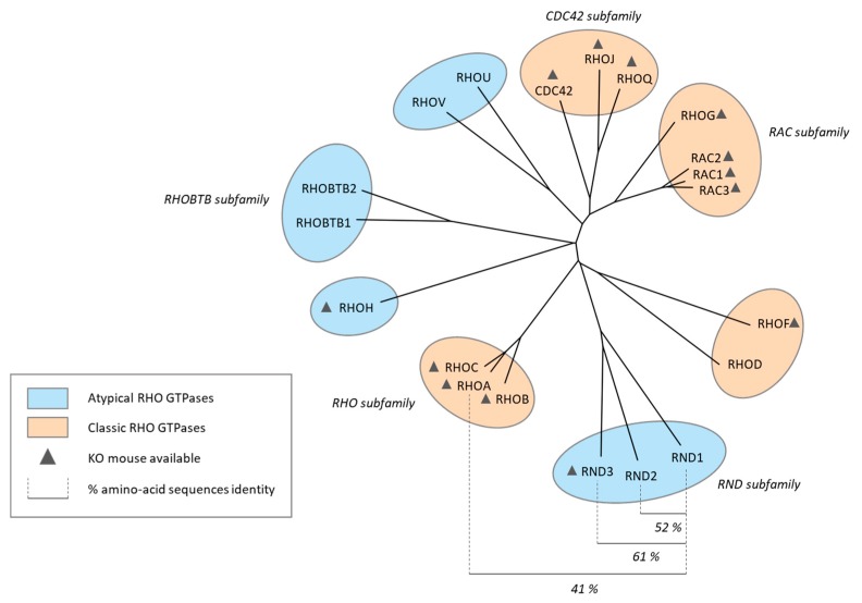

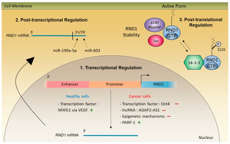
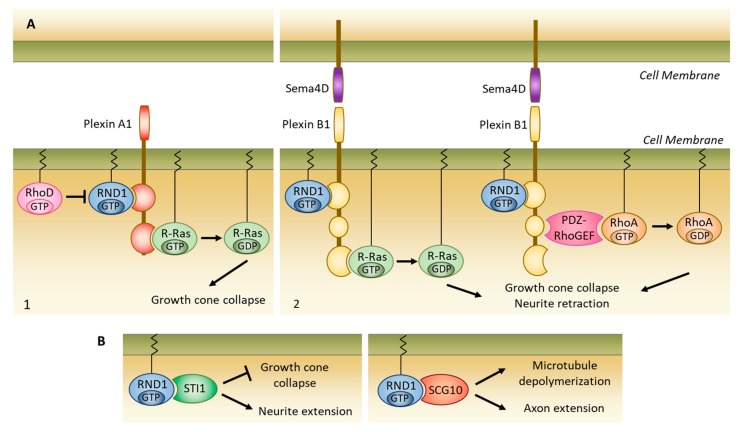
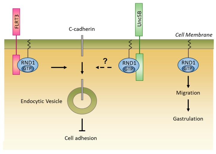
Verification
Claims the paper makes
The claims and statistics of this paper haven’t been checked yet.
Run the check — free, no account needed. It reads the open-access text and checks the sources it cites.
Claims other papers make about this one
Each is checked against this paper’s own text: does it say what the citing paper says it does?
In the citing paper
“RhoH is an unusual member of the Rho family of small GTP‐binding proteins in that it lacks GTPase activity. Since the RhoH protein is constantly bound by GTP, it is constitutively active and controlled predominantly by changes in quantitative expression.”
From Bimodal expression of RHOH during myelomonocytic differentiation: Implications for the expansion of AML differentiation therapy (2021) · cites this paper as [1]
Self-check
Percentages, sums and reported differences re-computed from the paper’s own numbers.
The self-check needs the paper’s full text.
Citations
Cites
No references could be resolved yet.
Research works printed but not linked: 62
- Heasman, 2008, Mammalian Rho GTPases: New insights into their functions from in vivo studies, Nat. Rev. Mol. Cell Biol., 9, 690, 10.1038/nrm2476 · DOI · Paper page
- Bustelo, 2018, RHO GTPases in cancer: Known facts, open questions, and therapeutic challenges, Biochem. Soc. Trans., 46, 741, 10.1042/BST20170531 · DOI
- Nobes, 1998, A New Member of the Rho Family, Rnd1, Promotes Disassembly of Actin Filament Structures and Loss of Cell Adhesion, J. Cell Boil., 141, 187, 10.1083/jcb.141.1.187 · DOI
- Shutes, 2004, Atypical Mechanism of Regulation of the Wrch-1 Rho Family Small GTPase, Curr. Boil., 14, 2052, 10.1016/j.cub.2004.11.011 · DOI
- Oinuma, 2012, Rnd1 and Rnd3 targeting to lipid raft is required for p190 RhoGAP activation, Mol. Biol. Cell, 23, 1593, 10.1091/mbc.e11-11-0900 · DOI
- Riou, 2010, Rnd proteins: Multifunctional regulators of the cytoskeleton and cell cycle progression, BioEssays, 32, 986, 10.1002/bies.201000060 · DOI
- Jaffe, 2005, RHO GTPASES: Biochemistry and Biology, Annu. Rev. Cell Dev. Boil., 21, 247, 10.1146/annurev.cellbio.21.020604.150721 · DOI · Paper page
- Ridley, 2015, Rho GTPase signalling in cell migration, Curr. Opin. Cell Biol., 36, 103, 10.1016/j.ceb.2015.08.005 · DOI
- Zandvakili, 2017, Rho GTPases: Anti- or pro-neoplastic targets?, Oncogene, 36, 3213, 10.1038/onc.2016.473 · DOI
- Okada, 2015, The Rho GTPase Rnd1 suppresses mammary tumorigenesis and EMT by restraining Ras-MAPK signalling, Nat. Cell Biol., 17, 81, 10.1038/ncb3082 · DOI
- Clarke, K., Daubon, T., Turan, N., Soulet, F., Mohd Zahari, M., Ryan, K.R., Durant, S., He, S., Herbert, J., and Ankers, J. (2015). Inference of Low and High-Grade Glioma Gene Regulatory Networks Delineates the Role of Rnd3 in Establishing Multiple Hallmarks of Cancer. PLoS Genet., 11. · DOI
- Grise, 2012, Rnd3/RhoE Is down-regulated in hepatocellular carcinoma and controls cellular invasion, Hepatology, 55, 1766, 10.1002/hep.25568 · DOI
- Paysan, 2016, Rnd3 in Cancer: A Review of the Evidence for Tumor Promoter or Suppressor, Mol. Cancer Res., 14, 1033, 10.1158/1541-7786.MCR-16-0164 · DOI
- Blitz, 1999, Involvement of the small GTPases XRhoA and XRnd1 in cell adhesion and head formation in early Xenopus development, Development, 126, 5339, 10.1242/dev.126.23.5339 · DOI
- Ishikawa, 2003, A Role of Rnd1 GTPase in Dendritic Spine Formation in Hippocampal Neurons, J. Neurosci., 23, 11065, 10.1523/JNEUROSCI.23-35-11065.2003 · DOI
- Kim, 2005, Differences in the gestational pattern of mRNA expression of the Rnd family in rat and human myometria, Comp. Biochem. Physiol. Part A Mol. Integr. Physiol., 142, 410, 10.1016/j.cbpa.2005.08.028 · DOI
- Kim, 2003, Up-regulation of Rnd1 during pregnancy serves as a negative-feedback control for Ca2+ sensitization of contractile elements in rat myometrium, Biochem. Biophys. Res. Commun., 311, 972, 10.1016/j.bbrc.2003.10.100 · DOI
- Katoh, 2002, Socius Is a Novel Rnd GTPase-Interacting Protein Involved in Disassembly of Actin Stress Fibers, Mol. Cell. Biol., 22, 2952, 10.1128/MCB.22.9.2952-2964.2002 · DOI
- Roberts, 2008, Rho Family GTPase modification and dependence on CAAX motif-signaled posttranslational modification, J. Biol. Chem., 283, 25150, 10.1074/jbc.M800882200 · DOI · Paper page
- Riou, 2013, 14-3-3 Proteins Interact with a Hybrid Prenyl-Phosphorylation Motif to Inhibit G Proteins, Cell, 153, 1164, 10.1016/j.cell.2013.05.003 · DOI
- Song, 2014, Inhibition of osteoclast bone resorption activity through osteoprotegerin-induced damage of the sealing zone, Int. J. Mol. Med., 34, 856, 10.3892/ijmm.2014.1846 · DOI
- Suehiro, 2014, Genome-wide Approaches Reveal Functional Vascular Endothelial Growth Factor (VEGF)-inducible Nuclear Factor of Activated T Cells (NFAT) c1 Binding to Angiogenesis-related Genes in the Endothelium, J. Biol. Chem., 289, 29044, 10.1074/jbc.M114.555235 · DOI
- Loirand, 1999, The Rho-related protein Rnd1 inhibits Ca2+ sensitization of rat smooth muscle, J. Physiol., 516, 825, 10.1111/j.1469-7793.1999.0825u.x · DOI
- Monastyrskaya, 2013, miR-199a-5p Regulates Urothelial Permeability and May Play a Role in Bladder Pain Syndrome, Am. J. Pathol., 182, 431, 10.1016/j.ajpath.2012.10.020 · DOI · Paper page
- Zhang, 2016, Primate-specific miR-603 is implicated in the risk and pathogenesis of Alzheimer’s disease, Aging (Albany N. Y.), 8, 272
- Hodge, 2016, Regulating Rho GTPases and their regulators, Nat. Rev. Mol. Cell Biol., 17, 496, 10.1038/nrm.2016.67 · DOI · Paper page
- Goh, 2012, The GTPase-deficient Rnd Proteins Are Stabilized by Their Effectors, J. Biol. Chem., 287, 31311, 10.1074/jbc.M111.327056 · DOI
- Wennerberg, 2003, Rnd Proteins Function as RhoA Antagonists by Activating p190 RhoGAP, Curr. Biol., 13, 1106, 10.1016/S0960-9822(03)00418-4 · DOI
- Mori, 2009, Rho-kinase contributes to sustained RhoA activation through phosphorylation of p190A RhoGAP, J. Biol. Chem., 284, 5067, 10.1074/jbc.M806853200 · DOI
- Goh, L.L., and Manser, E. (2010). The RhoA GEF Syx Is a Target of Rnd3 and Regulated via a Raf1-Like Ubiquitin-Related Domain. PLoS ONE, 5. · DOI
- Zalcman, 2000, Interaction of the Grb7 adapter protein with Rnd1, a new member of the Rho family, FEBS Lett., 467, 91, 10.1016/S0014-5793(99)01530-6 · DOI
- Villalobo, 2013, Deletion of the calmodulin-binding domain of Grb7 impairs cell attachment to the extracellular matrix and migration, Biochem. Biophys. Res. Commun., 436, 271, 10.1016/j.bbrc.2013.05.092 · DOI
- Prokop, 2013, Using fly genetics to dissect the cytoskeletal machinery of neurons during axonal growth and maintenance, J. Cell Sci., 126, 2331
- Zanata, 2002, Antagonistic Effects of Rnd1 and RhoD GTPases Regulate Receptor Activity in Semaphorin 3A-Induced Cytoskeletal Collapse, J. Neurosci., 22, 471, 10.1523/JNEUROSCI.22-02-00471.2002 · DOI
- Oinuma, 2004, The Semaphorin 4D Receptor Plexin-B1 Is a GTPase Activating Protein for R-Ras, Science, 305, 862, 10.1126/science.1097545 · DOI
- Oinuma, 2003, Direct Interaction of Rnd1 with Plexin-B1 Regulates PDZ-RhoGEF-mediated Rho Activation by Plexin-B1 and Induces Cell Contraction in COS-7 Cells, J. Biol. Chem., 278, 25671, 10.1074/jbc.M303047200 · DOI
- Oinuma, 2004, Molecular Dissection of the Semaphorin 4D Receptor Plexin-B1-Stimulated R-Ras GTPase-Activating Protein Activity and Neurite Remodeling in Hippocampal Neurons, J. Neurosci., 24, 11473, 10.1523/JNEUROSCI.3257-04.2004 · DOI
- Yukawa, 2010, Sema4A induces cell morphological changes through B-type plexin-mediated signaling, Int. J. Mol. Med., 25, 225
- Harada, 2005, Direct interaction of Rnd1 with FRS2 beta regulates Rnd1-induced down-regulation of RhoA activity and is involved in fibroblast growth factor-induced neurite outgrowth in PC12 cells, J. Biol. Chem., 280, 18418, 10.1074/jbc.M411356200 · DOI
- Chardin, 2006, Function and regulation of Rnd proteins, Nat. Rev. Mol. Cell Biol., 7, 54, 10.1038/nrm1788 · DOI
- Costa, 2014, STI1 antagonizes cytoskeleton collapse mediated by small GTPase Rnd1 and regulates neurite growth, Exp. Cell Res., 324, 84, 10.1016/j.yexcr.2014.03.017 · DOI
- Li, 2009, Rnd1 regulates axon extension by enhancing the microtubule destabilizing activity of SCG10, J. Biol. Chem., 284, 16060, 10.1074/jbc.A808126200 · DOI
- Ogata, 2007, TGF-beta signaling-mediated morphogenesis: Modulation of cell adhesion via cadherin endocytosis, Genes Dev., 21, 1817, 10.1101/gad.1541807 · DOI
- Karaulanov, E., Böttcher, R.T., Stannek, P., Wu, W., Rau, M., Ogata, S., Cho, K.W.Y., and Niehrs, C. (2009). Unc5B Interacts with FLRT3 and Rnd1 to Modulate Cell Adhesion in Xenopus Embryos. PLoS ONE, 4. · DOI
- Komatsu, 2017, Attenuated RND1 Expression Confers Malignant Phenotype and Predicts Poor Prognosis in Hepatocellular Carcinoma, Ann. Surg. Oncol., 24, 850, 10.1245/s10434-016-5573-9 · DOI
- Qin, 2018, The Rho GTPase Rnd1 inhibits epithelial–mesenchymal transition in hepatocellular carcinoma and is a favorable anti-metastasis target, Cell Death Dis., 9, 486, 10.1038/s41419-018-0517-x · DOI
- Boyrie, 2018, RND1 regulates migration of human glioblastoma stem-like cells according to their anatomical localization and defines a prognostic signature in glioblastoma, Oncotarget, 9, 33788, 10.18632/oncotarget.26082 · DOI
- Jiang, 2003, Prognostic value of rho GTPases and rho guanine nucleotide dissociation inhibitors in human breast cancers, Clin. Cancer Res., 9, 6432
- Nishigaki, 2005, Discovery of aberrant expression of R-RAS by cancer-linked DNA hypomethylation in gastric cancer using microarrays, Cancer Res., 65, 2115, 10.1158/0008-5472.CAN-04-3340 · DOI
- Xiang, 2016, RND1 is up-regulated in esophageal squamous cell carcinoma and promotes the growth and migration of cancer cells, Tumour Biol., 37, 773, 10.1007/s13277-015-3855-7 · DOI
- Aguirre, 2004, High-resolution characterization of the pancreatic adenocarcinoma genome, Proc. Natl. Acad. Sci. USA, 101, 9067, 10.1073/pnas.0402932101 · DOI
- Rutherford, 2005, Mapping of candidate tumor suppressor genes on chromosome 12 in adenoid cystic carcinoma, Lab. Investig., 85, 1076, 10.1038/labinvest.3700314 · DOI
- Mouly, 2018, PARP-1-dependent RND1 transcription induced by topoisomerase I cleavage complexes confers cellular resistance to camptothecin, Cell Death Dis., 9, 931, 10.1038/s41419-018-0981-3 · DOI
- Nishigaki, 2005, Proteomic identification of differentially-expressed genes in human gastric carcinomas, Proteomics, 5, 3205, 10.1002/pmic.200401307 · DOI
- Soheili, 2017, Distinctive expression pattern of OCT4 variants in different types of breast cancer, Cancer Biomark., 18, 69, 10.3233/CBM-160675 · DOI
- Kar, 2018, Overexpression of OCT4 induced by modulation of histone marks plays crucial role in breast cancer progression, Gene, 643, 35, 10.1016/j.gene.2017.11.077 · DOI
- Shen, 2014, Overexpression of Oct4 suppresses the metastatic potential of breast cancer cells via Rnd1 downregulation, Biochim. Biophys. Acta (BBA) Mol. Basis Dis., 1842, 2087, 10.1016/j.bbadis.2014.07.015 · DOI
- Li, W., Sun, M., Zang, C., Ma, P., He, J., Zhang, M., Huang, Z., Ding, Y., and Shu, Y. (2016). Upregulated long non-coding RNA AGAP2-AS1 represses LATS2 and KLF2 expression through interacting with EZH2 and LSD1 in non-small-cell lung cancer cells. Cell Death Dis., 7. · DOI
- Qi, 2017, Long noncoding AGAP2-AS1 is activated by SP1 and promotes cell proliferation and invasion in gastric cancer, J. Hematol. Oncol., 10, 11, 10.1186/s13045-017-0420-4 · DOI
- Chen, 2018, Inferring Novel Tumor Suppressor Genes with a Protein-Protein Interaction Network and Network Diffusion Algorithms, Mol. Ther. Methods Clin. Dev., 10, 57, 10.1016/j.omtm.2018.06.007 · DOI
- Bianchini, 2007, cDNA microarray study to identify expression changes relevant for apoptosis in K562 cells co-treated with amifostine and imatinib, Cancer Chemother. Pharmacol., 59, 349, 10.1007/s00280-006-0276-8 · DOI
- Turtoi, 2008, Early gene expression in human lymphocytes after gamma-irradiation—A genetic pattern with potential for biodosimetry, Int. J. Radiat. Biol., 84, 375, 10.1080/09553000802029886 · DOI
Cited by
Galiègue‐Zouitina, S., Fu, Q., Carton‐Latreche, C., Poret, N., Cheok, M., Leprêtre, F., Figeac, M., Quesnel, B., El Bouazzati, H., & Shelley, C. S. (2021) · eJHaem · doi:10.1002/jha2.128 · Cited by 7
Metadata
- Authors
- Mouly, L., Gilhodes, J., Lemarié, A., Cohen-Jonathan Moyal, E., Toulas, C., Favre, G., Sordet, O., & Monferran, S.
- Affiliations
- Cancer Research Center of Toulouse, INSERM UMR1037, 31037 Toulouse, France
- Institut Claudius Regaud, IUCT-O, 31059 Toulouse, France
- ORCID
- Lemarié, Anthony
- Venue
- International Journal of Molecular Sciences
- Publisher
- MDPI AG
- Published
- 2019-07-24
- Volume
- 20
- Issue
- 15
- Pages
- 3612
- ISSN
- 1422-0067
- PMC
- PMC6696182
- Cited by
- 19
- References
- 0
- Language
- en
- Funders
- Région Midi-Pyrénées INSERM (R14075BB)
About these checks
Registry, retraction, Cites and statistics results are computed, not judged, with one exception: a printed reference that no index lists is read once by a model before it counts as a citation error. Claims are extracted by a model and kept only when their quote appears verbatim in the text; each cited source is then checked, with a model’s help, for whether it says what the claim relies on. Those verdicts can be wrong — read the claim’s quote and the cited source before relying on one.
Statistics and arithmetic are recomputed, not judged.
Confidence bands are shown only for fields calibrated on our annotated test set. None is calibrated yet, so no band is shown.
What counts as a claim ›How we measure confidence ›How we check this ›
Are you an author, or do you see a problem with this page? Request a review
