

The DOI matches its Crossref record; 6 of the 6 references it cites were found in a registry or an index.
Cellular, Extracellular and Extracellular Vesicular miRNA Profiles of Pre-Ovulatory Follicles Indicate Signaling Disturbances in Polycystic Ovaries
Abstract
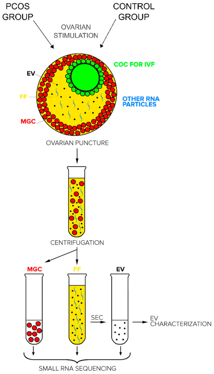
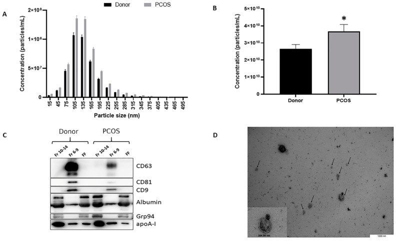
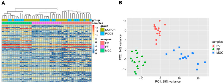
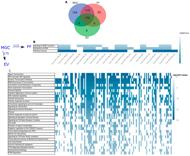
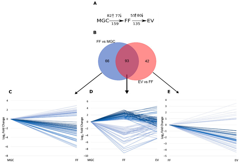
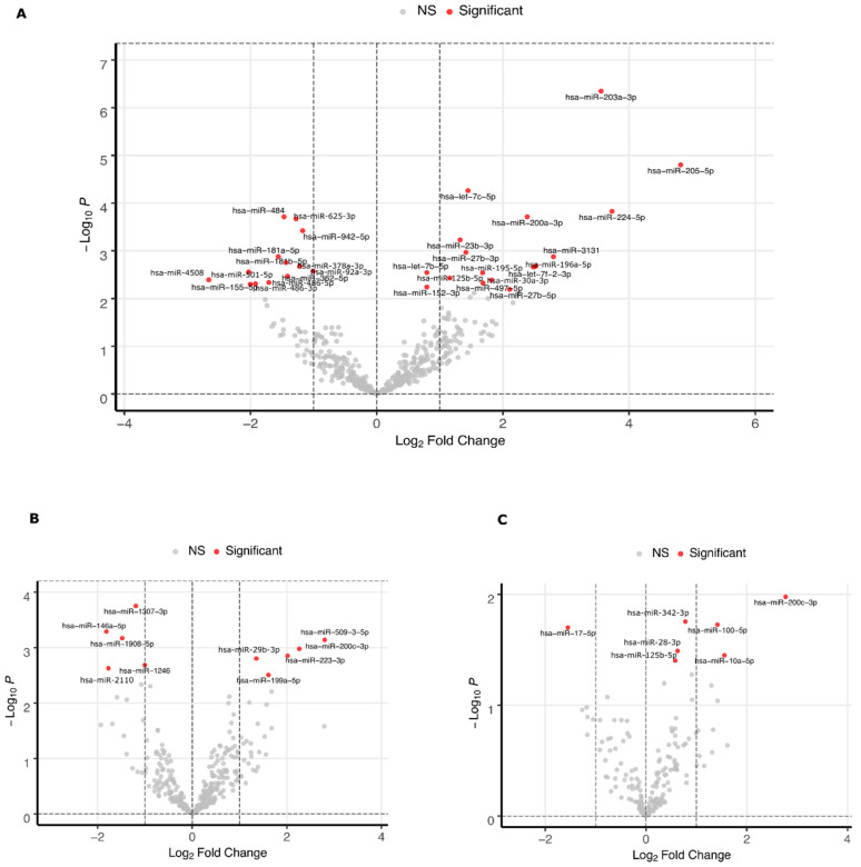
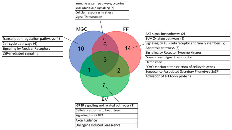
Cell-free RNAs have the potential to act as a means of gene expression regulation between cells and are therefore used as diagnostic markers describing the state of tissue environment. The origin and functions of such RNAs in human ovarian follicle, the environment of oocyte maturation, are unclear. The current study investigates the difference in the microRNA profiles of fertile women and polycystic ovary syndrome (PCOS) patients in three compartments from the same preovulatory follicle: mural granulosa cells (MGC), cell-free follicular fluid (FF), and extracellular vesicles (EV) of the FF by small RNA sequencing. In silico analysis was used for the prediction and over-representation of targeted pathways for the detected microRNAs. PCOS follicles were distinguished from normal tissue by the differential expression of 30 microRNAs in MGC and 10 microRNAs in FF (FDR < 0.1) that commonly regulate cytokine signaling pathways. The concentration of EV-s was higher in the FF of PCOS patients (p = 0.04) containing eight differentially expressed microRNAs (p < 0.05). In addition, we present the microRNA profiles of MGC, FF, and EV in the fertile follicle and demonstrate that microRNAs loaded into EVs target mRNAs of distinct signaling pathways in comparison to microRNAs in FF. To conclude, the three follicular compartments play distinct roles in the signaling disturbances associated with PCOS.
Overview
The claims and statistics of this paper haven’t been checked yet.
Run the check — free, no account needed. It reads the open-access text and checks the sources it cites.
Integrity signals
Registry
Cites
Statistics
Figures
Verification
Claims the paper makes
The claims and statistics of this paper haven’t been checked yet.
Run the check — free, no account needed. It reads the open-access text and checks the sources it cites.
Claims other papers make about this one
Each is checked against this paper’s own text: does it say what the citing paper says it does?
The source backs part of the claim, not all of it — or backs it for a different population, condition or comparison.
In the citing paper
“miRNAs have been detected from all body fluids, including FF [32, 56], and may be involved in cell-to-cell communication [33].”
From Hsa-mir-548 family expression in human reproductive tissues (2021) · cites this paper as [32]
Experimental — accuracy not yet measuredIn the citing paper
“Some miRNAs infiltrate into the FF from blood plasma [32], explaining the lack of their expression in the granulosa cells.”
From Hsa-mir-548 family expression in human reproductive tissues (2021) · cites this paper as [32]
Self-check
Percentages, sums and reported differences re-computed from the paper’s own numbers.
The self-check needs the paper’s full text.
Citations
Cites
miRDB: an online database for prediction of functional microRNA targets
Chen, Y., & Wang, X. (2020) · Nucleic Acids Research · doi:10.1093/nar/gkz757 · Cited by 3,277
Teede, H. J., Misso, M. L., Costello, M. F., Dokras, A., Laven, J., Moran, L., Piltonen, T., Norman, R. J., & the International PCOS Network (2018) · Clinical Endocrinology · doi:10.1111/cen.13795 · Cited by 1,823
RNA delivery by extracellular vesicles in mammalian cells and its applications
O’Brien, K., Breyne, K., Ughetto, S., Laurent, L. C., & Breakefield, X. O. (2020) · Nature Reviews Molecular Cell Biology · doi:10.1038/s41580-020-0251-y · Cited by 1,811
Yang, M., Chen, J., Su, F., Yu, B., Su, F., Lin, L., Liu, Y., Huang, J.-D., & Song, E. (2011) · Molecular Cancer · doi:10.1186/1476-4598-10-117 · Cited by 693
Physiological and Pathological Androgen Actions in the Ovary
Astapova, O., Minor, B. M. N., & Hammes, S. R. (2019) · Endocrinology · doi:10.1210/en.2019-00101 · Cited by 133
SMAD4 activates Wnt signaling pathway to inhibit granulosa cell apoptosis
Du, X., Li, Q., Yang, L., Liu, L., Cao, Q., & Li, Q. (2020) · Cell Death & Disease · doi:10.1038/s41419-020-2578-x · Cited by 76
Research works printed but not linked: 72
- Teede, H., Deeks, A., and Moran, L. (2010). Polycystic ovary syndrome: A complex condition with psychological, reproductive and metabolic manifestations that impacts on health across the lifespan. BMC Med., 8. · DOI · Paper page
- Ehrmann, 2005, Polycystic ovary syndrome, N. Engl. J. Med., 352, 1223, 10.1056/NEJMra041536 · DOI
- Goodman, 2015, American association of clinical endocrinologists, american college of endocrinology, and androgen excess and pcos society disease state clinical review: Guide to the best practices in the evaluation and treatment of polycystic ovary syndrome—Part 1, Endocr. Pract., 21, 1291, 10.4158/EP15748.DSC · DOI · Paper page
- Hennet, 2012, The antral follicle: A microenvironment for oocyte differentiation, Int. J. Dev. Biol., 56, 819, 10.1387/ijdb.120133cc · DOI
- Fritz, 2016, Sources and Functions of Extracellular Small RNAs in Human Circulation, Annu. Rev. Nutr., 36, 301, 10.1146/annurev-nutr-071715-050711 · DOI · Paper page
- Lamichhane, 2018, Production of Extracellular Vesicles Loaded with Therapeutic Cargo, Methods Mol. Biol., 1831, 37, 10.1007/978-1-4939-8661-3_4 · DOI
- Raposo, 2019, Extracellular vesicles: A new communication paradigm?, Nat. Rev. Mol. Cell Biol., 20, 509, 10.1038/s41580-019-0158-7 · DOI
- Sang, 2013, Identification of MicroRNAs in Human Follicular Fluid: Characterization of MicroRNAs That Govern Steroidogenesis in Vitro and Are Associated With Polycystic Ovary Syndrome in Vivo, J. Clin. Endocrinol. Metab., 98, 3068, 10.1210/jc.2013-1715 · DOI
- Santonocito, 2014, Molecular characterization of exosomes and their microRNA cargo in human follicular fluid: Bioinformatic analysis reveals that exosomal microRNAs control pathways involved in follicular maturation, Fertil. Steril., 102, 1751, 10.1016/j.fertnstert.2014.08.005 · DOI
- Sork, 2018, Heterogeneity and interplay of the extracellular vesicle small RNA transcriptome and proteome, Sci. Rep., 8, 10813, 10.1038/s41598-018-28485-9 · DOI · Paper page
- Valadi, 2007, Exosome-mediated transfer of mRNAs and microRNAs is a novel mechanism of genetic exchange between cells, Nat. Cell Biol., 9, 654, 10.1038/ncb1596 · DOI · Paper page
- da Silveira, J.C., Veeramachaneni, D.N.R., Winger, Q.A., Carnevale, E.M., and Bouma, G.J. (2012). Cell-Secreted Vesicles in Equine Ovarian Follicular Fluid Contain miRNAs and Proteins: A Possible New Form of Cell Communication Within the Ovarian Follicle. Biol. Reprod., 86. · DOI
- Bellingham, 2012, Small RNA deep sequencing reveals a distinct miRNA signature released in exosomes from prion-infected neuronal cells, Nucleic Acids Res., 40, 10937, 10.1093/nar/gks832 · DOI
- Driedonks, 2018, Immune stimuli shape the small non-coding transcriptome of extracellular vesicles released by dendritic cells, Cell. Mol. Life Sci., 75, 3857, 10.1007/s00018-018-2842-8 · DOI
- Raposo, 2013, Extracellular vesicles: Exosomes, microvesicles, and friends, J. Cell Biol., 200, 373, 10.1083/jcb.201211138 · DOI · Paper page
- Kowal, 2016, Proteomic comparison defines novel markers to characterize heterogeneous populations of extracellular vesicle subtypes, PNAS, 113, E968, 10.1073/pnas.1521230113 · DOI · Paper page
- Srinivasan, 2019, Small RNA Sequencing across Diverse Biofluids Identifies Optimal Methods for exRNA Isolation, Cell, 177, 446, 10.1016/j.cell.2019.03.024 · DOI
- Moreno, 2015, Follicular fluid and mural granulosa cells microRNA profiles vary in in vitro fertilization patients depending on their age and oocyte maturation stage, Fertil. Steril., 104, 1037, 10.1016/j.fertnstert.2015.07.001 · DOI
- Lammens, 2014, Age-associated differential microRNA levels in human follicular fluid reveal pathways potentially determining fertility and success of in vitro fertilization, Hum. Fertil., 17, 90, 10.3109/14647273.2014.897006 · DOI
- Xiao, G., Xia, C., Yang, J., Liu, J., Du, H., Kang, X., Lin, Y., Guan, R., Yan, P., and Tang, S. (2014). MiR-133b Regulates the Expression of the Actin Protein TAGLN2 during Oocyte Growth and Maturation: A Potential Target for Infertility Therapy. PLoS ONE, 9. · DOI
- Li, 2017, Upregulation of MiR-205 transcriptionally suppresses SMAD4 and PTEN and contributes to human ovarian cancer progression, Sci. Rep., 7, 1
- Woo, 2017, Distinct microrna suggest differential gene regulation in granulosa cells in young women with normal ovarian reserve compared to YOUNG WOMEN WITH Diminished ovarian reserve, Fertil. Steril., 107, e5, 10.1016/j.fertnstert.2017.02.009 · DOI
- Salilew-Wondim, D., Ahmad, I., Gebremedhn, S., Sahadevan, S., Hossain, M.M., Rings, F., Hoelker, M., Tholen, E., Neuhoff, C., and Looft, C. (2014). The Expression Pattern of microRNAs in Granulosa Cells of Subordinate and Dominant Follicles during the Early Luteal Phase of the Bovine Estrous Cycle. PLoS ONE, 9. · DOI
- Zhang, J., Xu, Y., Liu, H., and Pan, Z. (2019). MicroRNAs in ovarian follicular atresia and granulosa cell apoptosis. Reprod. Biol. Endocrinol., 17. · DOI · Paper page
- Simm, 2013, Research resource: Small RNA-seq of human granulosa cells reveals miRNAs in FSHR and aromatase genes, Mol. Endocrinol., 27, 1128, 10.1210/me.2013-1058 · DOI
- Zhang, Q., Sun, H., Jiang, Y., Ding, L., Wu, S., Fang, T., Yan, G., and Hu, Y. (2013). MicroRNA-181a Suppresses Mouse Granulosa Cell Proliferation by Targeting Activin Receptor IIA. PLoS ONE, 8. · DOI
- Zhang, 2017, miR-181a increases FoxO1 acetylation and promotes granulosa cell apoptosis via SIRT1 downregulation, Cell Death Dis., 8, e3088, 10.1038/cddis.2017.467 · DOI
- Butler, 2019, Expression of microRNA in follicular fluid in women with and without PCOS, Sci. Rep., 9, 16306, 10.1038/s41598-019-52856-5 · DOI
- He, T., Liu, Y., Jia, Y., Wang, H., Yang, X., Lu, G., Liu, H., and Shi, Y. (2018). MicroRNA-141 and MicroRNA-200c Are Overexpressed in Granulosa Cells of Polycystic Ovary Syndrome Patients. Front. Med. (Lausanne), 5. · DOI
- Yao, 2018, MiRNA-335-5p negatively regulates granulosa cell proliferation via SGK3 in PCOS, Reproduction, 156, 439
- Martinez, R.M., Liang, L., Racowsky, C., Dioni, L., Mansur, A., Adir, M., Bollati, V., Baccarelli, A.A., Hauser, R., and Machtinger, R. (2018). Extracellular microRNAs profile in human follicular fluid and IVF outcomes. Sci. Rep., 8. · DOI
- Xu, 2015, Characterization of microRNA profile in human cumulus granulosa cells: Identification of microRNAs that regulate Notch signaling and are associated with PCOS, Mol. Cell. Endocrinol., 404, 26, 10.1016/j.mce.2015.01.030 · DOI
- Teng, 2017, Low expression of microRNA-1908 predicts a poor prognosis for patients with ovarian cancer, Oncol. Lett., 14, 4277, 10.3892/ol.2017.6714 · DOI
- Andrei, 2019, Differential miRNA Expression Profiles in Cumulus and Mural Granulosa Cells from Human Pre-ovulatory Follicles, Microrna, 8, 61, 10.2174/2211536607666180912152618 · DOI
- Woo, 2018, Micro-RNAs involved in cellular proliferation have altered expression profiles in granulosa of young women with diminished ovarian reserve, J. Assist. Reprod. Genet., 35, 1777, 10.1007/s10815-018-1239-9 · DOI
- Liu, 2020, lncRNA PVT1/MicroRNA-17-5p/PTEN Axis Regulates Secretion of E2 and P4, Proliferation, and Apoptosis of Ovarian Granulosa Cells in PCOS, Mol. Ther. Nucleic Acids, 20, 205, 10.1016/j.omtn.2020.02.007 · DOI
- Witwer, 2018, Minimal information for studies of extracellular vesicles 2018 (MISEV2018): A position statement of the International Society for Extracellular Vesicles and update of the MISEV2014 guidelines, J. Extracell. Vesicles, 7, 1535750, 10.1080/20013078.2018.1535750 · DOI · Paper page
- Doyle, L.M., and Wang, M.Z. (2019). Overview of Extracellular Vesicles, Their Origin, Composition, Purpose, and Methods for Exosome Isolation and Analysis. Cells, 8. · DOI
- Benedikter, 2017, Ultrafiltration combined with size exclusion chromatography efficiently isolates extracellular vesicles from cell culture media for compositional and functional studies, Sci. Rep., 7, 15297, 10.1038/s41598-017-15717-7 · DOI
- Böing, A.N., van der Pol, E., Grootemaat, A.E., Coumans, F.A.W., Sturk, A., and Nieuwland, R. (2014). Single-step isolation of extracellular vesicles by size-exclusion chromatography. J. Extracell. Vesicles, 3. · DOI
- Hasan, M.M., Viil, J., Lättekivi, F., Ord, J., Reshi, Q.U.A., Jääger, K., Velthut-Meikas, A., Andronowska, A., Jaakma, Ü., and Salumets, A. (2020). Bovine Follicular Fluid and Extracellular Vesicles Derived from Follicular Fluid Alter the Bovine Oviductal Epithelial Cells Transcriptome. Int. J. Mol. Sci., 21. · DOI
- Simon, 2018, Extracellular Vesicles in Human Reproduction in Health and Disease, Endocr Rev., 39, 292, 10.1210/er.2017-00229 · DOI
- Koiou, 2013, Platelet-derived microparticles in overweight/obese women with the polycystic ovary syndrome, Gynecol. Endocrinol., 29, 250, 10.3109/09513590.2012.743005 · DOI
- Min, 2019, Evaluation of circulating small extracellular vesicles derived miRNAs as biomarkers of early colon cancer: A comparison with plasma total miRNAs, J. Extracell. Vesicles, 8, 1643670, 10.1080/20013078.2019.1643670 · DOI
- Endzeliņš, E., Berger, A., Melne, V., Bajo-Santos, C., Soboļevska, K., Ābols, A., Rodriguez, M., Šantare, D., Rudņickiha, A., and Lietuvietis, V. (2017). Detection of circulating miRNAs: Comparative analysis of extracellular vesicle-incorporated miRNAs and cell-free miRNAs in whole plasma of prostate cancer patients. BMC Cancer, 17. · DOI
- Jeppesen, 2019, Reassessment of Exosome Composition, Cell, 177, 428, 10.1016/j.cell.2019.02.029 · DOI · Paper page
- Baglio, 2015, Human bone marrow- and adipose-mesenchymal stem cells secrete exosomes enriched in distinctive miRNA and tRNA species, Stem Cell Res. Therapy, 6, 127, 10.1186/s13287-015-0116-z · DOI
- Hung, 2017, Stage-specific follicular extracellular vesicle uptake and regulation of bovine granulosa cell proliferation, Biol. Reprod., 97, 644, 10.1093/biolre/iox106 · DOI
- Sohel, M.M.H., Hoelker, M., Noferesti, S.S., Salilew-Wondim, D., Tholen, E., Looft, C., Rings, F., Uddin, M.J., Spencer, T.E., and Schellander, K. (2013). Exosomal and Non-Exosomal Transport of Extra-Cellular microRNAs in Follicular Fluid: Implications for Bovine Oocyte Developmental Competence. PLoS ONE, 8. · DOI
- Tang, Z.-R., Zhang, R., Lian, Z.-X., Deng, S.-L., and Yu, K. (2019). Estrogen-Receptor Expression and Function in Female Reproductive Disease. Cells, 8. · DOI
- Andrade, G.M., da Silveira, J.C., Perrini, C., Del Collado, M., Gebremedhn, S., Tesfaye, D., Meirelles, F.V., and Perecin, F. (2017). The role of the PI3K-Akt signaling pathway in the developmental competence of bovine oocytes. PLoS ONE, 12. · DOI
- McLaughlin, 2014, Inhibition of phosphatase and tensin homologue (PTEN) in human ovary in vitro results in increased activation of primordial follicles but compromises development of growing follicles, Mol. Hum. Reprod., 20, 736, 10.1093/molehr/gau037 · DOI
- Chevillet, 2014, Quantitative and stoichiometric analysis of the microRNA content of exosomes, PNAS, 111, 14888, 10.1073/pnas.1408301111 · DOI
- Cirillo, 2019, MiRNAs Regulating Insulin Sensitivity Are Dysregulated in Polycystic Ovary Syndrome (PCOS) Ovaries and Are Associated With Markers of Inflammation and Insulin Sensitivity, Front. Endocrinol. (Lausanne), 10, 879, 10.3389/fendo.2019.00879 · DOI
- Roth, 2014, Altered microRNA and gene expression in the follicular fluid of women with polycystic ovary syndrome, J. Assist. Reprod. Genet., 31, 355, 10.1007/s10815-013-0161-4 · DOI
- Yao, 2010, MicroRNA-224 Is Involved in Transforming Growth Factor-β-Mediated Mouse Granulosa Cell Proliferation and Granulosa Cell Function by Targeting Smad4, Mol. Endocrinol., 24, 540, 10.1210/me.2009-0432 · DOI
- Wiser, 2013, Age-related normogram for antral follicle count in women with polycystic ovary syndrome, Reprod. Biomed. Online, 27, 414, 10.1016/j.rbmo.2013.06.016 · DOI
- Ferraretti, 2011, ESHRE consensus on the definition of ‘poor response’ to ovarian stimulation for in vitro fertilization: The Bologna criteria, Hum. Reprod., 26, 1616, 10.1093/humrep/der092 · DOI
- Shi, 2015, miR-483–5p and miR-486–5p are down-regulated in cumulus cells of metaphase II oocytes from women with polycystic ovary syndrome, Reprod. Biomed. Online, 31, 565, 10.1016/j.rbmo.2015.06.023 · DOI
- Li, Y., Fang, Y., Liu, Y., and Yang, X. (2015). MicroRNAs in ovarian function and disorders. J. Ovarian Res., 8. · DOI
- Velthut, 2010, The differential transcriptome and ontology profiles of floating and cumulus granulosa cells in stimulated human antral follicles, Mol. Hum. Reprod., 16, 229, 10.1093/molehr/gap103 · DOI
- Rotterdam, 2004, ASRM-Sponsored PCOS Consensus Workshop Group Revised 2003 consensus on diagnostic criteria and long-term health risks related to polycystic ovary syndrome, Fertil. Steril., 81, 19, 10.1016/j.fertnstert.2003.10.004 · DOI · Paper page
- Bolger, 2014, Trimmomatic: A flexible trimmer for Illumina sequence data, Bioinformatics, 30, 2114, 10.1093/bioinformatics/btu170 · DOI · Paper page
- Mackowiak, 2012, miRDeep2 accurately identifies known and hundreds of novel microRNA genes in seven animal clades, Nucleic Acids Res., 40, 37, 10.1093/nar/gkr688 · DOI · Paper page
- Robinson, 2010, edgeR: A Bioconductor package for differential expression analysis of digital gene expression data, Bioinformatics, 26, 139, 10.1093/bioinformatics/btp616 · DOI · Paper page
- Love, 2014, Moderated estimation of fold change and dispersion for RNA-seq data with DESeq2, Genome Biol., 15, 550, 10.1186/s13059-014-0550-8 · DOI · Paper page
- Livak, 2001, Analysis of relative gene expression data using real-time quantitative PCR and the 2(-Delta Delta C(T)) Method, Methods, 25, 402, 10.1006/meth.2001.1262 · DOI · Paper page
- Raudvere, 2019, g:Profiler: A web server for functional enrichment analysis and conversions of gene lists (2019 update), Nucleic Acids Res., 47, W191, 10.1093/nar/gkz369 · DOI · Paper page
- Supek, F., Bošnjak, M., Škunca, N., and Šmuc, T. (2011). REVIGO Summarizes and Visualizes Long Lists of Gene Ontology Terms. PLoS ONE, 6. · DOI · Paper page
- Kern, F., Fehlmann, T., Solomon, J., Schwed, L., Grammes, N., Backes, C., Van Keuren-Jensen, K., Craig, D.W., Meese, E., and Keller, A. (2020). miEAA 2.0: Integrating multi-species microRNA enrichment analysis and workflow management systems. Nucleic Acids Res. · DOI
- Kehl, T., Kern, F., Backes, C., Fehlmann, T., Stöckel, D., Meese, E., Lenhof, H.P., and Keller, A. (2020). MiRPathDB 2.0: A novel release of the miRNA Pathway Dictionary Database. Nucleic Acids Res. · DOI
- Waterhouse, 2009, Jalview Version 2—A multiple sequence alignment editor and analysis workbench, Bioinformatics, 25, 1189, 10.1093/bioinformatics/btp033 · DOI
Cited by
Sorted by citations
Hsa-mir-548 family expression in human reproductive tissues
Rooda, I., Kaselt, B., Liivrand, M., Smolander, O.-P., Salumets, A., & Velthut-Meikas, A. (2021) · BMC Genomic Data · doi:10.1186/s12863-021-00997-w · Cited by 17
Self-citation
Metadata
- Authors
- Rooda, I., Hasan, M. M., Roos, K., Viil, J., Andronowska, A., Smolander, O.-P., Jaakma, Ü., Salumets, A., Fazeli, A., & Velthut-Meikas, A.
- Affiliations
- Department of Chemistry and Biotechnology, Tallinn University of Technology, Akadeemia tee 15, 12618 Tallinn, Estonia
- Department of Pathophysiology, Institute of Biomedicine and Translational Medicine, University of Tartu, Ravila 14B, 50411 Tartu, Estonia
- Institute of Animal Reproduction and Food Research, Polish Academy of Sciences, Tuwima St. 10, 10-748 Olsztyn, Poland
- Institute of Veterinary Medicine and Animal Sciences, Estonian University of Life Sciences, Fr. R. Kreutzwaldi 1, 51006 Tartu, Estonia
- Competence Centre on Health Technologies, Teaduspargi 13, 50411 Tartu, Estonia
- Venue
- International Journal of Molecular Sciences
- Publisher
- MDPI AG
- Published
- 2020-12-15
- Volume
- 21
- Issue
- 24
- Pages
- 9550
- ISSN
- 1422-0067
- PMC
- PMC7765449
- Cited by
- 38
- References
- 6
- Language
- en
- Funders
- Estonian Ministry of Education and Research (IUT34-16)
- H2020-WIDESPREAD-2018-04 (857418)
- European Commission Horizon 2020 research and innovation program (668989)
- Eesti Teadusagentuur (PSG433)
- Ettevõtluse Arendamise Sihtasutus (EU48695)
- Tallinna Tehnikaülikool (2014-2020.4.01.16-0032)
About these checks
Registry, retraction, Cites and statistics results are computed, not judged, with one exception: a printed reference that no index lists is read once by a model before it counts as a citation error. Claims are extracted by a model and kept only when their quote appears verbatim in the text; each cited source is then checked, with a model’s help, for whether it says what the claim relies on. Those verdicts can be wrong — read the claim’s quote and the cited source before relying on one.
Statistics and arithmetic are recomputed, not judged.
Confidence bands are shown only for fields calibrated on our annotated test set. None is calibrated yet, so no band is shown.
What counts as a claim ›How we measure confidence ›How we check this ›
Are you an author, or do you see a problem with this page? Request a review
How to cite
Cite CoChat for the checks on this page, and the paper for its own findings.
- This page
- CoChat. (2026). Citation check: Cellular, Extracellular and Extracellular Vesicular miRNA Profiles of Pre-Ovulatory Follicles Indicate Signaling Disturbances in Polycystic Ovaries. https://app.cochat.ai/papers/10.3390/ijms21249550
- The paper
- Rooda, I., Hasan, M. M., Roos, K., Viil, J., Andronowska, A., Smolander, O.-P., Jaakma, Ü., Salumets, A., Fazeli, A., & Velthut-Meikas, A. (2020). Cellular, Extracellular and Extracellular Vesicular miRNA Profiles of Pre-Ovulatory Follicles Indicate Signaling Disturbances in Polycystic Ovaries. International Journal of Molecular Sciences. https://doi.org/10.3390/ijms21249550
