

The DOI matches its Crossref record; 3 of the 3 references it cites were found in a registry or an index.
Identification of Adipose Tissue as a Reservoir of Macrophages after Acute Myocardial Infarction
Abstract
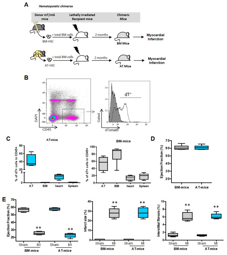
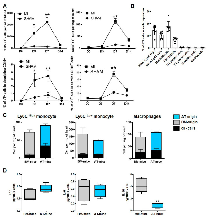
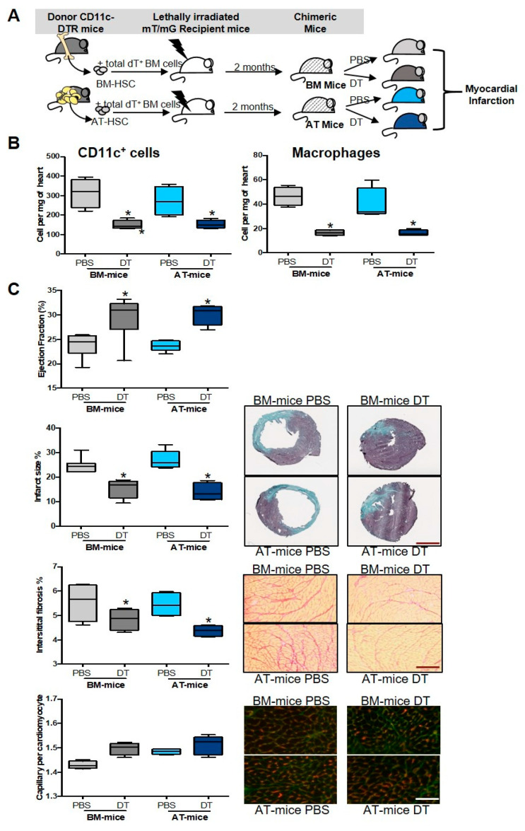
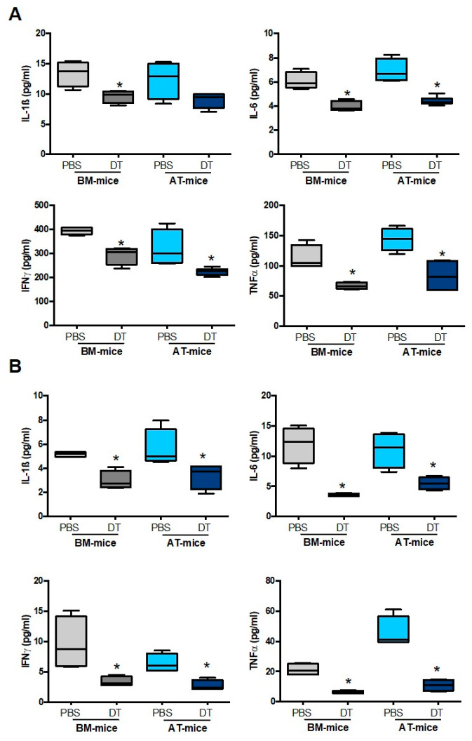
Medullary and extra-medullary hematopoiesis has been shown to govern inflammatory cell infiltration and subsequently cardiac remodeling and function after acute myocardial infarction (MI). Emerging evidence positions adipose tissue (AT) as an alternative source of immune cell production. We, therefore, hypothesized that AT could act as a reservoir of inflammatory cells that participate in cardiac homeostasis after MI. To reveal the distinct role of inflammatory cells derived from AT or bone marrow (BM), chimeric mice were generated using standard repopulation assays. We showed that AMI increased the number of AT-derived macrophages in the cardiac tissue. These macrophages exhibit pro-inflammatory characteristics and their specific depletion improved cardiac function as well as decreased infarct size and interstitial fibrosis. We then reasoned that the alteration of AT-immune compartment in type 2 diabetes could, thus, contribute to defects in cardiac remodeling. However, in these conditions, myeloid cells recruited in the infarcted heart mainly originate from the BM, and AT was no longer used as a myeloid cell reservoir. Altogether, we showed here that a subpopulation of cardiac inflammatory macrophages emerges from myeloid cells of AT origin and plays a detrimental role in cardiac remodeling and function after MI. Diabetes abrogates the ability of AT-derived myeloid cells to populate the infarcted heart.
Overview
The claims and statistics of this paper haven’t been checked yet.
Run the check — free, no account needed. It reads the open-access text and checks the sources it cites.
Integrity signals
Registry
Cites
Statistics
Figures
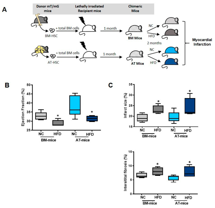
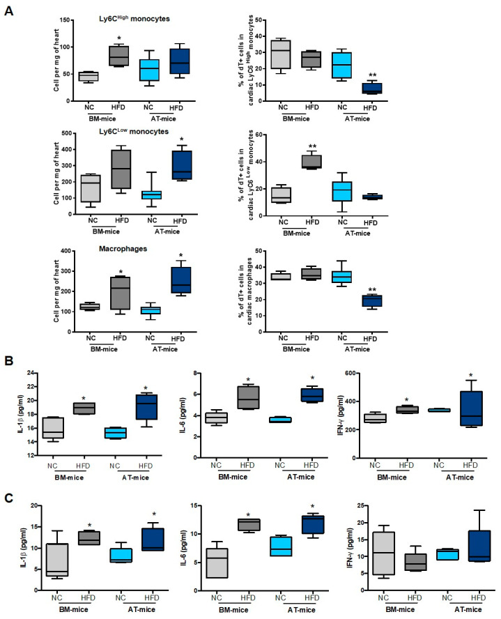
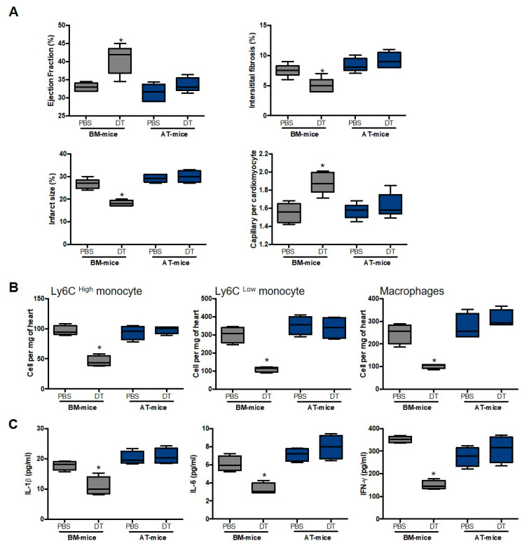
Verification
Claims the paper makes
The claims and statistics of this paper haven’t been checked yet.
Run the check — free, no account needed. It reads the open-access text and checks the sources it cites.
Claims other papers make about this one
Each is checked against this paper’s own text: does it say what the citing paper says it does?
In the citing paper
“Indeed, mast cell progenitors are also present in the spleen 104 and murine adipose tissues, 105 to which the latter are mobilized upon experimental myocardial infarction. 106”
From Mast cell ontogeny: From fetal development to life‐long health and disease (2023) · cites this paper as [106]
Self-check
Percentages, sums and reported differences re-computed from the paper’s own numbers.
The self-check needs the paper’s full text.
Citations
Cites
Transcriptome-Based Network Analysis Reveals a Spectrum Model of Human Macrophage Activation
Xue, J., Schmidt, S. V., Sander, J., Draffehn, A., Krebs, W., Quester, I., De Nardo, D., Gohel, T. D., Emde, M., Schmidleithner, L., et al. (2014) · Immunity · doi:10.1016/j.immuni.2014.01.006 · Cited by 2,210
The human heart contains distinct macrophage subsets with divergent origins and functions
Bajpai, G., Schneider, C., Wong, N., Bredemeyer, A., Hulsmans, M., Nahrendorf, M., Epelman, S., Kreisel, D., Liu, Y., Itoh, A., et al. (2018) · Nature Medicine · doi:10.1038/s41591-018-0059-x · Cited by 712
Iron regulator hepcidin impairs macrophage-dependent cardiac repair after injury
Zlatanova, I., Pinto, C., Bonnin, P., Mathieu, J. R. R., Bakker, W., Vilar, J., Lemitre, M., Voehringer, D., Vaulont, S., Peyssonnaux, C., et al. (2019) · Circulation · doi:10.1161/circulationaha.118.034545 · Cited by 81
Self-citation
Research works printed but not linked: 29
- Han, 2010, Adipose tissue is an extramedullary reservoir for functional hematopoietic stem and progenitor cells, Blood, 115, 957, 10.1182/blood-2009-05-219923 · DOI
- Poglio, 2012, In situ production of innate immune cells in murine white adipose tissue, Blood, 120, 4952, 10.1182/blood-2012-01-406959 · DOI
- Cousin, 2016, Immuno-metabolism and adipose tissue: The key role of hematopoietic stem cells, Biochimie, 124, 21, 10.1016/j.biochi.2015.06.012 · DOI
- Rabiller, 2021, Driving regeneration, instead of healing, in adult mammals: The decisive role of resident macrophages through efferocytosis, NPJ Regen. Med., 6, 1, 10.1038/s41536-021-00151-1 · DOI
- Poglio, 2010, Adipose tissue as a dedicated reservoir of functional mast cell progenitors, Stem Cells, 28, 2065, 10.1002/stem.523 · DOI · Paper page
- Ngkelo, 2016, Mast cells regulate myofilament calcium sensitization and heart function after myocardial infarction, J. Exp. Med., 213, 1353, 10.1084/jem.20160081 · DOI
- Luche, 2017, Corrupted adipose tissue endogenous myelopoiesis initiates diet-induced metabolic disease, eLife, 6, e23194, 10.7554/eLife.23194 · DOI
- Peet, 2020, Cardiac monocytes and macrophages after myocardial infarction, Cardiovasc. Res., 116, 1101, 10.1093/cvr/cvz336 · DOI
- Nahrendorf, 2007, The healing myocardium sequentially mobilizes two monocyte subsets with divergent and complementary functions, J. Exp. Med., 204, 3037, 10.1084/jem.20070885 · DOI · Paper page
- Swirski, 2009, Identification of splenic reservoir monocytes and their deployment to inflammatory sites, Science, 325, 612, 10.1126/science.1175202 · DOI · Paper page
- Leuschner, 2012, Rapid monocyte kinetics in acute myocardial infarction are sustained by extramedullary monocytopoiesis, J. Exp. Med., 209, 123, 10.1084/jem.20111009 · DOI
- Sica, 2012, Macrophage plasticity and polarization: In vivo veritas, J. Clin. Investig., 122, 787, 10.1172/JCI59643 · DOI · Paper page
- Howangyin, 2016, Myeloid-Epithelial-Reproductive Receptor Tyrosine Kinase and Milk Fat Globule Epidermal Growth Factor 8 Coordinately Improve Remodeling After Myocardial Infarction via Local Delivery of Vascular Endothelial Growth Factor, Circulation, 133, 826, 10.1161/CIRCULATIONAHA.115.020857 · DOI
- Singla, D.K., Singla, R.D., Abdelli, L.S., and Glass, C. (2015). Fibroblast growth factor-9 enhances M2 macrophage differentiation and attenuates adverse cardiac remodeling in the infarcted diabetic heart. PLoS ONE, 10. · DOI
- Fadini, 2013, An unbalanced monocyte polarisation in peripheral blood and bone marrow of patients with type 2 diabetes has an impact on microangiopathy, Diabetologia, 56, 1856, 10.1007/s00125-013-2918-9 · DOI
- Jadhav, 2013, The heme oxygenase system selectively enhances the anti-inflammatory macrophage-M2 phenotype, reduces pericardial adiposity, and ameliorated cardiac injury in diabetic cardiomyopathy in Zucker diabetic fatty rats, J. Pharmacol. Exp. Ther., 345, 239, 10.1124/jpet.112.200808 · DOI
- Nagareddy, 2013, Hyperglycemia promotes myelopoiesis and impairs the resolution of atherosclerosis, Cell Metab., 17, 695, 10.1016/j.cmet.2013.04.001 · DOI
- Miller, 2008, Characterization of mouse hematopoietic stem and progenitor cells, Curr. Protoc. Immunol., 22, 22B.2
- Burcelin, 2002, Heterogeneous metabolic adaptation of C57BL/6J mice to high-fat diet, Am. J. Physiol. Endocrinol. Metab., 282, E834, 10.1152/ajpendo.00332.2001 · DOI
- Zaman, 2021, Exploring cardiac macrophage heterogeneity in the healthy and diseased myocardium, Curr. Opin. Immunol., 68, 54, 10.1016/j.coi.2020.09.005 · DOI · Paper page
- Guilliams, 2020, Establishment and Maintenance of the Macrophage Niche, Immunity, 52, 434, 10.1016/j.immuni.2020.02.015 · DOI · Paper page
- Frantz, 2014, Cardiac macrophages and their role in ischaemic heart disease, Cardiovasc. Res., 102, 240, 10.1093/cvr/cvu025 · DOI
- Dick, 2022, Three tissue resident macrophage subsets coexist across organs with conserved origins and life cycles, Sci. Immunol., 7, eabf777, 10.1126/sciimmunol.abf7777 · DOI · Paper page
- Kuppe, 2022, Spatial multi-omic map of human myocardial infarction, Nature, 608, 766, 10.1038/s41586-022-05060-x · DOI · Paper page
- Yan, 2018, Diabetes impairs wound healing by Dnmt1-dependent dysregulation of hematopoietic stem cells differentiation towards macrophages, Nat. Commun., 9, 33, 10.1038/s41467-017-02425-z · DOI
- Barman, 2020, Macrophage Dysregulation and Impaired Skin Wound Healing in Diabetes, Front. Cell Dev. Biol., 8, 528, 10.3389/fcell.2020.00528 · DOI
- Ritchie, 2020, Basic Mechanisms of Diabetic Heart Disease, Circ. Res., 126, 1501, 10.1161/CIRCRESAHA.120.315913 · DOI
- Lopaschuk, 2002, Metabolic Abnormalities in the Diabetic Heart, Heart Fail. Rev., 7, 149, 10.1023/A:1015328625394 · DOI
- Gomez, 2021, Extracellular vesicles from human cardiovascular progenitors trigger a reparative immune response in infarcted hearts, Cardiovasc. Res., 117, 292, 10.1093/cvr/cvaa028 · DOI
Cited by
Sorted by citations
Mast cell ontogeny: From fetal development to life‐long health and disease
Chia, S. L., Kapoor, S., Carvalho, C., Bajénoff, M., & Gentek, R. (2023) · Immunological Reviews · doi:10.1111/imr.13191 · Cited by 60
Metadata
- Authors
- Gomez, I., Robert, V., Alayrac, P., Arlat, A., Duval, V., Renoud, M.-L., Vilar, J., Lemitre, M., Silvestre, J.-S., & Cousin, B.
- Affiliations
- PARCC, INSERM, Université Paris Cité, 75015 Paris, France
- RESTORE Research Center, Université de Toulouse, INSERM 1301, CNRS 5070, EFS, ENVT, 31100 Toulouse, France
- ORCID
- Venue
- International Journal of Molecular Sciences
- Publisher
- MDPI AG
- Published
- 2022-09-10
- Volume
- 23
- Issue
- 18
- Pages
- 10498
- ISSN
- 1422-0067
- PMC
- PMC9499676
- Cited by
- 3
- References
- 3
- Language
- en
- Funders
- French National Research Agency (ANR), Project «WAT-HEART» (ANR 16-CE14-0006-01, ANR 16-CE14-0006-02)
About these checks
Registry, retraction, Cites and statistics results are computed, not judged, with one exception: a printed reference that no index lists is read once by a model before it counts as a citation error. Claims are extracted by a model and kept only when their quote appears verbatim in the text; each cited source is then checked, with a model’s help, for whether it says what the claim relies on. Those verdicts can be wrong — read the claim’s quote and the cited source before relying on one.
Statistics and arithmetic are recomputed, not judged.
Confidence bands are shown only for fields calibrated on our annotated test set. None is calibrated yet, so no band is shown.
What counts as a claim ›How we measure confidence ›How we check this ›
Are you an author, or do you see a problem with this page? Request a review
How to cite
Cite CoChat for the checks on this page, and the paper for its own findings.
- This page
- CoChat. (2026). Citation check: Identification of Adipose Tissue as a Reservoir of Macrophages after Acute Myocardial Infarction. https://app.cochat.ai/papers/10.3390/ijms231810498
- The paper
- Gomez, I., Robert, V., Alayrac, P., Arlat, A., Duval, V., Renoud, M.-L., Vilar, J., Lemitre, M., Silvestre, J.-S., & Cousin, B. (2022). Identification of Adipose Tissue as a Reservoir of Macrophages after Acute Myocardial Infarction. International Journal of Molecular Sciences. https://doi.org/10.3390/ijms231810498
