

The DOI matches its Crossref record; 6 of the 6 references it cites were found in a registry or an index.
Comprehensive Analysis of the Potential Toxicity of Magnetic Iron Oxide Nanoparticles for Medical Applications: Cellular Mechanisms and Systemic Effects
Abstract
Owing to recent advancements in nanotechnology, magnetic iron oxide nanoparticles (MNPs), particularly magnetite (Fe3O4) and maghemite (γ-Fe2O3), are currently widely employed in the field of medicine. These MNPs, characterized by their large specific surface area, potential for diverse functionalization, and magnetic properties, have found application in various medical domains, including tumor imaging (MRI), radiolabelling, internal radiotherapy, hyperthermia, gene therapy, drug delivery, and theranostics. However, ensuring the non-toxicity of MNPs when employed in medical practices is paramount. Thus, ongoing research endeavors are essential to comprehensively understand and address potential toxicological implications associated with their usage. This review aims to present the latest research and findings on assessing the potential toxicity of magnetic nanoparticles. It meticulously delineates the primary mechanisms of MNP toxicity at the cellular level, encompassing oxidative stress, genotoxic effects, disruption of the cytoskeleton, cell membrane perturbation, alterations in the cell cycle, dysregulation of gene expression, inflammatory response, disturbance in ion homeostasis, and interference with cell migration and mobility. Furthermore, the review expounds upon the potential impact of MNPs on various organs and systems, including the brain and nervous system, heart and circulatory system, liver, spleen, lymph nodes, skin, urinary, and reproductive systems.
Overview
The claims and statistics of this paper haven’t been checked yet.
Run the check — free, no account needed. It reads the open-access text and checks the sources it cites.
Integrity signals
Registry
Cites
Statistics
Figures
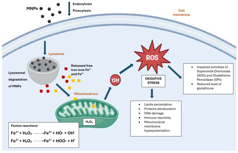
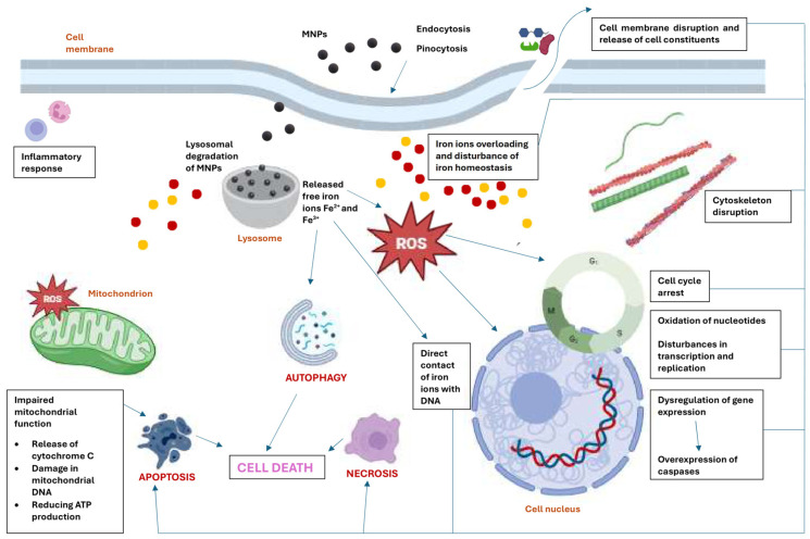
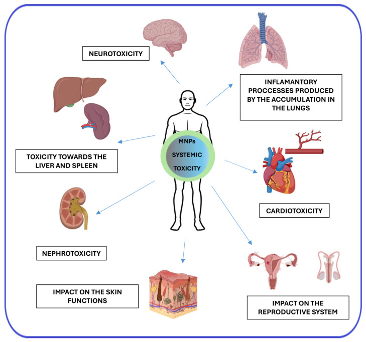
Verification
Claims the paper makes
The claims and statistics of this paper haven’t been checked yet.
Run the check — free, no account needed. It reads the open-access text and checks the sources it cites.
Claims other papers make about this one
Each is checked against this paper’s own text: does it say what the citing paper says it does?
In the citing paper
“Biocompatibility is one of the most important criteria for clinical use of MNP. Since these particles intend to be used in biological systems, their interaction with cells, tissues, and organs should not induce side effects, such as inflammation, toxicity, or immune rejection [69].”
From Advances in green-synthesized magnetic nanoparticles for targeted cancer therapy: mechanisms, applications, and future perspectives (2026) · cites this paper as [69]
In the citing paper
“While MNPs are often considered biocompatible, their long-term accumulation and degradation products (e.g., free iron ions) may induce oxidative stress or interfere with normal cellular processes.”
From Advances in green-synthesized magnetic nanoparticles for targeted cancer therapy: mechanisms, applications, and future perspectives (2026) · cites this paper as [69]
In the citing paper
“Toxicity and clearance remain partially unresolved.”
From Advances in green-synthesized magnetic nanoparticles for targeted cancer therapy: mechanisms, applications, and future perspectives (2026) · cites this paper as [69]
In the citing paper
“Comprehensive toxicological evaluations are necessary to ensure safety over extended periods, especially for repeated dosing regimens [69, 98].”
From Advances in green-synthesized magnetic nanoparticles for targeted cancer therapy: mechanisms, applications, and future perspectives (2026) · cites this paper as [69]
Self-check
Percentages, sums and reported differences re-computed from the paper’s own numbers.
The self-check needs the paper’s full text.
Citations
Cites
Wang, Y.-X. J., Hussain, S. M., & Krestin, G. P. (2001) · European Radiology · doi:10.1007/s003300100908 · Cited by 1,381
Mitochondria and Mitochondrial Cascades in Alzheimer’s disease
Swerdlow, R. H. (2018) · Journal of Alzheimer’s Disease · doi:10.3233/jad-170585 · Cited by 850
Parameters influencing the stealthiness of colloidal drug delivery systems
Vonarbourg, A., Passirani, C., Saulnier, P., & Benoit, J.-P. (2006) · Biomaterials · doi:10.1016/j.biomaterials.2006.03.039 · Cited by 740
Iron Oxide Nanoparticles for Magnetically-Guided and Magnetically-Responsive Drug Delivery
Estelrich, J., Escribano, E., Queralt, J., & Busquets, M. (2015) · International Journal of Molecular Sciences · doi:10.3390/ijms16048070 · Cited by 532
Cappellini, M. D., Comin‐Colet, J., de Francisco, A., Dignass, A., Doehner, W., Lam, C. S., Macdougall, I. C., Rogler, G., Camaschella, C., Kadir, R., et al. (2017) · American Journal of Hematology · doi:10.1002/ajh.24820 · Cited by 502
Remya, N. S., Syama, S., Sabareeswaran, A., & Mohanan, P. V. (2016) · International Journal of Pharmaceutics · doi:10.1016/j.ijpharm.2016.06.119 · Cited by 97 · cited 1×
“Further modification with various phytochemicals has been observed to augment the antioxidant activity of MNPs. Conversely, studies by Hohnholt [40], Linde-mann et al. [41], and Remya et al. [42] reported no signi fi cant ROS formation after treat-ment with MNPs.”
Research works printed but not linked: 204
- Avval, 2020, Introduction of Magnetic and Supermagnetic Nanoparticles in New Approach of Targeting Drug Delivery and Cancer Therapy Application, Drug Metab. Rev., 52, 157, 10.1080/03602532.2019.1697282 · DOI
- Foy, 2010, Optical Imaging and Magnetic Field Targeting of Magnetic Nanoparticles in Tumors, ACS Nano, 4, 5217, 10.1021/nn101427t · DOI
- Stanicki, 2022, An Update on the Applications and Characteristics of Magnetic Iron Oxide Nanoparticles for Drug Delivery, Expert. Opin. Drug Deliv., 19, 321, 10.1080/17425247.2022.2047020 · DOI
- Palzer, J., Eckstein, L., Slabu, I., Reisen, O., Neumann, U.P., and Roeth, A.A. (2021). Iron Oxide Nanoparticle-Based Hyperthermia as a Treatment Option in Various Gastrointestinal Malignancies. Nanomaterials, 11. · DOI
- Obaidat, I.M., Narayanaswamy, V., Alaabed, S., Sambasivam, S., and Muralee Gopi, C.V.V. (2019). Principles of Magnetic Hyperthermia: A Focus on Using Multifunctional Hybrid Magnetic Nanoparticles. Magnetochemistry, 5. · DOI
- Mu, 2018, siRNA Delivery with Stem Cell Membrane-Coated Magnetic Nanoparticles for Imaging-Guided Photothermal Therapy and Gene Therapy, ACS Biomater. Sci. Eng., 4, 3895, 10.1021/acsbiomaterials.8b00858 · DOI
- Russell, 2021, Impact of Superparamagnetic Iron Oxide Nanoparticles on in Vitro and in Vivo Radiosensitisation of Cancer Cells, Radiat. Oncol., 16, 104, 10.1186/s13014-021-01829-y · DOI
- Rosseto, 2018, Iron Oxide Magnetic Nanoparticles as Antimicrobials for Therapeutics, Pharm. Dev. Technol., 23, 316, 10.1080/10837450.2017.1337793 · DOI
- Rodrigues, 2019, Antimicrobial Magnetic Nanoparticles Based-Therapies for Controlling Infectious Diseases, Int. J. Pharm., 555, 356, 10.1016/j.ijpharm.2018.11.043 · DOI
- Li, Z., Xue, L., Wang, P., Ren, X., Zhang, Y., Wang, C., and Sun, J. (2023). Biological Scaffolds Assembled with Magnetic Nanoparticles for Bone Tissue Engineering: A Review. Materials, 16. · DOI
- Skotland, 2010, New Metal-Based Nanoparticles for Intravenous Use: Requirements for Clinical Success with Focus on Medical Imaging, Nanomed. Nanotechnol. Biol. Med., 6, 730, 10.1016/j.nano.2010.05.002 · DOI
- Wallace, 2016, The Regulation of Iron Absorption and Homeostasis, Clin. Biochem. Rev., 37, 51
- Mai, 2017, Magnetic Nanoparticles: Reactive Oxygen Species Generation and Potential Therapeutic Applications, J. Nanopart Res., 19, 253, 10.1007/s11051-017-3943-2 · DOI
- Ahmad, 2020, Comparative Acute Intravenous Toxicity Study of Triple Polymer-Layered Magnetic Nanoparticles with Bare Magnetic Nanoparticles in Swiss Albino Mice, Nanotoxicology, 14, 1362, 10.1080/17435390.2020.1829144 · DOI
- Mahmoudi, 2011, Toxicity Evaluations of Superparamagnetic Iron Oxide Nanoparticles: Cell “Vision” versus Physicochemical Properties of Nanoparticles, ACS Nano, 5, 7263, 10.1021/nn2021088 · DOI
- Malvindi, M.A., De Matteis, V., Galeone, A., Brunetti, V., Anyfantis, G.C., Athanassiou, A., Cingolani, R., and Pompa, P.P. (2014). Toxicity Assessment of Silica Coated Iron Oxide Nanoparticles and Biocompatibility Improvement by Surface Engineering. PLoS ONE, 9. · DOI
- Shukla, 2015, In Vitro Toxicity Assessment of Chitosan Oligosaccharide Coated Iron Oxide Nanoparticles, Toxicol. Rep., 2, 27, 10.1016/j.toxrep.2014.11.002 · DOI
- Abakumov, M.A., Semkina, A.S., Skorikov, A.S., Vishnevskiy, D.A., Ivanova, A.V., Mironova, E., Davydova, G.A., Majouga, A.G., and Chekhonin, V.P. (2018). Toxicity of Iron Oxide Nanoparticles: Size and Coating Effects. J. Biochem. Mol. Toxicol., 32. · DOI
- Yang, 2013, Difference between Toxicities of Iron Oxide Magnetic Nanoparticles with Various Surface-Functional Groups against Human Normal Fibroblasts and Fibrosarcoma Cells, Materials, 6, 4689, 10.3390/ma6104689 · DOI
- Malhotra, N., Lee, J.-S., Liman, R.A.D., Ruallo, J.M.S., Villaflores, O.B., Ger, T.-R., and Hsiao, C.-D. (2020). Potential Toxicity of Iron Oxide Magnetic Nanoparticles: A Review. Molecules, 25. · DOI
- Owensiii, 2006, Opsonization, Biodistribution, and Pharmacokinetics of Polymeric Nanoparticles, Int. J. Pharm., 307, 93, 10.1016/j.ijpharm.2005.10.010 · DOI · Paper page
- Manzanares, D., and Ceña, V. (2020). Endocytosis: The Nanoparticle and Submicron Nanocompounds Gateway into the Cell. Pharmaceutics, 12. · DOI
- Swanson, 1995, Phagocytosis by Zippers and Triggers, Trends Cell Biol., 5, 89, 10.1016/S0962-8924(00)88956-4 · DOI
- Wu, 2014, Reactive Oxygen Species-Related Activities of Nano-Iron Metal and Nano-Iron Oxides, J. Food Drug Anal., 22, 86, 10.1016/j.jfda.2014.01.007 · DOI
- Sengul, 2020, Toxicity of Metal and Metal Oxide Nanoparticles: A Review, Environ. Chem. Lett., 18, 1659, 10.1007/s10311-020-01033-6 · DOI
- Valdiglesias, 2015, Effects of Iron Oxide Nanoparticles: Cytotoxicity, Genotoxicity, Developmental Toxicity, and Neurotoxicity: Effects of Iron Oxide Nanoparticles, Environ. Mol. Mutagen., 56, 125, 10.1002/em.21909 · DOI
- Zhang, 2010, Integration of ER Stress, Oxidative Stress and the Inflammatory Response in Health and Disease, Int. J. Clin. Exp. Med., 3, 33
- Pongrac, 2016, Oxidative Stress Response in Neural Stem Cells Exposed to Different Superparamagnetic Iron Oxide Nanoparticles, Int. J. Nanomed., 11, 1701
- Wu, 2022, Ultrasmall Iron Oxide Nanoparticles Cause Significant Toxicity by Specifically Inducing Acute Oxidative Stress to Multiple Organs, Part. Fibre Toxicol., 19, 24, 10.1186/s12989-022-00465-y · DOI
- Ying, 2022, Iron Oxide Nanoparticles Size-Dependently Activate Mouse Primary Macrophages via Oxidative Stress and Endoplasmic Reticulum Stress, Int. Immunopharmacol., 105, 108533, 10.1016/j.intimp.2022.108533 · DOI
- Ansari, M.O., Parveen, N., Ahmad, M.F., Wani, A.L., Afrin, S., Rahman, Y., Jameel, S., Khan, Y.A., Siddique, H.R., and Tabish, M. (2019). Evaluation of DNA Interaction, Genotoxicity and Oxidative Stress Induced by Iron Oxide Nanoparticles Both in Vitro and in Vivo: Attenuation by Thymoquinone. Sci. Rep., 9. · DOI
- Kenzaoui, 2012, Evaluation of Uptake and Transport of Ultrasmall Superparamagnetic Iron Oxide Nanoparticles by Human Brain-Derived Endothelial Cells, Nanomedicine, 7, 39, 10.2217/nnm.11.85 · DOI
- Zhang, 2012, Evaluation on Cytotoxicity and Genotoxicity of the L-Glutamic Acid Coated Iron Oxide Nanoparticles, J. Nanosci. Nanotechnol., 12, 2866, 10.1166/jnn.2012.5763 · DOI
- Du, 2017, Overendocytosis of Superparamagnetic Iron Oxide Particles Increases Apoptosis and Triggers Autophagic Cell Death in Human Osteosarcoma Cell under a Spinning Magnetic Field, Oncotarget, 8, 9410, 10.18632/oncotarget.14114 · DOI
- Petters, 2016, Lysosomal Iron Liberation Is Responsible for the Vulnerability of Brain Microglial Cells to Iron Oxide Nanoparticles: Comparison with Neurons and Astrocytes, Nanotoxicology, 10, 332, 10.3109/17435390.2015.1071445 · DOI · Paper page
- Hanot, C.C., Choi, Y.S., Anani, T.B., Soundarrajan, D., and David, A.E. (2015). Effects of Iron-Oxide Nanoparticle Surface Chemistry on Uptake Kinetics and Cytotoxicity in CHO-K1 Cells. Int. J. Mol. Sci., 17. · DOI
- Watanabe, 2013, Effects of Fe3O4 Magnetic Nanoparticles on A549 Cells, Int. J. Mol. Sci., 14, 15546, 10.3390/ijms140815546 · DOI
- Hohnholt, 2011, Treatment with Iron Oxide Nanoparticles Induces Ferritin Synthesis but Not Oxidative Stress in Oligodendroglial Cells, Acta Biomater., 7, 3946, 10.1016/j.actbio.2011.06.052 · DOI
- Lindemann, 2014, Biological Impact of Superparamagnetic Iron Oxide Nanoparticles for Magnetic Particle Imaging of Head and Neck Cancer Cells, Int. J. Nanomed., 9, 5025, 10.2147/IJN.S63873 · DOI
- Repar, N., Jovičić, E.J., Kump, A., Birarda, G., Vaccari, L., Erman, A., Kralj, S., Nemec, S., Petan, T., and Drobne, D. (2022). Oleic Acid Protects Endothelial Cells from Silica-Coated Superparamagnetic Iron Oxide Nanoparticles (SPIONs)-Induced Oxidative Stress and Cell Death. Int. J. Mol. Sci., 23. · DOI
- Ahamed, 2013, Selective Killing of Cancer Cells by Iron Oxide Nanoparticles Mediated through Reactive Oxygen Species via P53 Pathway, J. Nanopart Res., 15, 1225, 10.1007/s11051-012-1225-6 · DOI
- Flieger, J., Franus, W., Panek, R., Szymańska-Chargot, M., Flieger, W., Flieger, M., and Kołodziej, P. (2021). Green Synthesis of Silver Nanoparticles Using Natural Extracts with Proven Antioxidant Activity. Molecules, 26. · DOI
- Khalil, I., Yehye, W.A., Etxeberria, A.E., Alhadi, A.A., Dezfooli, S.M., Julkapli, N.B.M., Basirun, W.J., and Seyfoddin, A. (2019). Nanoantioxidants: Recent Trends in Antioxidant Delivery Applications. Antioxidants, 9. · DOI
- Samrot, 2022, Nanoparticles, a Double-Edged Sword with Oxidant as Well as Antioxidant Properties—A Review, Oxygen, 2, 591, 10.3390/oxygen2040039 · DOI
- Chavan, 2020, Characterization, Antioxidant, Antimicrobial and Cytotoxic Activities of Green Synthesized Silver and Iron Nanoparticles Using Alcoholic Blumea Eriantha DC Plant Extract, Mater. Today Commun., 24, 101320, 10.1016/j.mtcomm.2020.101320 · DOI
- Valavanidis, 2009, 8-Hydroxy-2′ -Deoxyguanosine (8-OHdG): A Critical Biomarker of Oxidative Stress and Carcinogenesis, J. Environ. Sci. Health Part C, 27, 120, 10.1080/10590500902885684 · DOI
- Ayala, 2014, Lipid Peroxidation: Production, Metabolism, and Signaling Mechanisms of Malondialdehyde and 4-Hydroxy-2-Nonenal, Oxidative Med. Cell. Longev., 2014, 360438, 10.1155/2014/360438 · DOI
- Jahanbani, J., Ghotbi, M., Shahsavari, F., Seydi, E., Rahimi, S., and Pourahmad, J. (2020). Selective Anticancer Activity of Superparamagnetic Iron Oxide Nanoparticles (SPIONs) against Oral Tongue Cancer Using in Vitro Methods: The Key Role of Oxidative Stress on Cancerous Mitochondria. J. Biochem. Mol. Toxicol., 34. · DOI
- Shi, Y., Wang, J., Liu, J., Lin, G., Xie, F., Pang, X., Pei, Y., Cheng, Y., Zhang, Y., and Lin, Z. (2020). Oxidative Stress-Driven DR5 Upregulation Restores TRAIL/Apo2L Sensitivity Induced by Iron Oxide Nanoparticles in Colorectal Cancer. Biomaterials, 233. · DOI
- Alvarez, 2015, Reactive Oxygen Species and Mitochondria: A Nexus of Cellular Homeostasis, Redox Biol., 6, 472, 10.1016/j.redox.2015.09.005 · DOI
- Saquib, 2018, Cellular and Molecular Toxicity of Iron Oxide Nanoparticles, Cellular and Molecular Toxicology of Nanoparticles, Volume 1048, 199
- Yarjanli, Z., Ghaedi, K., Esmaeili, A., Rahgozar, S., and Zarrabi, A. (2017). Iron Oxide Nanoparticles May Damage to the Neural Tissue through Iron Accumulation, Oxidative Stress, and Protein Aggregation. BMC Neurosci., 18. · DOI
- Zhang, Z., Huang, Q., Zhao, D., Lian, F., Li, X., and Qi, W. (2023). The Impact of Oxidative Stress-Induced Mitochondrial Dysfunction on Diabetic Microvascular Complications. Front. Endocrinol., 14. · DOI
- Guo, 2013, Oxidative Stress, Mitochondrial Damage and Neurodegenerative Diseases, Neural Regen. Res., 8, 2003
- Mancuso, 2009, Mitochondria, Cognitive Impairment, and Alzheimer’s Disease, Int. J. Alzheimer’s Dis., 2009, 951548
- D’Errico, M., Parlanti, E., Pascucci, B., Filomeni, G., Mastroberardino, P.G., and Dogliotti, E. (2021). The Interplay between Mitochondrial Functionality and Genome Integrity in the Prevention of Human Neurologic Diseases. Arch. Biochem. Biophys., 710. · DOI
- Sharma, C., Kim, S., Nam, Y., Jung, U.J., and Kim, S.R. (2021). Mitochondrial Dysfunction as a Driver of Cognitive Impairment in Alzheimer’s Disease. Int. J. Mol. Sci., 22. · DOI
- Shukla, R.K., Badiye, A., Vajpayee, K., and Kapoor, N. (2021). Genotoxic Potential of Nanoparticles: Structural and Functional Modifications in DNA. Front. Genet., 12. · DOI
- Bourrinet, 2006, Preclinical Safety and Pharmacokinetic Profile of Ferumoxtran-10, an Ultrasmall Superparamagnetic Iron Oxide Magnetic Resonance Contrast Agent, Investig. Radiol., 41, 313, 10.1097/01.rli.0000197669.80475.dd · DOI
- Zou, 2015, Iron Nanoparticles Significantly Affect the In Vitro and In Vivo Expression of Id Genes, Chem. Res. Toxicol., 28, 373, 10.1021/tx500333q · DOI
- Shahabadi, 2016, Functionalization of Fe3O4@SiO2 Magnetic Nanoparticles with Nicotinamide and in Vitro DNA Interaction, J. Mol. Liq., 224, 227, 10.1016/j.molliq.2016.09.103 · DOI
- Shokrollahi, 2023, Evaluation of the Effect of Iron Oxide Nanoparticles Functionalized by Glucose and Conjugated with Coumarin (Fe3O4@Glu-Coumarin NPs) on the Expression of CASP8, CASP9, P53, mTOR1, and MAPK1 Genes in Liver Cancer Cell Line, Gene Rep., 33, 101818, 10.1016/j.genrep.2023.101818 · DOI
- Riedl, 2004, Molecular Mechanisms of Caspase Regulation during Apoptosis, Nat. Rev. Mol. Cell Biol., 5, 897, 10.1038/nrm1496 · DOI
- Fridman, 2003, Control of Apoptosis by P53, Oncogene, 22, 9030, 10.1038/sj.onc.1207116 · DOI · Paper page
- Siddiqui, M.A., Wahab, R., Saquib, Q., Ahmad, J., Farshori, N.N., Al-Sheddi, E.S., Al-Oqail, M.M., Al-Massarani, S.M., and Al-Khedhairy, A.A. (2023). Iron Oxide Nanoparticles Induced Cytotoxicity, Oxidative Stress, Cell Cycle Arrest, and DNA Damage in Human Umbilical Vein Endothelial Cells. J. Trace Elem. Med. Biol., 80. · DOI
- Kozics, 2014, The Role of Reactive Oxygen Species in the Genotoxicity of Surface-Modified Magnetite Nanoparticles, Toxicol. Lett., 226, 303, 10.1016/j.toxlet.2014.02.025 · DOI
- Robinson, J.M. (2008). Reactive Oxygen Species in Phagocytic Leukocytes. Histochem. Cell Biol., 130. · DOI
- Hamelin, 2008, Cytoskeleton and Apoptosis, Biochem. Pharmacol., 76, 11, 10.1016/j.bcp.2008.03.016 · DOI
- Askri, 2019, Investigating the Toxic Effects Induced by Iron Oxide Nanoparticles on Neuroblastoma Cell Line: An Integrative Study Combining Cytotoxic, Genotoxic and Proteomic Tools, Nanotoxicology, 13, 1021, 10.1080/17435390.2019.1621399 · DOI
- Master, A.M., Williams, P.N., Pothayee, N., Pothayee, N., Zhang, R., Vishwasrao, H.M., Golovin, Y.I., Riffle, J.S., Sokolsky, M., and Kabanov, A.V. (2016). Remote Actuation of Magnetic Nanoparticles For Cancer Cell Selective Treatment Through Cytoskeletal Disruption. Sci. Rep., 6. · DOI
- Connord, 2015, Real-Time Analysis of Magnetic Hyperthermia Experiments on Living Cells under a Confocal Microscope, Small, 11, 2437, 10.1002/smll.201402669 · DOI
- Zhang, 2014, Dynamic Magnetic Fields Remote-Control Apoptosis via Nanoparticle Rotation, ACS Nano, 8, 3192, 10.1021/nn406302j · DOI
- Královec, K., Melounková, L., Slováková, M., Mannová, N., Sedlák, M., Bartáček, J., and Havelek, R. (2020). Disruption of Cell Adhesion and Cytoskeletal Networks by Thiol-Functionalized Silica-Coated Iron Oxide Nanoparticles. Int. J. Mol. Sci., 21. · DOI
- Shin, 2013, Membrane Potential Mediates the Cellular Binding of Nanoparticles, Nanoscale, 5, 5879, 10.1039/c3nr01667f · DOI
- Pongrac, 2016, Improved Biocompatibility and Efficient Labeling of Neural Stem Cells with Poly(L-Lysine)-Coated Maghemite Nanoparticles, Beilstein J. Nanotechnol., 7, 926, 10.3762/bjnano.7.84 · DOI
- Gonnissen, 2016, Comparison of Cellular Effects of Starch-Coated SPIONs and Poly(Lactic-Co-Glycolic Acid) Matrix Nanoparticles on Human Monocytes, Int. J. Nanomed., 11, 5221, 10.2147/IJN.S106540 · DOI
- Yan, 2015, Fe2O3 Nanoparticles Suppress Kv1.3 Channels via Affecting the Redox Activity of Kv β 2 Subunit in Jurkat T Cells, Nanotechnology, 26, 505103, 10.1088/0957-4484/26/50/505103 · DOI
- Gualdani, 2019, Superparamagnetic Iron Oxide Nanoparticles (SPIONs) Modulate hERG Ion Channel Activity, Nanotoxicology, 13, 1197, 10.1080/17435390.2019.1650969 · DOI
- Hernandes, E.P., Lazarin-Bidóia, D., Bini, R.D., Nakamura, C.V., Cótica, L.F., and De Oliveira Silva Lautenschlager, S. (2023). Doxorubicin-Loaded Iron Oxide Nanoparticles Induce Oxidative Stress and Cell Cycle Arrest in Breast Cancer Cells. Antioxidants, 12. · DOI
- Rostami, 2022, High Efficacy of Tamoxifen-Loaded L-Lysine Coated Magnetic Iron Oxide Nanoparticles in Cell Cycle Arrest and Anti-Cancer Activity for Breast Cancer Therapy, Bioimpacts, 12, 301, 10.34172/bi.2021.23337 · DOI
- Majeed, 2023, In Vitro Apoptosis and Molecular Response of Engineered Green Iron Oxide Nanoparticles with L-Arginine in MDA-MB-231 Breast Cancer Cells, J. Drug Deliv. Sci. Technol., 80, 104185, 10.1016/j.jddst.2023.104185 · DOI
- Grosse, 2016, Iron Oxide Nanoparticles Modulate Lipopolysaccharide-Induced Inflammatory Responses in Primary Human Monocytes, Int. J. Nanomed., 11, 4625, 10.2147/IJN.S113425 · DOI
- Soares, 2010, The Role of Lipopolysaccharide/Toll-like Receptor 4 Signaling in Chronic Liver Diseases, Hepatol. Int., 4, 659, 10.1007/s12072-010-9219-x · DOI
- Gerogianni, A., Bal, M., Mohlin, C., Woodruff, T.M., Lambris, J.D., Mollnes, T.E., Sjöström, D.J., and Nilsson, P.H. (2023). In Vitro Evaluation of Iron Oxide Nanoparticle-Induced Thromboinflammatory Response Using a Combined Human Whole Blood and Endothelial Cell Model. Front. Immunol., 14. · DOI
- Chauhan, 2023, Vitamin K3-Loaded Magnetic Nanoparticle-Mediated Synergistic Magnetothermodynamic Therapy Evokes Massive ROS and Immune Modulation for Augmented Antitumor Potential, ACS Appl. Mater. Interfaces, 15, 27515, 10.1021/acsami.3c01702 · DOI
- Knutson, 2019, Non-Transferrin-Bound Iron Transporters, Free. Radic. Biol. Med., 133, 101, 10.1016/j.freeradbiomed.2018.10.413 · DOI · Paper page
- Scaramellini, N., Fischer, D., Agarvas, A.R., Motta, I., Muckenthaler, M.U., and Mertens, C. (2023). Interpreting Iron Homeostasis in Congenital and Acquired Disorders. Pharmaceuticals, 16. · DOI
- Chrishtop, 2021, Organ-Specific Toxicity of Magnetic Iron Oxide-Based Nanoparticles, Nanotoxicology, 15, 167, 10.1080/17435390.2020.1842934 · DOI · Paper page
- Yang, 2010, Superparamagnetic Iron Oxide Nanoparticles May Affect Endothelial Progenitor Cell Migration Ability and Adhesion Capacity, Cytotherapy, 12, 251, 10.3109/14653240903446910 · DOI
- Mulens-Arias, V., Rojas, J.M., Sanz-Ortega, L., Portilla, Y., Pérez-Yagüe, S., and Barber, D.F. (2019). Polyethylenimine-Coated Superparamagnetic Iron Oxide Nanoparticles Impair in Vitro and in Vivo Angiogenesis. Nanomed. Nanotechnol. Biol. Med., 21. · DOI
- Kshitiz, 2013, Cell Motility of Neural Stem Cells Is Reduced after SPIO-Labeling, Which Is Mitigated after Exocytosis: Inhibition of Cell Motility and SPIO Exocytosis, Magn. Reson. Med., 69, 255, 10.1002/mrm.24216 · DOI
- Rojas, 2016, Superparamagnetic Iron Oxide Nanoparticle Uptake Alters M2 Macrophage Phenotype, Iron Metabolism, Migration and Invasion, Nanomed. Nanotechnol. Biol. Med., 12, 1127, 10.1016/j.nano.2015.11.020 · DOI
- Mohsin, 2022, Recent Advances of Magnetic Nanomaterials for Bioimaging, Drug Delivery, and Cell Therapy, ACS Appl. Nano Mater., 5, 10118, 10.1021/acsanm.2c02014 · DOI
- Petters, 2014, Uptake and Metabolism of Iron Oxide Nanoparticles in Brain Cells, Neurochem. Res., 39, 1648, 10.1007/s11064-014-1380-5 · DOI
- D’Agata, F., Ruffinatti, F., Boschi, S., Stura, I., Rainero, I., Abollino, O., Cavalli, R., and Guiot, C. (2017). Magnetic Nanoparticles in the Central Nervous System: Targeting Principles, Applications and Safety Issues. Molecules, 23. · DOI
- Thomsen, 2013, Uptake and Transport of Superparamagnetic Iron Oxide Nanoparticles through Human Brain Capillary Endothelial Cells, ACS Chem. Neurosci., 4, 1352, 10.1021/cn400093z · DOI
- Shin, 2024, Reduced Lysosomal Activity and Increased Amyloid Beta Accumulation in Silica-Coated Magnetic Nanoparticles-Treated Microglia, Arch. Toxicol., 98, 121, 10.1007/s00204-023-03612-2 · DOI
- Fahmy, 2020, Neurotoxicity of Green- Synthesized Magnetic Iron Oxide Nanoparticles in Different Brain Areas of Wistar Rats, NeuroToxicology, 77, 80, 10.1016/j.neuro.2019.12.014 · DOI
- Kim, 2013, In Vivo Nanoneurotoxicity Screening Using Oxidative Stress and Neuroinflammation Paradigms, Nanomed. Nanotechnol. Biol. Med., 9, 1057, 10.1016/j.nano.2013.05.002 · DOI
- Kim, 2006, Toxicity and Tissue Distribution of Magnetic Nanoparticles in Mice, Toxicol. Sci., 89, 338, 10.1093/toxsci/kfj027 · DOI
- Mabrouk, 2022, Neuroprotective Potential of Intranasally Delivered Sulforaphane-Loaded Iron Oxide Nanoparticles Against Cisplatin-Induced Neurotoxicity, Neurotox. Res., 40, 1479, 10.1007/s12640-022-00555-x · DOI
- Shen, 2019, Co-Loading Antioxidant N-Acetylcysteine Attenuates Cytotoxicity of Iron Oxide Nanoparticles in Hypoxia/Reoxygenation Cardiomyocytes, Int. J. Nanomed., 14, 6103, 10.2147/IJN.S209820 · DOI
- Kumfu, 2018, A Combination of an Iron Chelator with an Antioxidant Exerts Greater Efficacy on Cardioprotection than Monotherapy in Iron-Overload Thalassemic Mice, Free. Radic. Res., 52, 70, 10.1080/10715762.2017.1414208 · DOI
- Wongjaikam, S., Kumfu, S., Khamseekaew, J., Sripetchwandee, J., Srichairatanakool, S., Fucharoen, S., Chattipakorn, S.C., and Chattipakorn, N. (2016). Combined Iron Chelator and Antioxidant Exerted Greater Efficacy on Cardioprotection Than Monotherapy in Iron-Overloaded Rats. PLoS ONE, 11. · DOI
- Mahmoudi, 2012, Assessing the In Vitro and In Vivo Toxicity of Superparamagnetic Iron Oxide Nanoparticles, Chem. Rev., 112, 2323, 10.1021/cr2002596 · DOI
- Iversen, 2013, Superparamagnetic Iron Oxide Polyacrylic Acid Coated γ-Fe2O3 Nanoparticles Do Not Affect Kidney Function but Cause Acute Effect on the Cardiovascular Function in Healthy Mice, Toxicol. Appl. Pharmacol., 266, 276, 10.1016/j.taap.2012.10.014 · DOI
- Edge, 2016, Pharmacokinetics and Bio-Distribution of Novel Super Paramagnetic Iron Oxide Nanoparticles (SPIONs) in the Anaesthetized Pig, Clin. Exp. Pharmacol. Physiol., 43, 319, 10.1111/1440-1681.12533 · DOI
- Manickam, 2017, Recurrent Exposure to Ferric Oxide Nanoparticles Alters Myocardial Oxidative Stress, Apoptosis and Necrotic Markers in Male Mice, Chem. Biol. Interact., 278, 54, 10.1016/j.cbi.2017.10.003 · DOI
- Nemmar, 2015, Ultrasmall Superparamagnetic Iron Oxide Nanoparticles Acutely Promote Thrombosis and Cardiac Oxidative Stress and DNA Damage in Mice, Part. Fibre Toxicol., 13, 22, 10.1186/s12989-016-0132-x · DOI
- Shetake, N.G., Ali, M., Kumar, A., Bellare, J., and Pandey, B.N. (2022). Theranostic Magnetic Nanoparticles Enhance DNA Damage and Mitigate Doxorubicin-Induced Cardio-Toxicity for Effective Multi-Modal Tumor Therapy. Biomater. Adv., 142. · DOI
- Ichikawa, 2014, Cardiotoxicity of Doxorubicin Is Mediated through Mitochondrial Iron Accumulation, J. Clin. Investig., 124, 617, 10.1172/JCI72931 · DOI
- Jain, 2000, Cardiotoxicity of Doxorubicin and Other Anthracycline Derivatives, J. Nucl. Cardiol., 7, 53, 10.1067/mnc.2000.103324 · DOI
- Namdari, 2017, Cardioprotective Effects of Curcumin-Loaded Magnetic Hydrogel Nanocomposite (Nanocurcumin) against Doxorubicin-Induced Cardiac Toxicity in Rat Cardiomyocyte Cell Lines, Artif. CellsNanomed. Biotechnol., 45, 731, 10.1080/21691401.2016.1261033 · DOI
- Yang, 2016, Doxorubicin-Conjugated Heparin-Coated Superparamagnetic Iron Oxide Nanoparticles for Combined Anticancer Drug Delivery and Magnetic Resonance Imaging, J. Biomed. Nanotechnol., 12, 1963, 10.1166/jbn.2016.2298 · DOI
- Xiong, F., Wang, H., Feng, Y., Li, Y., Hua, X., Pang, X., Zhang, S., Song, L., Zhang, Y., and Gu, N. (2015). Cardioprotective Activity of Iron Oxide Nanoparticles. Sci. Rep., 5. · DOI
- Machnicka, 2023, In Vivo Biodistribution and Clearance of Magnetic Iron Oxide Nanoparticles for Medical Applications, Int. J. Nanomed., 18, 4067, 10.2147/IJN.S415063 · DOI
- Wolf, 1999, Biochemical Diagnosis of Liver Disease, Indian. J. Clin. Biochem., 14, 59, 10.1007/BF02869152 · DOI
- Askri, 2018, Intranasal Instillation of Iron Oxide Nanoparticles Induces Inflammation and Perturbation of Trace Elements and Neurotransmitters, but Not Behavioral Impairment in Rats, Environ. Sci. Pollut. Res., 25, 16922, 10.1007/s11356-018-1854-0 · DOI
- Kazemipour, 2018, Hepatotoxicity and Nephrotoxicity of Quercetin, Iron Oxide Nanoparticles, and Quercetin Conjugated with Nanoparticles in Rats, Comp. Clin. Pathol., 27, 1621, 10.1007/s00580-018-2783-5 · DOI
- Salimi, 2018, Biodistribution, Pharmacokinetics, and Toxicity of Dendrimer-Coated Iron Oxide Nanoparticles in BALB/c Mice, Int. J. Nanomed., 13, 1483, 10.2147/IJN.S157293 · DOI
- Yaremenko, A.V., Zelepukin, I.V., Ivanov, I.N., Melikov, R.O., Pechnikova, N.A., Dzhalilova, D.S., Mirkasymov, A.B., Bragina, V.A., Nikitin, M.P., and Deyev, S.M. (2022). Influence of Magnetic Nanoparticle Biotransformation on Contrasting Efficiency and Iron Metabolism. J. Nanobiotechnol., 20. · DOI
- Paulini, F., Marangon, A.R.M., Azevedo, C.L., Brito, J.L.M., Lemos, M.S., Sousa, M.H., Veiga-Souza, F.H., Souza, P.E.N., Lucci, C.M., and Azevedo, R.B. (2022). In Vivo Evaluation of DMSA-Coated Magnetic Nanoparticle Toxicity and Biodistribution in Rats: A Long-Term Follow-Up. Nanomaterials, 12. · DOI
- Fakhri, Z., Karimi, N., Saba, F., and Zhaleh, M. (2023). Biocompatibility of Magnetic Nanoparticles Synthesized through Green Routed with a Focus on Hematological and Histological Analysis. Bioorganic Chem., 137. · DOI
- Rahman, 2023, Magnetic Resonance Imaging and Iron-Oxide Nanoparticles in the Era of Personalized Medicine, Nanotheranostics, 7, 424, 10.7150/ntno.86467 · DOI · Paper page
- Zhou, 2023, Nanotheranostics in Cancer Lymph Node Metastasis: The Long Road Ahead, Pharmacol. Res., 198, 106989, 10.1016/j.phrs.2023.106989 · DOI
- Yoo, 2011, Targeted Delivery of Mannan-Coated Superparamagnetic Iron Oxide Nanoparticles to Antigen-Presenting Cells for Magnetic Resonance-Based Diagnosis of Metastatic Lymph Nodes in Vivo, Acta Biomater., 7, 3935, 10.1016/j.actbio.2011.06.044 · DOI
- Sekino, M., Kuwahata, A., Ookubo, T., Shiozawa, M., Ohashi, K., Kaneko, M., Saito, I., Inoue, Y., Ohsaki, H., and Takei, H. (2018). Handheld Magnetic Probe with Permanent Magnet and Hall Sensor for Identifying Sentinel Lymph Nodes in Breast Cancer Patients. Sci. Rep., 8. · DOI
- Hu, 2023, Polyacrylic Acid-Modified Superparamagnetic Iron Oxide Nanoparticles Differentiate Between Hyperplastic and Metastatic Breast Cancer Lymph Nodes, J. Biomed. Nanotechnol., 19, 2085, 10.1166/jbn.2023.3721 · DOI
- Kubovcikova, M., Sobotova, R., Zavisova, V., Antal, I., Khmara, I., Lisnichuk, M., Bednarikova, Z., Jurikova, A., Strbak, O., and Vojtova, J. (2023). N-Acetylcysteine-Loaded Magnetic Nanoparticles for Magnetic Resonance Imaging. Int. J. Mol. Sci., 24. · DOI
- Tenório, M.C.D.S., Graciliano, N.G., Moura, F.A., Oliveira, A.C.M.D., and Goulart, M.O.F. (2021). N-Acetylcysteine (NAC): Impacts on Human Health. Antioxidants, 10. · DOI · Paper page
- Moacă, E.-A., Watz, C., Faur, A.-C., Lazăr, D., Socoliuc, V., Păcurariu, C., Ianoș, R., Rus, C.-I., Minda, D., and Barbu-Tudoran, L. (2022). Biologic Impact of Green Synthetized Magnetic Iron Oxide Nanoparticles on Two Different Lung Tumorigenic Monolayers and a 3D Normal Bronchial Model—EpiAirwayTM Microtissue. Pharmaceutics, 15. · DOI
- Sadhukha, 2013, Inhalable Magnetic Nanoparticles for Targeted Hyperthermia in Lung Cancer Therapy, Biomaterials, 34, 5163, 10.1016/j.biomaterials.2013.03.061 · DOI
- Abdelaziz, 2022, Respirable Spray Dried Vancomycin Coated Magnetic Nanoparticles for Localized Lung Delivery, Int. J. Pharm., 611, 121318, 10.1016/j.ijpharm.2021.121318 · DOI
- Ruiz, 2014, Magnetic Nanoparticles Coated with Dimercaptosuccinic Acid: Development, Characterization, and Application in Biomedicine, J. Nanopart Res., 16, 2589, 10.1007/s11051-014-2589-6 · DOI
- Chaves, 2005, Interleukin-1 and Interleukin-6 Production in Mice’s Lungs Induced by 2, 3 Meso-Dimercaptosuccinic-Coated Magnetic Nanoparticles, J. Appl. Phys., 97, 10Q915, 10.1063/1.1854531 · DOI
- Zhang, Q., Yin, R., Guan, G., Liu, H., and Song, G. (2024). Renal Clearable Magnetic Nanoparticles for Magnetic Resonance Imaging and Guided Therapy. WIREs Nanomed. Nanobiotechnol., 16. · DOI
- Choi, 2009, Tissue- and Organ-Selective Biodistribution of NIR Fluorescent Quantum Dots, Nano Lett., 9, 2354, 10.1021/nl900872r · DOI
- Zhou, 2020, Biodegradable Inorganic Nanoparticles for Cancer Theranostics: Insights into the Degradation Behavior, Bioconjugate Chem., 31, 315, 10.1021/acs.bioconjchem.9b00699 · DOI
- Wei, 2017, Exceedingly Small Iron Oxide Nanoparticles as Positive MRI Contrast Agents, Proc. Natl. Acad. Sci. USA, 114, 2325, 10.1073/pnas.1620145114 · DOI
- Liu, 2020, Renal-Clearable Zwitterionic Conjugated Hollow Ultrasmall Fe3O4 Nanoparticles for T1-Weighted MR Imaging in Vivo, J. Mater. Chem. B, 8, 3087, 10.1039/D0TB00086H · DOI
- Zhou, 2024, Highly Sensitive Early Diagnosis of Kidney Damage Using Renal Clearable Zwitterion-Coated Ferrite Nanoprobe via Magnetic Resonance Imaging In Vivo, Adv. Healthc. Mater., 13, 2304577, 10.1002/adhm.202304577 · DOI
- Alrubeii, 2023, Effect of Iron Oxide Nanoparticles Prepared by Chemical Method on the Kidneys, Liver and Brain of Male Mice, IOP Conf. Ser. Earth Environ. Sci., 1252, 012132, 10.1088/1755-1315/1252/1/012132 · DOI
- Alrawaiq, 2014, A Review of Flavonoid Quercetin: Metabolism, Bioactivity and Antioxidant Properties, Int. J. PharmTech Res., 6, 933
- Attia, 2023, Determination of the Effect of Oral Dosage of Labna Product Supplemented with Fe3O4 Conjugated with Chitosan Nanoparticles on Growth Parameters, Liver and Kidney Parameters in Anaemic Rats Induced by Phenylhydrazine, IOP Conf. Ser. Earth Environ. Sci., 1262, 062049, 10.1088/1755-1315/1262/6/062049 · DOI
- Odhiambo, 2014, Increased Conception Rates in Beef Cattle Inseminated with Nanopurified Bull Semen1, Biol. Reprod., 91, 1, 10.1095/biolreprod.114.121897 · DOI
- Feugang, 2019, Treatment of Boar Sperm with Nanoparticles for Improved Fertility, Theriogenology, 137, 75, 10.1016/j.theriogenology.2019.05.040 · DOI
- Rateb, 2021, Purification of Cryopreserved Camel Spermatozoa Following Protease-based Semen Liquefaction by Lectin-functionalized DNA-defrag Magnetic Nanoparticles, Reprod. Domest. Anim., 56, 183, 10.1111/rda.13863 · DOI
- Kim, 2015, Use of Nanoparticles to Monitor Human Mesenchymal Stem Cells Transplanted into Penile Cavernosum of Rats with Erectile Dysfunction, Korean J. Urol., 56, 280, 10.4111/kju.2015.56.4.280 · DOI
- Sundarraj, 2017, Repeated Exposure to Iron Oxide Nanoparticles Causes Testicular Toxicity in Mice, Environ. Toxicol., 32, 594, 10.1002/tox.22262 · DOI
- Noori, 2011, Effect of Magnetic Iron Oxide Nanoparticles on Pregnancy and Testicular Development of Mice, Afr. J. Biotechnol., 10, 1221
- 2023, Influence of Magnetic Iron Oxide Nanoparticles in Reproductive Efficiency of Adult Male Rats, Iraqi J. Vet. Sci., 37, 507, 10.33899/ijvs.2022.133978.2326 · DOI
- Zhao, 2014, Nanotoxicology in the Skin: How Deep Is the Issue?, Nano LIFE, 4, 1440004, 10.1142/S1793984414400042 · DOI
- Silva, 2017, An Overview about Oxidation in Clinical Practice of Skin Aging, An. Bras. Dermatol., 92, 367, 10.1590/abd1806-4841.20175481 · DOI · Paper page
- Amin, 2014, Cytotoxicity of Magnetic Nanoparticles on Normal and Malignant Human Skin Cells, Nano LIFE, 04, 1440002, 10.1142/S1793984414400029 · DOI
- Duval, 2019, Immunogenetic Effects of Low Dose (CEM43 30) Magnetic Nanoparticle Hyperthermia and Radiation in Melanoma Cells, Int. J. Hyperth., 36, 37, 10.1080/02656736.2019.1627433 · DOI
- Amatya, 2022, Iron Oxide Nanoparticles-Loaded Hydrogels for Effective Topical Photothermal Treatment of Skin Cancer, J. Pharm. Investig., 52, 775, 10.1007/s40005-022-00593-9 · DOI
- Iacovita, C., Florea, A., Dudric, R., Pall, E., Moldovan, A., Tetean, R., Stiufiuc, R., and Lucaciu, C. (2016). Small versus Large Iron Oxide Magnetic Nanoparticles: Hyperthermia and Cell Uptake Properties. Molecules, 21. · DOI · Paper page
- Jarockyte, G., Dangelaite, E., Stasys, M., Statkute, U., Poderys, V., Tseng, T.-C., Hsu, S.-H., Karabanovas, V., and Rotomskis, R. (2016). Accumulation and Toxicity of Superparamagnetic Iron Oxide Nanoparticles in Cells and Experimental Animals. Int. J. Mol. Sci., 17. · DOI
- Patsula, 2019, Toxicity Evaluation of Monodisperse PEGylated Magnetic Nanoparticles for Nanomedicine, Nanotoxicology, 13, 510, 10.1080/17435390.2018.1555624 · DOI
- Zhang, 2016, Magnetic Ferroferric Oxide Nanoparticles Induce Vascular Endothelial Cell Dysfunction and Inflammation by Disturbing Autophagy, J. Hazard. Mater., 304, 186, 10.1016/j.jhazmat.2015.10.041 · DOI
- Jiang, Y., Xu, X., Lu, J., Yin, C., Li, G., Bai, L., Zhang, T., Mo, J., Wang, X., and Shi, Q. (2023). Development of ε-Poly(L-Lysine) Carbon Dots-Modified Magnetic Nanoparticles and Their Applications as Novel Antibacterial Agents. Front. Chem., 11. · DOI
- Nosrati, H., Salehiabar, M., Fridoni, M., Abdollahifar, M.-A., Kheiri Manjili, H., Davaran, S., and Danafar, H. (2019). New Insight about Biocompatibility and Biodegradability of Iron Oxide Magnetic Nanoparticles: Stereological and In Vivo MRI Monitor. Sci. Rep., 9. · DOI
- Ayubi, 2019, Magnetic Nanoparticles Decorated with PEGylated Curcumin as Dual Targeted Drug Delivery: Synthesis, Toxicity and Biocompatibility Study, Mater. Sci. Eng. C, 104, 109810, 10.1016/j.msec.2019.109810 · DOI
- Chandekar, 2021, One-Spot Fabrication and in-Vivo Toxicity Evaluation of Core-Shell Magnetic Nanoparticles, Mater. Sci. Eng. C, 122, 111898, 10.1016/j.msec.2021.111898 · DOI
- Wang, Q., Shen, M., Zhao, T., Xu, Y., Lin, J., Duan, Y., and Gu, H. (2015). Low Toxicity and Long Circulation Time of Polyampholyte-Coated Magnetic Nanoparticles for Blood Pool Contrast Agents. Sci. Rep., 5. · DOI
- Qi, 2022, Synthesis of Silver/Fe3O4@chitosan@polyvinyl Alcohol Magnetic Nanoparticles as an Antibacterial Agent for Accelerating Wound Healing, Int. J. Biol. Macromol., 221, 1404, 10.1016/j.ijbiomac.2022.09.030 · DOI
- Agotegaray, 2017, Magnetic Nanoparticles for Drug Targeting: From Design to Insights into Systemic Toxicity. Preclinical Evaluation of Hematological, Vascular and Neurobehavioral Toxicology, Biomater. Sci., 5, 772, 10.1039/C6BM00954A · DOI
- Yang, 2015, Size Dependent Biodistribution and Toxicokinetics of Iron Oxide Magnetic Nanoparticles in Mice, Nanoscale, 7, 625, 10.1039/C4NR05061D · DOI
- Chen, 2016, Azo-Functionalized Fe3O4 Nanoparticles: A near-infrared Light Triggered Drug Delivery System for Combined Therapy of Cancer with Low Toxicity, J. Mater. Chem. B, 4, 3660, 10.1039/C5TB02704G · DOI
- Rozhina, 2021, Biocompatibility of Magnetic Nanoparticles Coating with Polycations Using A549 Cells, J. Biotechnol., 325, 25, 10.1016/j.jbiotec.2020.12.003 · DOI
- Caro, 2019, Comprehensive Toxicity Assessment of PEGylated Magnetic Nanoparticles for in Vivo Applications, Colloid. Surface B, 177, 253, 10.1016/j.colsurfb.2019.01.051 · DOI
- Agotegaray, 2016, Influence of Chitosan Coating on Magnetic Nanoparticles in Endothelial Cells and Acute Tissue Biodistribution, J. Biomater. Sci. Polym. Ed., 27, 1069, 10.1080/09205063.2016.1170417 · DOI
- Hohnholt, 2011, Iron-Dependent Formation of Reactive Oxygen Species and Glutathione Depletion after Accumulation of Magnetic Iron Oxide Nanoparticles by Oligodendroglial Cells, J. Nanopart Res., 13, 6761, 10.1007/s11051-011-0585-7 · DOI
- Obireddy, 2022, ROS-Generating Amine-Functionalized Magnetic Nanoparticles Coupled with Carboxymethyl Chitosan for pH-Responsive Release of Doxorubicin, Int. J. Nanomed., 17, 589, 10.2147/IJN.S338897 · DOI
- Bhandari, 2016, Single Step Synthesis, Characterization and Applications of Curcumin Functionalized Iron Oxide Magnetic Nanoparticles, Mater. Sci. Eng. C Mater. Biol. Appl., 67, 59, 10.1016/j.msec.2016.04.093 · DOI
- Feng, Q., Liu, Y., Huang, J., Chen, K., Huang, J., and Xiao, K. (2018). Uptake, Distribution, Clearance, and Toxicity of Iron Oxide Nanoparticles with Different Sizes and Coatings. Sci. Rep., 8. · DOI
- Do, X.-H., Nguyen, T.D., Le, T.T.H., To, T.T., Bui, T.V.K., Pham, N.H., Lam, K., Hoang, T.M.N., and Ha, P.T. (2023). High Biocompatibility, MRI Enhancement, and Dual Chemo- and Thermal-Therapy of Curcumin-Encapsulated Alginate/Fe3O4 Nanoparticles. Pharmaceutics, 15. · DOI
- 2019, Evaluation of the in Vivo Toxicity of Green Magnetic Nanoparticles Using Caenorhabditis Elegans as a Biological Model, Environ. Nanotechnol. Monit. Manag., 12, 100253
- Farid, 2020, Chemotherapeutic Potential of L-Carnosine from Stimuli-Responsive Magnetic Nanoparticles against Breast Cancer Model, Nanomedicine, 15, 891, 10.2217/nnm-2019-0428 · DOI
- Buchman, 2019, Understanding Nanoparticle Toxicity Mechanisms To Inform Redesign Strategies To Reduce Environmental Impact, Acc. Chem. Res., 52, 1632, 10.1021/acs.accounts.9b00053 · DOI
- Nowak-Jary, J., Płóciennik, A., and Machnicka, B. (2024). Functionalized Magnetic Fe3O4 Nanoparticles for Targeted Methotrexate Delivery in Ovarian Cancer Therapy. Int. J. Mol. Sci., 25. · DOI
- Muharemagic, 2012, Anti-Fab Aptamers for Shielding Virus from Neutralizing Antibodies, J. Am. Chem. Soc., 134, 17168, 10.1021/ja306856y · DOI
- Kolovskaya, 2014, DNA-Aptamer/Protein Interaction as a Cause of Apoptosis and Arrest of Proliferation in Ehrlich Ascites Adenocarcinoma Cells, Biochem. Moscow Suppl. Ser. A, 8, 60, 10.1134/S1990747813050061 · DOI
- Zamay, 2015, Aptamers Selected to Postoperative Lung Adenocarcinoma Detect Circulating Tumor Cells in Human Blood, Mol. Ther., 23, 1486, 10.1038/mt.2015.108 · DOI
- Yang, 2011, Aptamer-Conjugated Nanomaterials and Their Applications, Adv. Drug Deliv. Rev., 63, 1361, 10.1016/j.addr.2011.10.002 · DOI
- Misra, 2012, The Complexity of Nanoparticle Dissolution and Its Importance in Nanotoxicological Studies, Sci. Total Environ., 438, 225, 10.1016/j.scitotenv.2012.08.066 · DOI
- Ucar, 2022, Magnetic Nanoparticles-Induced Neurotoxicity and Oxidative Stress in Brain of Rainbow Trout: Mitigation by Ulexite through Modulation of Antioxidant, Anti-Inflammatory, and Antiapoptotic Activities, Sci. Total Environ., 838, 155718, 10.1016/j.scitotenv.2022.155718 · DOI
- Bardestani, A., Ebrahimpour, S., Esmaeili, A., and Esmaeili, A. (2021). Quercetin Attenuates Neurotoxicity Induced by Iron Oxide Nanoparticles. J. Nanobiotechnol., 19. · DOI
- Olas, 2013, Influence of Naturally Occurring Antioxidants on Magnetic Nanoparticles: Risks, Benefits, and Possible Therapeutic Applications, Gen. Physiol. Biophys., 32, 173, 10.4149/gpb_2013039 · DOI
- Singh, 2014, Magnetic Nanoparticles: A Novel Platform for Cancer Theranostics, Drug Discov. Today, 19, 474, 10.1016/j.drudis.2013.10.005 · DOI
- Thiesen, 2008, Clinical Applications of Magnetic Nanoparticles for Hyperthermia, Int. J. Hyperth., 24, 467, 10.1080/02656730802104757 · DOI · Paper page
- Mahmoudi, 2018, Magnetic Hyperthermia Therapy for the Treatment of Glioblastoma: A Review of the Therapy’s History, Efficacy and Application in Humans, Int. J. Hyperth., 34, 1316, 10.1080/02656736.2018.1430867 · DOI
- Liu, 2020, Comprehensive Understanding of Magnetic Hyperthermia for Improving Antitumor Therapeutic Efficacy, Theranostics, 10, 3793, 10.7150/thno.40805 · DOI
- Southern, 2018, Commentary on the Clinical and Preclinical Dosage Limits of Interstitially Administered Magnetic Fluids for Therapeutic Hyperthermia Based on Current Practice and Efficacy Models, Int. J. Hyperth., 34, 671, 10.1080/02656736.2017.1365953 · DOI
- Onishi, 2009, Safety of Ferucarbotran in MR Imaging of the Liver: A Pre- and Postexamination Questionnaire-based Multicenter Investigation, Magn. Reson. Imaging, 29, 106, 10.1002/jmri.21608 · DOI
- Hudgins, 2002, Ferumoxtran-10, a Superparamagnetic Iron Oxide as a Magnetic Resonance Enhancement Agent for Imaging Lymph Nodes: A Phase 2 Dose Study, AJNR Am. J. Neuroradiol., 23, 649
- Zamecnik, 2022, Ferumoxtran-10-Enhanced 3-T Magnetic Resonance Angiography of Pelvic Arteries: Initial Experience, Eur. Urol. Focus, 8, 1802, 10.1016/j.euf.2022.03.001 · DOI
- Lecesne, 1998, Contribution of Abdoscan in MRI cholangio-pancreatography and MRI urography, J. Radiol., 79, 573
- Pratt, 2019, FDA-Approved Ferumoxytol Displays Anti-Leukaemia Efficacy against Cells with Low Ferroportin Levels, Nat. Nanotechnol., 14, 616, 10.1038/s41565-019-0406-1 · DOI
- Gerb, 2021, Ferumoxytol for the Treatment of Iron Deficiency and Iron-Deficiency Anemia of Pregnancy, Ther. Adv. Hematol., 12, 20406207211018042, 10.1177/20406207211018042 · DOI
200 of 204 shown
Cited by
Sorted by citations
El-khawaga, A. M., Youssef, O. I., El-dydamoni, O. A., Ragab, W. M., & Mohammed, R. A. E. (2026) · European Journal of Medical Research · doi:10.1186/s40001-025-03804-9 · Cited by 14
Metadata
- Authors
- Nowak-Jary, J., & Machnicka, B.
- Venue
- International Journal of Molecular Sciences
- Publisher
- MDPI AG
- Published
- 2024-11-08
- Volume
- 25
- Issue
- 22
- Pages
- 12013
- ISSN
- 1422-0067
- PMC
- PMC11594039
- Cited by
- 74
- References
- 6
- Language
- en
About these checks
Registry, retraction, Cites and statistics results are computed, not judged, with one exception: a printed reference that no index lists is read once by a model before it counts as a citation error. Claims are extracted by a model and kept only when their quote appears verbatim in the text; each cited source is then checked, with a model’s help, for whether it says what the claim relies on. Those verdicts can be wrong — read the claim’s quote and the cited source before relying on one.
Statistics and arithmetic are recomputed, not judged.
Confidence bands are shown only for fields calibrated on our annotated test set. None is calibrated yet, so no band is shown.
What counts as a claim ›How we measure confidence ›How we check this ›
Are you an author, or do you see a problem with this page? Request a review
