

The DOI matches its Crossref record; 49 of the 49 references it cites were found in a registry or an index.
Apocynin: Chemical and Biophysical Properties of a NADPH Oxidase Inhibitor
Open accessAbstract
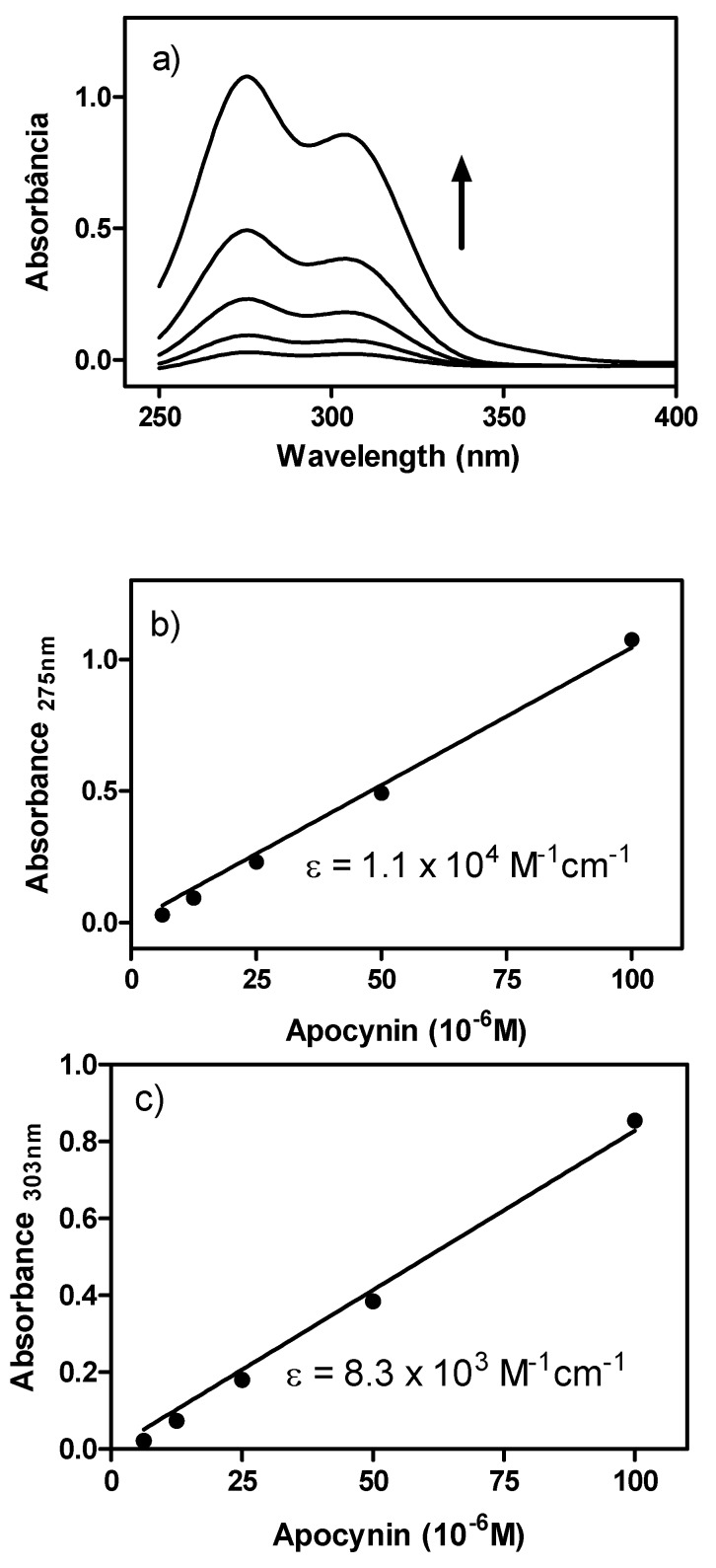
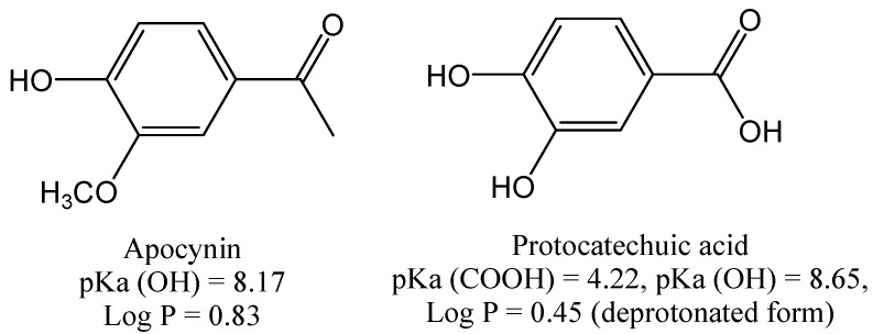
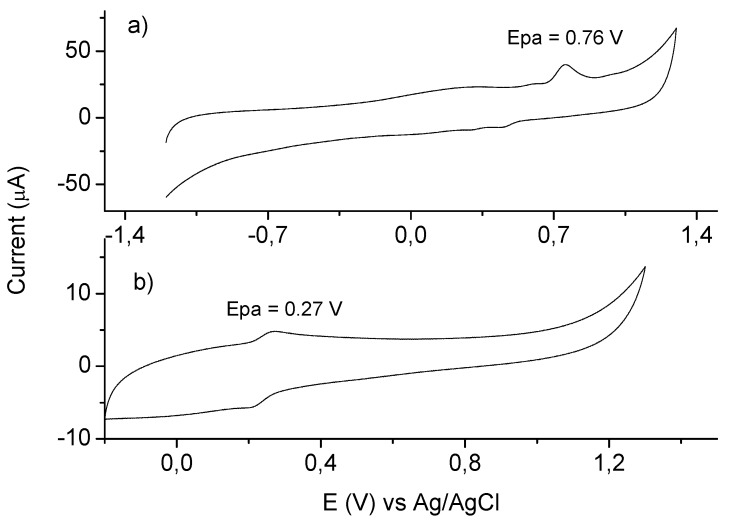
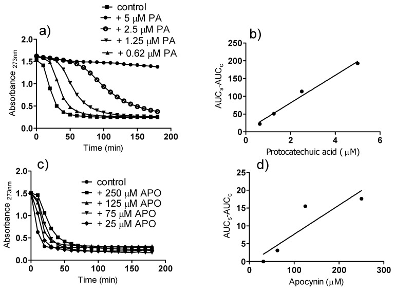
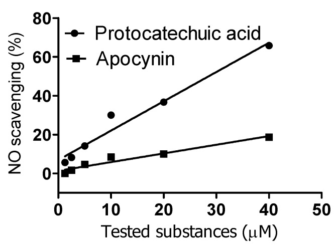
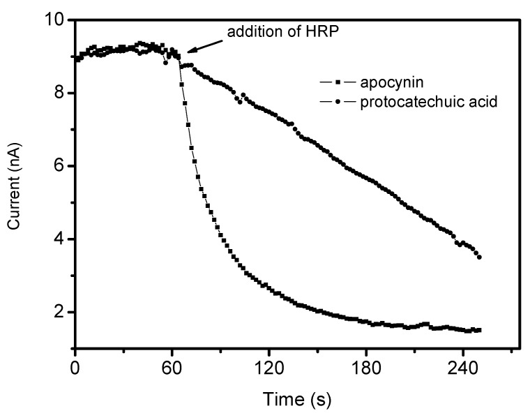
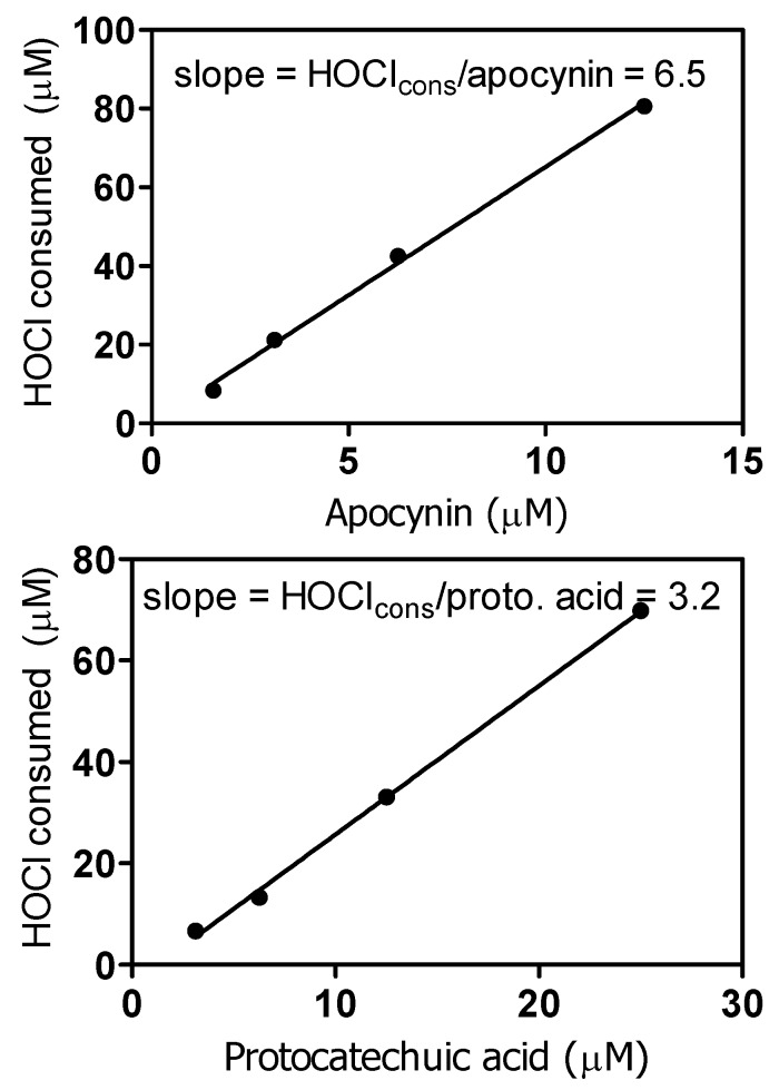
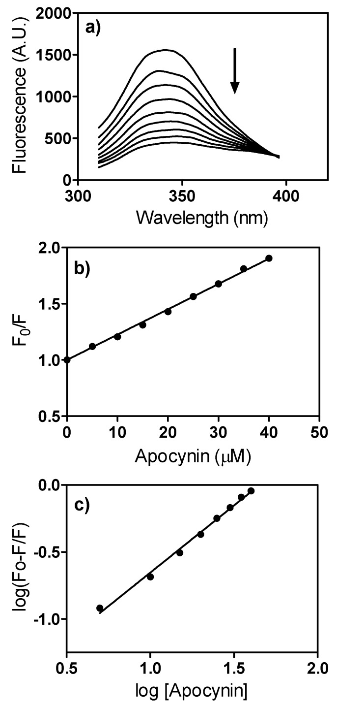
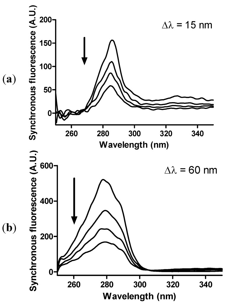
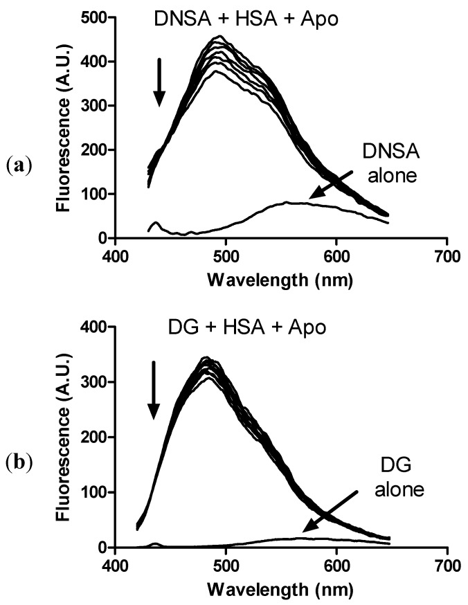
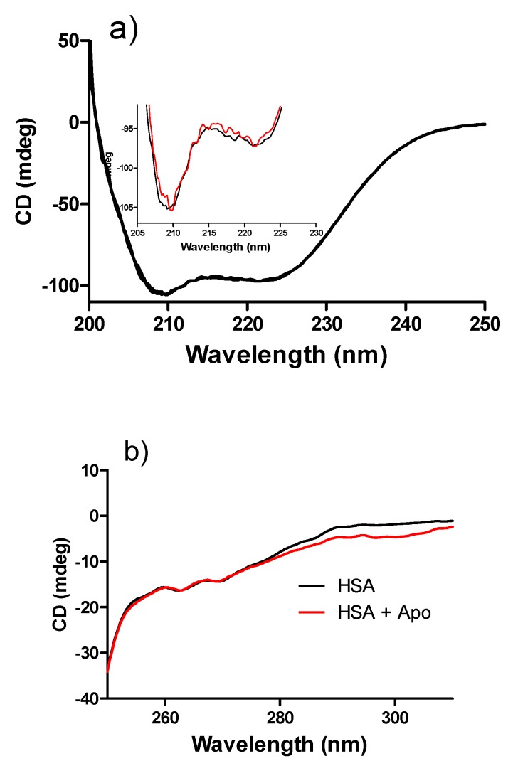
Apocynin is the most employed inhibitor of NADPH oxidase (NOX), a multienzymatic complex capable of catalyzing the one-electron reduction of molecular oxygen to the superoxide anion. Despite controversies about its selectivity, apocynin has been used as one of the most promising drugs in experimental models of inflammatory and neurodegenerative diseases. Here, we aimed to study the chemical and biophysical properties of apocynin. The oxidation potential was determined by cyclic voltammetry (Epa = 0.76V), the hydrophobicity index was calculated (logP = 0.83) and the molar absorption coefficient was determined (e275nm = 1.1 × 10⁴ M−1 cm−1). Apocynin was a weak free radical scavenger (as measured using the DPPH, peroxyl radical and nitric oxide assays) when compared to protocatechuic acid, used here as a reference antioxidant. On the other hand, apocynin was more effective than protocatechuic acid as scavenger of the non-radical species hypochlorous acid. Apocynin reacted promptly with the non-radical reactive species H2O2 only in the presence of peroxidase. This finding is relevant, since it represents a new pathway for depleting H2O2 in cellular experimental models, besides the direct inhibition of NADPH oxidase. This could be relevant for its application as an inhibitor of NOX4, since this isoform produces H2O2 and not superoxide anion. The binding parameters calculated by fluorescence quenching showed that apocynin binds to human serum albumin (HSA) with a binding affinity of 2.19 × 10⁴ M−1. The association did not alter the secondary and tertiary structure of HSA, as verified by synchronous fluorescence and circular dichroism. The displacement of fluorescent probes suggested that apocynin binds to site I and site II of HSA. Considering the current biomedical applications of this phytochemical, the dissemination of these chemical and biophysical properties can be very helpful for scientists and physicians interested in the use of apocynin.
Overview
The claims and statistics of this paper haven’t been checked yet.
Run the check — free, no account needed. It reads the open-access text and checks the sources it cites.
Integrity signals
Registry
Cites
Statistics
Figures
Verification
Claims the paper makes
The claims and statistics of this paper haven’t been checked yet.
Run the check — free, no account needed. It reads the open-access text and checks the sources it cites.
Claims other papers make about this one
Each is checked against this paper’s own text: does it say what the citing paper says it does?
In the citing paper
“Finally, it must be noted that arginases play an important role in the regulation of l-arginine levels (144, 259, 734, 804).”
From Sources of Vascular Nitric Oxide and Reactive Oxygen Species and Their Regulation (2019) · cites this paper as [734]
Self-check
Percentages, sums and reported differences re-computed from the paper’s own numbers.
The self-check needs the paper’s full text.
Citations
Cites
Principles of Fluorescence Spectroscopy
Lakowicz, J. R. (1999) · Springer eBooks · doi:10.1007/978-1-4757-3061-6 · Cited by 26,948
Use of a Free Radical Method to Evaluate Antioxidant Activity
Brand-Williams, W., Cuvelier, M. E., & Berset, C. (1995) · LWT - Food Science and Technology · doi:10.1016/s0023-6438(95)80008-5 · Cited by 25,994
Principles of Fluorescence Spectroscopy
Lakowicz, J. R. (2006) · Springer eBooks · doi:10.1007/978-0-387-46312-4 · Cited by 19,795
How to measure and predict the molar absorption coefficient of a protein
Pace, C., Vajdos, F., Fee, L. R., Grimsley, G., & Gray, T. (1995) · Protein Science · doi:10.1002/pro.5560041120 · Cited by 3,924
Quenching of fluorescence by oxygen. Probe for structural fluctuations in macromolecules
Lakowicz, J. R., & Weber, G. (1973) · Biochemistry · doi:10.1021/bi00745a020 · Cited by 2,795
Human serum albumin: From bench to bedside
Fanali, G., di Masi, A., Trezza, V., Marino, M., Fasano, M., & Ascenzi, P. (2011) · Molecular Aspects of Medicine · doi:10.1016/j.mam.2011.12.002 · Cited by 2,008
FLUORESCENCE AND THE LOCATION OF TRYPTOPHAN RESIDUES IN PROTEIN MOLECULES
Burstein, E. A., Vedenkina, N. S., & Mn, I. (1973) · Photochemistry and Photobiology · doi:10.1111/j.1751-1097.1973.tb06422.x · Cited by 1,187
Ghose, A. K., & Crippen, G. M. (1987) · Journal of Chemical Information and Computer Sciences · doi:10.1021/ci00053a005 · Cited by 804
Apocynin Is Not an Inhibitor of Vascular NADPH Oxidases but an Antioxidant
Heumüller, S., Wind, S., Barbosa-Sicard, E., Schmidt, H. H. H. W., Busse, R., Schröder, K., & Brandes, R. P. (2008) · Hypertension · doi:10.1161/hypertensionaha.107.100214 · Cited by 735
Brown, R., James, C., Henderson, L. A., & Macefield, V. G. (2012) · Frontiers in Physiology · doi:10.3389/fphys.2012.00394 · Cited by 730
Cited by
Sorted by citations
The Immunomodulatory and Anti-Inflammatory Role of Polyphenols
Yahfoufi, N., Alsadi, N., Jambi, M., & Matar, C. (2018) · Nutrients · doi:10.3390/nu10111618 · Cited by 1,737
Sources of Vascular Nitric Oxide and Reactive Oxygen Species and Their Regulation
Tejero, J., Shiva, S., & Gladwin, M. T. (2019) · Physiological Reviews · doi:10.1152/physrev.00036.2017 · Cited by 541
NADPH oxidase in brain injury and neurodegenerative disorders
Ma, M. W., Wang, J., Zhang, Q., Wang, R., Dhandapani, K. M., Vadlamudi, R. K., & Brann, D. W. (2017) · Molecular Neurodegeneration · doi:10.1186/s13024-017-0150-7 · Cited by 432
Endothelial NADPH oxidases: which NOX to target in vascular disease?
Drummond, G. R., & Sobey, C. G. (2014) · Trends in Endocrinology & Metabolism · doi:10.1016/j.tem.2014.06.012 · Cited by 315
Rochael, N. C., Guimarães-Costa, A. B., Nascimento, M. T. C., DeSouza-Vieira, T. S., Oliveira, M., Souza, L. F. G. E., de Oliveira, M. F., & Saraiva, E. M. (2015) · Scientific Reports · doi:10.1038/srep18302 · Cited by 252
Metadata
- Authors
- Petrônio, M., Zeraik, M., Fonseca, L., & Ximenes, V.
- Affiliations
- Universidade Estadual Paulista (Unesp) (BR)
- Corresponding author
- Ximenes, Valdecir
- Venue
- Molecules
- Publisher
- MDPI AG
- Published
- 2013-03-01
- Volume
- 18
- Issue
- 3
- Pages
- 2821-2839
- ISSN
- 1420-3049 (ISSN-L)
- PMC
- PMC6269682
- OpenAlex
- W2036509107
- Cited by
- 149
- References
- 49
- Citations by year
- 2026: 8 · 2025: 12 · 2024: 8 · 2023: 11 · 2022: 14 · 2021: 14 · 2020: 9 · 2019: 12 · 2018: 11 · 2017: 11 · 2016: 6 · 2015: 12 · 2014: 14 · 2013: 7
- FWCI
- 5.91
- Type
- journal article
- Language
- en
- Open access
- gold
- doi.org
- Licence
- Locations
- Molecules (publishedVersion, cc-by)
- Europe PMC (PubMed Central) (publishedVersion, cc-by)
- PubMed Central (publishedVersion, cc-by)
- PubMed (publishedVersion)
- Acervo Digital da Universidade Estadual Paulista (Universidade Estadual Paulista) (submittedVersion, other-oa)
- Americanae (AECID Library) (submittedVersion, other-oa)
- DOAJ (DOAJ: Directory of Open Access Journals) (submittedVersion, cc-by-sa)
- LA Referencia (Red Federada de Repositorios Institucionales de Publicaciones Científicas) (submittedVersion, other-oa)
- Funders
- Fundação de Amparo à Pesquisa do Estado de São Paulo
- Conselho Nacional de Desenvolvimento Científico e Tecnológico
- Topics
- Physical Sciences › Chemistry › Organic Chemistry › Free Radicals and Antioxidants
- Life Sciences › Pharmacology, Toxicology and Pharmaceutics › Toxicology › Bioactive Compounds and Antitumor Agents
- Life Sciences › Biochemistry, Genetics and Molecular Biology › Molecular Biology › Redox biology and oxidative stress
- Keywords
- apocynin, NADPH oxidase inhibitors, NOX4, superoxide, hydrogen peroxide scavenging, free radical scavenging, hypochlorous acid, protocatechuic acid, human serum albumin binding, fluorescence quenching, antioxidant activity, physicochemical properties
- MeSH terms
- Acetophenones, Enzyme Inhibitors, Hydrogen Peroxide, Hydrogen-Ion Concentration, Hypochlorous Acid, Kinetics, Oxidation-Reduction, Free Radical Scavengers, NADPH Oxidases, Hydrophobic and Hydrophilic Interactions
About these checks
Registry, retraction, Cites and statistics results are computed, not judged, with one exception: a printed reference that no index lists is read once by a model before it counts as a citation error. Claims are extracted by a model and kept only when their quote appears verbatim in the text; each cited source is then checked, with a model’s help, for whether it says what the claim relies on. Those verdicts can be wrong — read the claim’s quote and the cited source before relying on one.
Statistics and arithmetic are recomputed, not judged.
Confidence bands are shown only for fields calibrated on our annotated test set. None is calibrated yet, so no band is shown.
What counts as a claim ›How we measure confidence ›How we check this ›
Are you an author, or do you see a problem with this page? Request a review
