

The DOI matches its Crossref record; 304 of the 305 references it cites were found in a registry or an index.
Pineal Calcification, Melatonin Production, Aging, Associated Health Consequences and Rejuvenation of the Pineal Gland
Open accessAbstract
The pineal gland is a unique organ that synthesizes melatonin as the signaling molecule of natural photoperiodic environment and as a potent neuronal protective antioxidant. An intact and functional pineal gland is necessary for preserving optimal human health. Unfortunately, this gland has the highest calcification rate among all organs and tissues of the human body. Pineal calcification jeopardizes melatonin’s synthetic capacity and is associated with a variety of neuronal diseases. In the current review, we summarized the potential mechanisms of how this process may occur under pathological conditions or during aging. We hypothesized that pineal calcification is an active process and resembles in some respects of bone formation. The mesenchymal stem cells and melatonin participate in this process. Finally, we suggest that preservation of pineal health can be achieved by retarding its premature calcification or even rejuvenating the calcified gland.
Overview
The claims and statistics of this paper haven’t been checked yet.
Run the check — free, no account needed. It reads the open-access text and checks the sources it cites.
Integrity signals
Registry
Cites
Statistics
Figures
Verification
Claims the paper makes
The claims and statistics of this paper haven’t been checked yet.
Run the check — free, no account needed. It reads the open-access text and checks the sources it cites.
Claims other papers make about this one
Each is checked against this paper’s own text: does it say what the citing paper says it does?
In the citing paper
“It is present in nearly all bodily fluids: cerebrospinal fluid, saliva, bile, synovial fluid, amniotic fluid, urine, feces, semen, and breast milk [16,19,20].”
From Is Melatonin the “Next Vitamin D”?: A Review of Emerging Science, Clinical Uses, Safety, and Dietary Supplements (2022) · cites this paper as [16]
In the citing paper
“In fact, it has been suggested to be one of the most potent antioxidants because of its ability to scavenge up to 10 reactive oxygen (ROS) and nitrogen species (RNS) with its metabolites compared with most antioxidants, which may only be able to quench a few ROS [16,23,24].”
From Is Melatonin the “Next Vitamin D”?: A Review of Emerging Science, Clinical Uses, Safety, and Dietary Supplements (2022) · cites this paper as [16]
Self-check
Percentages, sums and reported differences re-computed from the paper’s own numbers.
The self-check needs the paper’s full text.
Citations
Cites
3 retracted
Zhou, H., Du, W., Li, Y., Chen, S., Hu, N., Ma, S., Wang, W., & Ren, J. T. (2017) · Journal of Pineal Research · doi:10.1111/jpi.12450 · Cited by 292
RetractedMortezaee, K., Khanlarkhani, N., Sabbaghziarani, F., Nekoonam, S., Majidpoor, J., Hosseini, A., Pasbakhsh, P., Kashani, I. R., & Zendedel, A. (2017) · Cell and Tissue Research · doi:10.1007/s00441-017-2604-1 · Cited by 70
RetractedKashani, I. R., Rajabi, Z., Akbari, M., Hassanzadeh, G., Mohseni, A., Eramsadati, M. K., Rafiee, K., Beyer, C., Kipp, M., & Zendedel, A. (2014) · Experimental Brain Research · doi:10.1007/s00221-014-3946-5 · Cited by 56
RetractedPineal Melatonin: Cell Biology of Its Synthesis and of Its Physiological Interactions*
REITER, R. J. (1991) · Endocrine Reviews · doi:10.1210/edrv-12-2-151 · Cited by 2,192
Self-citationMelatonin as an antioxidant: under promises but over delivers
Reiter, R. J., Mayo, J. C., Tan, D. X., Sainz, R. M., Alatorre-Jiménez, M. A., & Qin, L. (2016) · Journal of Pineal Research · doi:10.1111/jpi.12360 · Cited by 1,777
Self-citationISOLATION OF MELATONIN, THE PINEAL GLAND FACTOR THAT LIGHTENS MELANOCYTES 1
Lerner, A. B., Case, J. D., TAKAHASHI, Y., Lee, T. H., & Mori, W. (1958) · Journal of the American Chemical Society · doi:10.1021/ja01543a060 · Cited by 1,759
Melatonin : a potent, endogenous hydroxyl radical scavenger
Tan, D. (1993) · Endocrine Journal · Cited by 1,659
Self-citationExtrapineal melatonin: sources, regulation, and potential functions
Acuña-Castroviejo, D., Escames, G., Venegas, C., Díaz-Casado, M. E., Lima‐Cabello, E., López, L. C., Rosales‐Corral, S., Tan, D., & Reiter, R. J. (2014) · Cellular and Molecular Life Sciences · doi:10.1007/s00018-014-1579-2 · Cited by 1,133
Self-citationKhan, S. R., Pearle, M. S., Robertson, W., Gambaro, G., Canales, B. K., Doizi, S., Traxer, O., & Tiselius, H. (2016) · Nature Reviews Disease Primers · doi:10.1038/nrdp.2016.8 · Cited by 1,103
Hattori, A., Migitaka, H., Iigo, M., Itoh, M., Yamamoto, K., Ohtani‐Kaneko, R., Hara, M., Suzuki, T., & Reiter, R. J. (1995) · PubMed · Cited by 827
Self-citation
Research works printed but not linked: 4
- Commentz, 1986, Pineal calcification does not affect melatonin production, J. Neural Transm. Suppl., 21, 481
- Welsh, 1985, Pineal calcification: Structural and functional aspects, Pineal Res. Rev., 3, 41
- Welsh, 1977, Effects of superior cervical gangalinectomy, constant light and bliding on the gerbil pineal gland: An ultrastructural analysis, Anat. Rec., 187, 746
- Krstic, 1976, Human pineal acervuli: Ultrastructural and X-ray energy dispensive microprobe analysis, Experientia, 32, 800
Cited by
Sorted by citations
Principles of fluoride toxicity and the cellular response: a review
Johnston, N. R., & Strobel, S. A. (2020) · Archives of Toxicology · doi:10.1007/s00204-020-02687-5 · Cited by 356
Biological Rhythm and Chronotype: New Perspectives in Health
Montaruli, A., Castelli, L., Mulè, A., Scurati, R., Esposito, F., Galasso, L., & Roveda, E. (2021) · Biomolecules · doi:10.3390/biom11040487 · Cited by 400
Melatonin: Awakening the Defense Mechanisms during Plant Oxidative Stress
Khan, A., Numan, M., Khan, A. L., Lee, I., Imran, M., Asaf, S., & Al‐Harrasi, A. (2020) · Plants · doi:10.3390/plants9040407 · Cited by 256
Melatonin Mediates Enhancement of Stress Tolerance in Plants
Debnath, B., Islam, W., Li, M., Sun, Y., Lu, X., Mitra, S., Hussain, M., Liu, S., & Qiu, D. (2019) · International Journal of Molecular Sciences · doi:10.3390/ijms20051040 · Cited by 250
Melatonin and Its Effects on Plant Systems
Sharif, R., Xie, C., Zhang, H., Arnao, M. B., Ali, M., Ali, Q., Muhammad, I., Shalmani, A., Nawaz, M. A., Chen, P., et al. (2018) · Molecules · doi:10.3390/molecules23092352 · Cited by 238
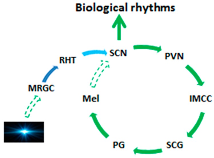
“The pineal gland is an organ present in animals’ bodies, responsible for the production of melatonin in order to control the behavior of the body toward changing photoperiod and also serve as a neuronal protective antioxidant [3].”
Background
Metadata
- Authors
- Tan, D., Xu, B., Zhou, X., & Reiter, R.
- Affiliations
- The University of Texas at San Antonio Health Science Center (US)
- Corresponding author
- Tan, Dun
- Reiter, Russel
- Venue
- Molecules
- Publisher
- MDPI AG
- Published
- 2018-01-31
- Volume
- 23
- Issue
- 2
- Pages
- 301
- ISSN
- 1420-3049 (ISSN-L)
- PMC
- PMC6017004
- OpenAlex
- W2790796192
- Cited by
- 220
- References
- 305
- Citations by year
- 2026: 17 · 2025: 30 · 2024: 30 · 2023: 33 · 2022: 30 · 2021: 32 · 2020: 24 · 2019: 19 · 2018: 5
- FWCI
- 2.80
- Type
- journal article
- Language
- en
- Open access
- gold
- doi.org
- Licence
- Locations
- Molecules (publishedVersion, cc-by)
- Europe PMC (PubMed Central) (publishedVersion, cc-by)
- PubMed Central (publishedVersion, cc-by)
- PubMed (publishedVersion)
- DOAJ (DOAJ: Directory of Open Access Journals) (submittedVersion, cc-by-sa)
- Topics
- Life Sciences › Neuroscience › Endocrine and Autonomic Systems › Circadian rhythm and melatonin
- Life Sciences › Biochemistry, Genetics and Molecular Biology › Cell Biology › melanin and skin pigmentation
- Health Sciences › Medicine › Nephrology › Parathyroid Disorders and Treatments
- Keywords
- pineal gland calcification, melatonin, pineal gland, aging, neuroprotection, antioxidant signaling, photoperiod, mesenchymal stem cells, neuronal diseases
- MeSH terms
- Aging, Animals, Calcinosis, Humans, Melatonin, Pineal Gland, Rejuvenation
About these checks
Registry, retraction, Cites and statistics results are computed, not judged, with one exception: a printed reference that no index lists is read once by a model before it counts as a citation error. Claims are extracted by a model and kept only when their quote appears verbatim in the text; each cited source is then checked, with a model’s help, for whether it says what the claim relies on. Those verdicts can be wrong — read the claim’s quote and the cited source before relying on one.
Statistics and arithmetic are recomputed, not judged.
Confidence bands are shown only for fields calibrated on our annotated test set. None is calibrated yet, so no band is shown.
What counts as a claim ›How we measure confidence ›How we check this ›
Are you an author, or do you see a problem with this page? Request a review
